. 2013 Jan;21(1):210-6.
doi: 10.1038/mt.2012.236. Epub 2012 Nov 20.

Polyethylenimine-modified pluronics (PCMs) improve morpholino oligomer delivery in cell culture and dystrophic mdx mice

Affiliations

Polyethylenimine-modified pluronics (PCMs) improve morpholino oligomer delivery in cell culture and dystrophic mdx mice

Mingxing Wang et al. Mol Ther. 2013 Jan.

Abstract

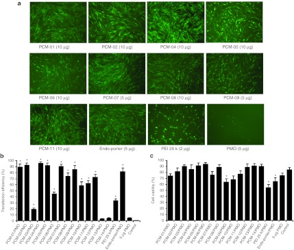
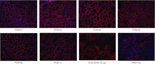
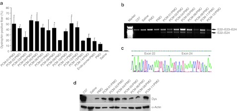
We investigated a series of small-sized polyethylenimine (PEI, 0.8/1.2 k)-conjugated pluronic copolymers (PCMs) for their potential to enhance delivery of an antisense phosphorodiamidate morpholino oligomer (PMO) in vitro and in dystrophic mdx mice. PCM polymers containing pluronics of molecular weight (Mw) ranging 2-6 k, with hydrophilic-lipophilic balance (HLB) 7-23, significantly enhanced PMO-induced exon-skipping in a green fluorescent protein (GFP) reporter-based myoblast culture system. Application of optimized formulations of PCMs with PMO targeted to dystrophin exon 23 demonstrated a significant increase in exon-skipping efficiency in dystrophic mdx mice. Consistent with our observations in vitro, optimization of molecular size and the HLB of pluronics are important factors for PCMs to achieve enhanced PMO delivery in vivo. Observed cytotoxicity of the PCMs was lower than Endo-porter and PEI 25 k. Tissue toxicity of PCMs in muscle was not clearly detected with the concentrations used, indicating the potential of the PCMs as effective and safe PMO carriers for treating diseases such as muscular dystrophy.


Figures

Figure 1
Figure 1
Cell viability of C2C12E50 cell line after treatment with polymers at three doses (4, 10, 20 µg/ml from left to right for each polymer). Cells were seeded in 96-well plates at an initial density of 1 × 10⁴ cells/well in 0.2 ml growth media. Cell viability was determined by MTS assay. n = 3, two-tailed t-test, *P ≤ 0.05 compared with untreated cell as control. MTS, [3-(4,5-dimethylthiazol-2-yl)-5-(3-carboxymethoxyphenyl)- 2-(4-sulfophenyl)-2H-tetrazolium] PCM, pluronic copolymer; PEI, polyethylenimine.
Figure 2
Figure 2
Dose-dependent PMO delivery in C2C12E50 cells. Polymers were used at the doses of 2, 5, 10, 20 µg together with 5 µg PMOE50 in 500 µl medium. Original magnification, ×100. PCM, pluronic copolymer; PMO, phosphorodiamidate morpholino oligomer.
Figure 3
Figure 3
Delivery efficiency of PMOE50 and toxicity of PCMs in C2C12E50 cell line determined by fluorescence microscope and FACS analysis. (a) Representative fluorescence images of PMOE50 induced exon skipping. PMOE50 (5 µg) formulated with polymers (5, 10 µg in 500 µl medium). Original magnification, ×100. (b) Transfection efficiency of PMOE50 (5 µg) formulated with 10 µg PCM (n = 3, two-tailed t-test, *P ≤ 0.05 compared with 5 µg PMO only). (c) Cell viability was tested for PCM (10 µg), Endo-porter (5 µg) or PEI 25 k (2 µg) formulated with PMOE50 (n = 3, two-tailed t-test, *P ≤ 0.05 compared with untreated cell as control). FACS, fluorescence-activated cell sorting; PCM, pluronic copolymer; PEI, polyethylenimine; PMO, phosphorodiamidate morpholino oligomer.
Figure 4
Figure 4
GFP expression induced by PMOE50 (5 µg) formulated with amphiphilic polymers PCM-11, PCM-12 and hydrophilic polymers PCM-13, PCM-14 (10 µg) in C2C12E50 cells. Original magnification, ×100. GFP, green fluorescent protein; PCM, pluronic copolymer; PMO, phosphorodiamidate morpholino oligomer.
Figure 5
Figure 5
Restoration of dystrophin in TA muscles of mdx mice (age 4–5 weeks) 2 weeks after i.m. injection. Dystrophin was detected by immunohistochemistry with rabbit polyclonal antibody P7 against dystrophin. Blue nuclear staining with DAPI. Muscles treated with PMOE23 (2 µg) only and Endo-porter (2 µg)-formulated PMOE23 (2 µg) were used as controls. All other samples were from muscles treated with 5 µg polymer and 2 µg PMOE23 in 40 µl saline. Original magnification, ×100. DAPI, 4′,6-diamidino-2-phenylindole; i.m., intramuscular; PCM, pluronic copolymer; PMO, phosphorodiamidate morpholino oligomer; TA, tibialis anterior.
Figure 6
Figure 6
Dystrophin expression in TA muscles of mdx mice after 2 weeks treatment. (a) The percentage of dystrophin-positive fibers in muscles treated with 2 µg PMOE23 with and without polymers (5 µg). The maximum numbers of dystrophin-positive fibers were counted in a single cross-section (n = 5, two-tailed t-test, *P ≤ 0.05 compared with 2 µg PMO). (b) Detection of exon 23 skipping by RT-PCR and sequence confirmation. Total RNA of 100 ng from each sample was used for amplification of dystrophin mRNA from exon 20 to exon 26. The upper 1,093-bp bands (indicated by E22+E23+E24) correspond to the normal mRNA, and the lower 880-bp bands (indicated by E22+E24) correspond to the mRNA with exon E23 skipped. (c) Sequencing of the 880-bp RT-PCR product confirmed the skipping of the exon 23. (d) Western blots demonstrate the expression of dystrophin protein. Dys, dystrophin detected with monoclonal antibody Dys 1. α-Actin was used as the loading control. PCM, pluronic copolymer; PMO, phosphorodiamidate morpholino oligomer; RT-PCR, reverse transcription-PCR.
Figure 7
Figure 7
Dystrophin expression in diaphragm and heart muscles of mdx mice (aged 4–5 weeks) 2 weeks after systemic administration of PMO with polymers. Each mouse was injected wit+h 2 mg PMOE23 with and without polymer (0.4 mg). Left panel, immunohistochemistry with antibody P7 for the detection of dystrophin. Original magnification, ×100. Right panel, percentage of dystrophin-positive fibers in three muscle tissues (n = 5, two-tailed t-test, *P ≤ 0.05 compared with 2 mg PMO). PCM, pluronic copolymer; PMO, phosphorodiamidate morpholino oligomer; TA, tibialis anterior.

References

    1. Hoffman EP, Brown RH., Jr, and, Kunkel LM. Dystrophin: the protein product of the Duchenne muscular dystrophy locus. Cell. 1987;51:919–928. - PubMed
    1. Koenig M, Beggs AH, Moyer M, Scherpf S, Heindrich K, Bettecken T.et al. (1989The molecular basis for Duchenne versus Becker muscular dystrophy: correlation of severity with type of deletion Am J Hum Genet 45498–506. - PMC - PubMed
    1. Wu B, Moulton HM, Iversen PL, Jiang J, Li J, Li J.et al. (2008Effective rescue of dystrophin improves cardiac function in dystrophin-deficient mice by a modified morpholino oligomer Proc Natl Acad Sci USA 10514814–14819. - PMC - PubMed
    1. Yin H, Moulton HM, Seow Y, Boyd C, Boutilier J, Iverson P.et al. (2008Cell-penetrating peptide-conjugated antisense oligonucleotides restore systemic muscle and cardiac dystrophin expression and function Hum Mol Genet 173909–3918. - PMC - PubMed
    1. Wu B, Lu P, Benrashid E, Malik S, Ashar J, Doran TJ.et al. (2010Dose-dependent restoration of dystrophin expression in cardiac muscle of dystrophic mice by systemically delivered morpholino Gene Ther 17132–140. - PubMed

Publication types

LinkOut - more resources