. 2012 Dec 15;11(24):4570-8.
doi: 10.4161/cc.22843. Epub 2012 Nov 19.

Transcriptional regulation of thymine DNA glycosylase (TDG) by the tumor suppressor protein p53

Affiliations

Transcriptional regulation of thymine DNA glycosylase (TDG) by the tumor suppressor protein p53

Nathalia Meireles da Costa et al. Cell Cycle. .

Abstract

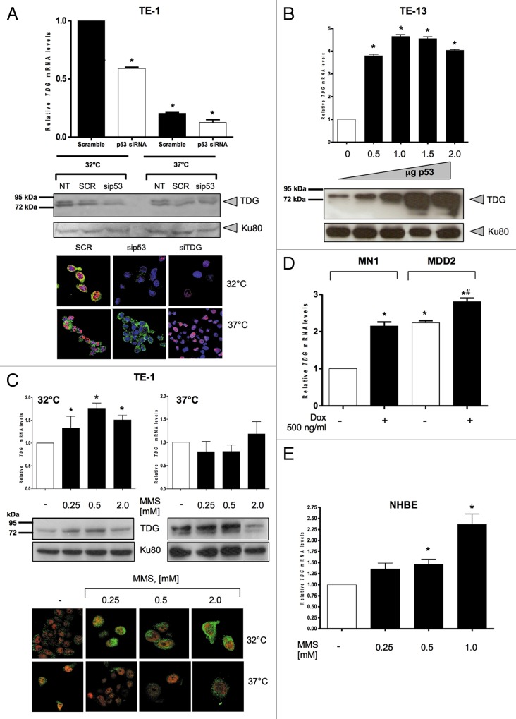
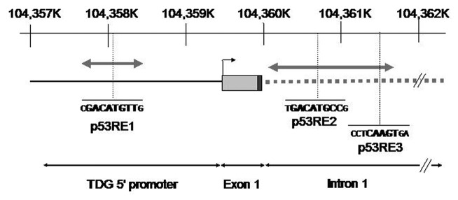
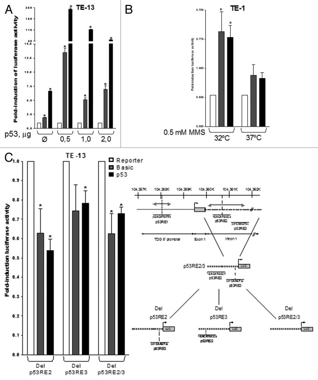
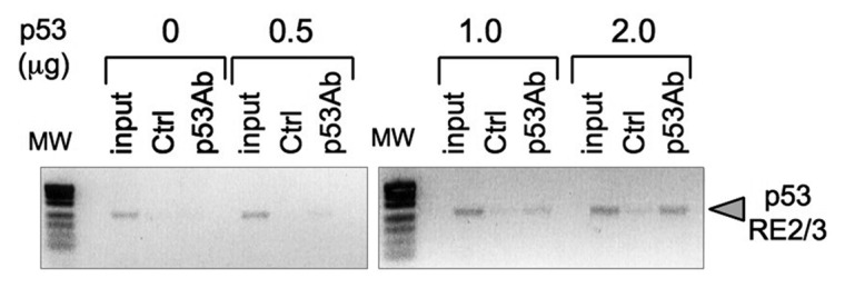
Thymine DNA glycosylase (TDG) belongs to the superfamily of uracil DNA glycosylases (UDG) and is the first enzyme in the base-excision repair pathway (BER) that removes thymine from G:T mismatches at CpG sites. This glycosylase activity has also been found to be critical for active demethylation of genes involved in embryonic development. Here we show that wild-type p53 transcriptionally regulates TDG expression. Chromatin immunoprecipitation (ChIP) and luciferase assays indicate that wild-type p53 binds to a domain of TDG promoter containing two p53 consensus response elements (p53RE) and activates its transcription. Next, we have used a panel of cell lines with different p53 status to demonstrate that TDG mRNA and protein expression levels are induced in a p53-dependent manner under different conditions. This panel includes isogenic breast and colorectal cancer cell lines with wild-type or inactive p53, esophageal squamous cell carcinoma cell lines lacking p53 or expressing a temperature-sensitive p53 mutant and normal human bronchial epithelial cells. Induction of TDG mRNA expression is accompanied by accumulation of TDG protein in both nucleus and cytoplasm, with nuclear re-localization occurring upon DNA damage in p53-competent, but not -incompetent, cells. These observations suggest a role for p53 activity in TDG nuclear translocation. Overall, our results show that TDG expression is directly regulated by p53, suggesting that loss of p53 function may affect processes mediated by TDG, thus negatively impacting on genetic and epigenetic stability.


Figures

None
Figure 1. Wild-type p53 regulates thymine DNA glycosylase (TDG) expression. (A) siRNA targeting p53 (p53 siRNA) or TDG (TDG siRNA) or control siRNA (Scramble, SCR) was transfected in TE-1 cells which express the temperature-sensitive p53 mutant, V272M. Cells maintained at 32°C (permissive temperature) present most of the p53 in an active form, and those maintained at 37°C (restrictive temperature) present most of the p53 in an inactive form. Upper panel: detection of TDG mRNA levels by reverse-transcription quantitative PCR (RT-qPCR). Middle panel: western blotting analysis of TDG, using Ku-80 as loading control. Lower panel: detection of TDG and p53 by confocal microscopy. sip53 and siTDG: siRNA to p53 and TDG, respectively. Green fluorescence, TDG; red fluorescence, p53; blue fluorescence, nucleus (ToPro). (B) The ESCC p53-null cell line TE-13 was transfected with either 0.5, 1.0, 1.5 or 2.0 μg of DNA of an expression vector for p53 protein (p53wt-pcDNA3) or with an empty vector (pCDNA3-empty), used as control (0). Upper panel: detection of TDG mRNA levels by RT-qPCR. Lower panel: western blotting analysis of TDG, using Ku-80 as loading control. (C) TE-1 cells, cultured at either 32°C or 37°C, were treated with 0.25, 0.5, 1.0 or 2.0 mM of MMS for 3 h or not (-). Upper panel: detection of TDG mRNA levels by RT-qPCR. Middle panel: western blotting analysis of TDG, using Ku-80 as loading control. Lower panel: detection of TDG and p53 by confocal microscopy. Green fluorescence, TDG; red fluorescence, p53. (D) The isogenic breast cancer cell lines, MN1 and MDD2, were treated with 500 ng/mL doxorubicin (Dox) for 24 h andTDG mRNA levels were detected by RT-qPCR. (E) The non-transformed bronchial cells, NHBE, were treated with 0.25, 0.5 or 1.0 mM of MMS for 3 h or not (-) and TDG mRNA expression levels were assessed by RT-qPCR. Stars indicate statistical significance: *p < 0.05 (Student’s t-test using with software GraphPad Prism 4, GraphPad Software Inc.).
None
Figure 2. Thymine DNA glycosylase promoter. Schematic representation of the position of putative p53 response elements (p53RE) in the 5′ regulatory region of TDG. The interval 104,357K‒104,362K on Chr 12p is represented (reference: GRCh37.p5). The position of TDG transcription initiation site and of exon 1 are shown. The proximal part of intron 1 is represented by a gray hatched bar. The motif p53RE1 is located about 1.4 kb upstream of transcription initiation site. The p53R2 and p53R3 motifs are located in intron 1. The size and position of fragments used for cloning in luciferase vectors are shown by gray double-arrowed bars. p53R1 fragment, 0.84 kb; p53R2/R3 fragment, 1.5 kb.
None
Figure 3. p53 transcriptional regulation of TDG. (A) TE-13 cells were co-transfected with TDG-luciferase reporters and either increasing amounts of the expression vector for p53 (p53wt-pcDNA3) or with 2 μg of the control (empty) vector pcDNA3 (Ø). (B) TE-1 cells cultured at either 32°C or 37°C were treated with 0.25, 0.5 or 1.0 mM of methyl methanesulfonate (MMS) for 3 h and transfected with TDG-luciferase reporters and the luciferase assays were performed. Basic, promoterless luciferase reporter (pGL3 basic); p53RE1, luciferase reporter under the control of the 838 bp segment of TDG promoter containing the first p53RE; p53RE2/3, luciferase reporter under the control of the 1.5 kb segment of TDG promoter containing the second and the third p53REs. (C) The TDG-luciferase construct containing p53RE2/p53RE3 had either p53RE2, p53RE3 or both deleted by site-directed mutagenesis (as schematically shown in right panel) and mutated plasmids were co-transfected with 2 μg of expression vector for p53 (p53wt-pcDNA3) in TE-13 cells and the luciferase assays performed. Del p53RE2, plasmids harboring the deletion of p53RE2; Del p53RE3, plasmids harboring the deletion of p53RE3; Del p53RE2/3, plasmids harboring the deletion of p53RE2 and 3. Stars indicate statistical significance: *p < 0.05 (Student’s t-test was performed with software GraphPad Prism 4, GraphPad Software Inc.).
None
Figure 4. Binding of p53 to TDG promoter region. TE-13 cells were transfected with increasing amounts p53 expression vector (p53wt-pcDNA3) as indicated. Cross-linked chromatin was immunoprecipited with anti-p53 antibody CM1 (p53Ab) or control antibody (Ctrl) and analyzed by polymerase chain reaction (PCR) using primers specific for TDG promoter region encompassing p53RE2/3. MW, molecular weight markers; input, non-precipitated cross-linked chromatin.

Similar articles

Cited by

References

    1. Krawczak M, Ball EV, Cooper DN. Neighboring-nucleotide effects on the rates of germ-line single-base-pair substitution in human genes. Am J Hum Genet. 1998;63:474–88. doi: 10.1086/301965. - DOI - PMC - PubMed
    1. Tomso DJ, Bell DA. Sequence context at human single nucleotide polymorphisms: overrepresentation of CpG dinucleotide at polymorphic sites and suppression of variation in CpG islands. J Mol Biol. 2003;327:303–8. doi: 10.1016/S0022-2836(03)00120-7. - DOI - PubMed
    1. Waters TR, Swann PF. Thymine-DNA glycosylase and G to A transition mutations at CpG sites. Mutat Res. 2000;462:137–47. doi: 10.1016/S1383-5742(00)00031-4. - DOI - PubMed
    1. Kumar S, Subramanian S. Mutation rates in mammalian genomes. Proc Natl Acad Sci USA. 2002;99:803–8. doi: 10.1073/pnas.022629899. - DOI - PMC - PubMed
    1. Wiebauer K, Jiricny J. In vitro correction of G.T mispairs to G.C pairs in nuclear extracts from human cells. Nature. 1989;339:234–6. doi: 10.1038/339234a0. - DOI - PubMed

Publication types

MeSH terms

Substances