

Separated at birth? The functional and molecular divergence of OLIG1 and OLIG2
- PMID: 23165259
- PMCID: PMC3733228
- DOI: 10.1038/nrn3386
Separated at birth? The functional and molecular divergence of OLIG1 and OLIG2
Abstract
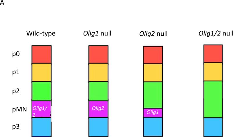
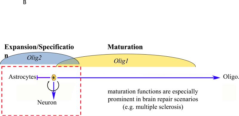
The basic helix-loop-helix transcription factors oligodendrocyte transcription factor 1 (OLIG1) and OLIG2 are structurally similar and, to a first approximation, coordinately expressed in the developing CNS and postnatal brain. Despite these similarities, it was apparent from early on after their discovery that OLIG1 and OLIG2 have non-overlapping developmental functions in patterning, neuron subtype specification and the formation of oligodendrocytes. Here, we summarize more recent insights into the separate roles of these transcription factors in the postnatal brain during repair processes and in neurological disease states, including multiple sclerosis and malignant glioma. We discuss how the unique functions of OLIG1 and OLIG2 may reflect their distinct genetic targets, co-regulator proteins and/or post-translational modifications.
Figures


Similar articles
-
Enhancing oligodendrocyte differentiation by transient transcription activation via DNA nanoparticle-mediated transfection.Acta Biomater. 2017 May;54:249-258. doi: 10.1016/j.actbio.2017.03.032. Epub 2017 Mar 23. Acta Biomater. 2017. PMID: 28344151 Free PMC article.
-
Olig transcription factors are expressed in oligodendrocyte and neuronal cells in human fetal CNS.J Neurosci. 2005 Nov 2;25(44):10064-73. doi: 10.1523/JNEUROSCI.2324-05.2005. J Neurosci. 2005. PMID: 16267213 Free PMC article.
-
Stage-specific deletion of Olig2 conveys opposing functions on differentiation and maturation of oligodendrocytes.J Neurosci. 2013 May 8;33(19):8454-62. doi: 10.1523/JNEUROSCI.2453-12.2013. J Neurosci. 2013. PMID: 23658182 Free PMC article.
-
Olig gene function in CNS development and disease.Glia. 2006 Jul;54(1):1-10. doi: 10.1002/glia.20273. Glia. 2006. PMID: 16652341 Review.
-
The Effects of the Olig Family on the Regulation of Spinal Cord Development and Regeneration.Neurochem Res. 2021 Nov;46(11):2776-2782. doi: 10.1007/s11064-021-03383-1. Epub 2021 Jul 6. Neurochem Res. 2021. PMID: 34228233 Review.
Cited by
-
Oligodendroglia and Myelin in Neurodegenerative Diseases: More Than Just Bystanders?Mol Neurobiol. 2016 Jul;53(5):3046-3062. doi: 10.1007/s12035-015-9205-3. Epub 2015 May 13. Mol Neurobiol. 2016. PMID: 25966971 Free PMC article. Review.
-
A Novel Three-Dimensional Culture Device Favors a Myelinating Morphology of Neural Stem Cell-Derived Oligodendrocytes.Front Cell Dev Biol. 2021 Oct 1;9:759982. doi: 10.3389/fcell.2021.759982. eCollection 2021. Front Cell Dev Biol. 2021. PMID: 34660610 Free PMC article.
-
Extrinsic Factors Driving Oligodendrocyte Lineage Cell Progression in CNS Development and Injury.Neurochem Res. 2020 Mar;45(3):630-642. doi: 10.1007/s11064-020-02967-7. Epub 2020 Jan 29. Neurochem Res. 2020. PMID: 31997102 Free PMC article. Review.
-
Combining Hypothermia and Oleuropein Subacutely Protects Subcortical White Matter in a Swine Model of Neonatal Hypoxic-Ischemic Encephalopathy.J Neuropathol Exp Neurol. 2021 Jan 20;80(2):182-198. doi: 10.1093/jnen/nlaa132. J Neuropathol Exp Neurol. 2021. PMID: 33212486 Free PMC article.
-
Identification of Qk as a Glial Precursor Cell Marker that Governs the Fate Specification of Neural Stem Cells to a Glial Cell Lineage.Stem Cell Reports. 2020 Oct 13;15(4):883-897. doi: 10.1016/j.stemcr.2020.08.010. Epub 2020 Sep 24. Stem Cell Reports. 2020. PMID: 32976762 Free PMC article.
References
-
- Charcot JM. Histologie de la sclerose en plaques. Gazette des hopitaux Paris. 1868;41:554–555.
-
- Marburg O. Die sogenannte akute multiple Sklerose. Jahrb Psychiatre. 1906;27:211–312.
-
- Prineas JW, Connell F. Remyelination in multiple sclerosis. Annals of neurology. 1979;5:22–31. - PubMed
-
- Lucchinetti C, et al. A quantitative analysis of oligodendrocytes in multiple sclerosis lesions. A study of 113 cases. Brain : a journal of neurology. 1999;122(Pt 12):2279–95. - PubMed
Publication types
MeSH terms
Substances
Grants and funding
LinkOut - more resources
Full Text Sources
Other Literature Sources
Molecular Biology Databases
