. 2013 Feb 1;374(1):198-209.
doi: 10.1016/j.ydbio.2012.11.007. Epub 2012 Nov 17.

Evolutionary conservation of Nkx2.5 autoregulation in the second heart field

Affiliations

Evolutionary conservation of Nkx2.5 autoregulation in the second heart field

Christopher D Clark et al. Dev Biol. .

Abstract

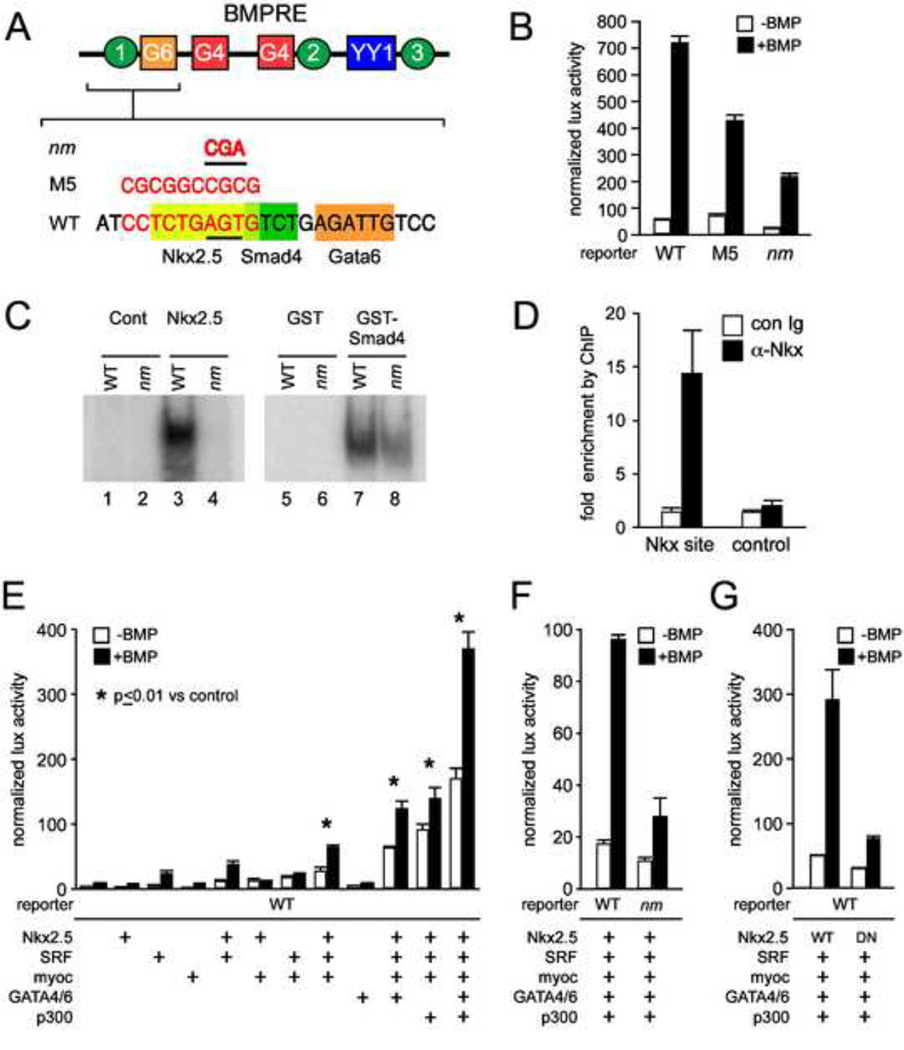
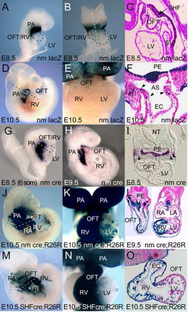
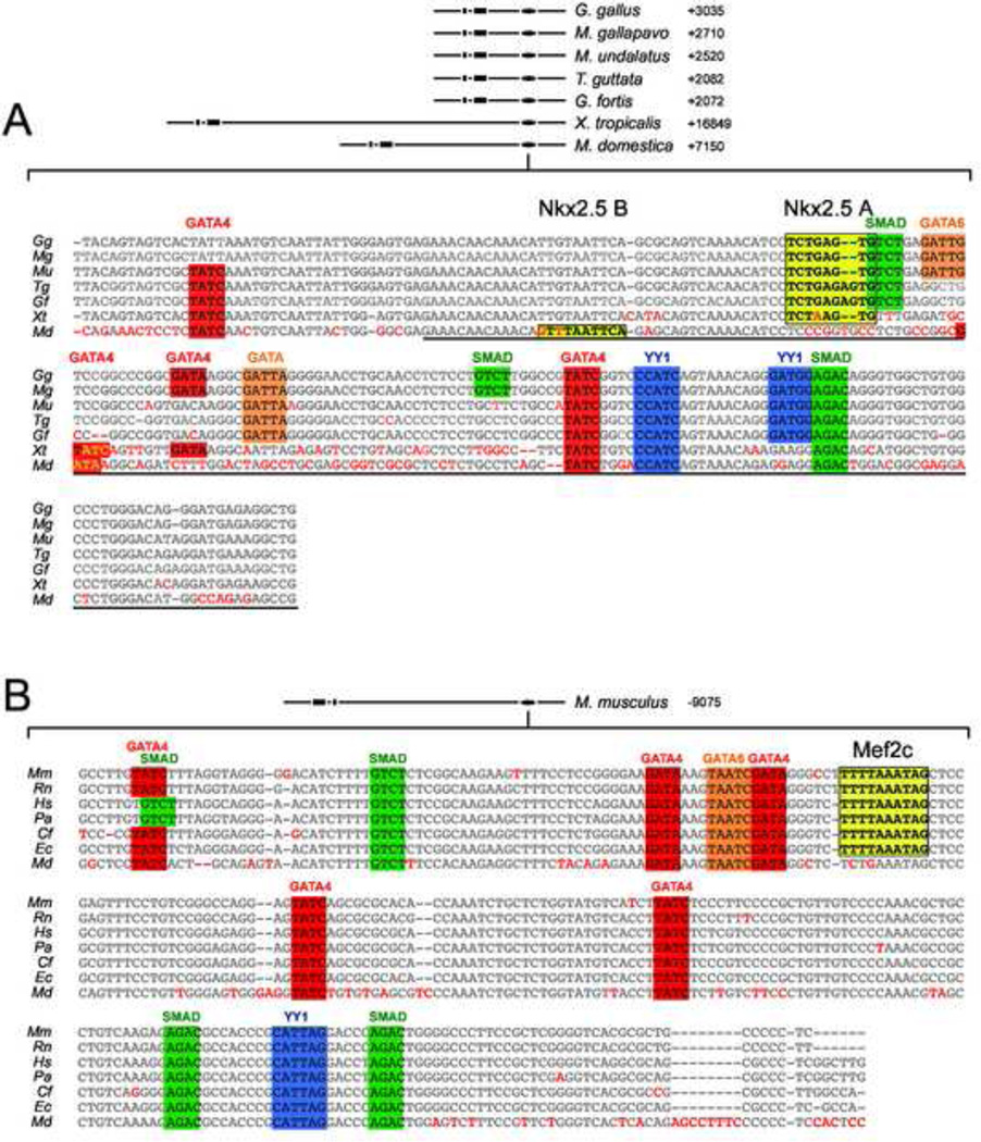
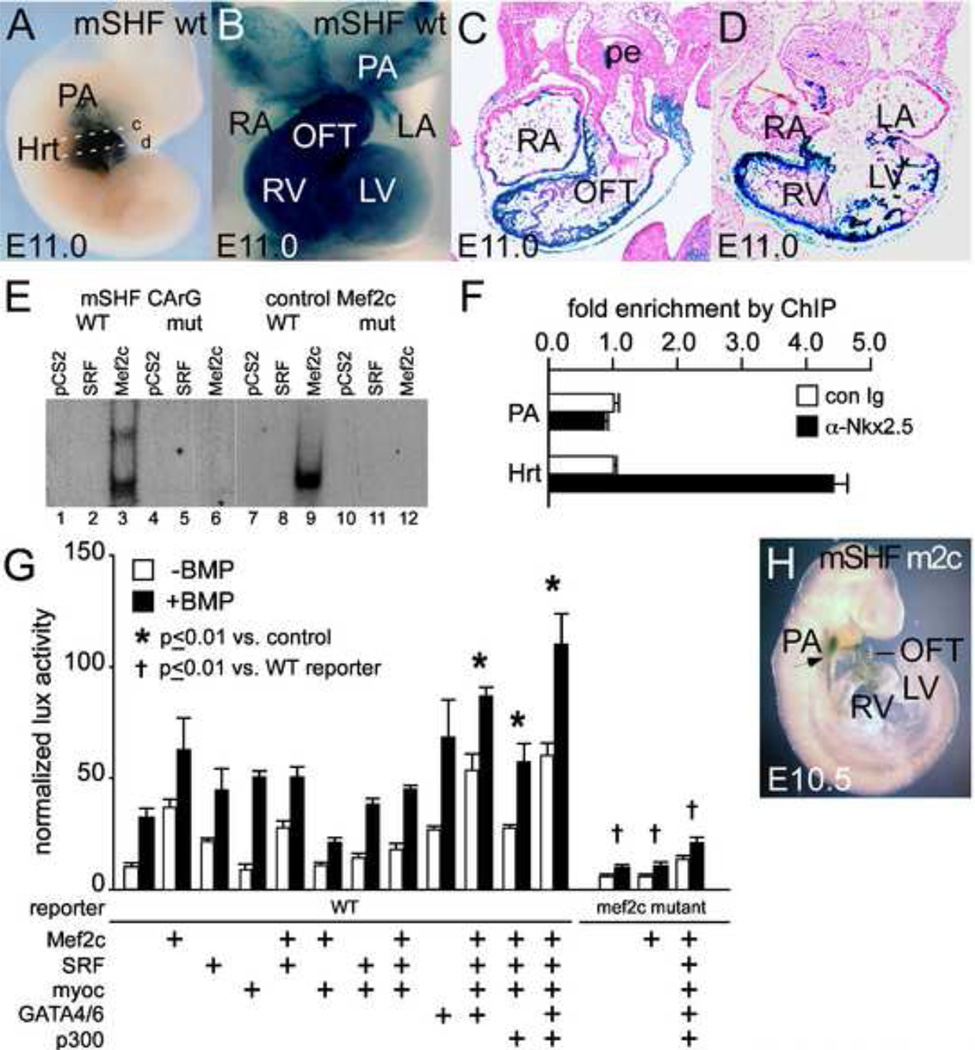
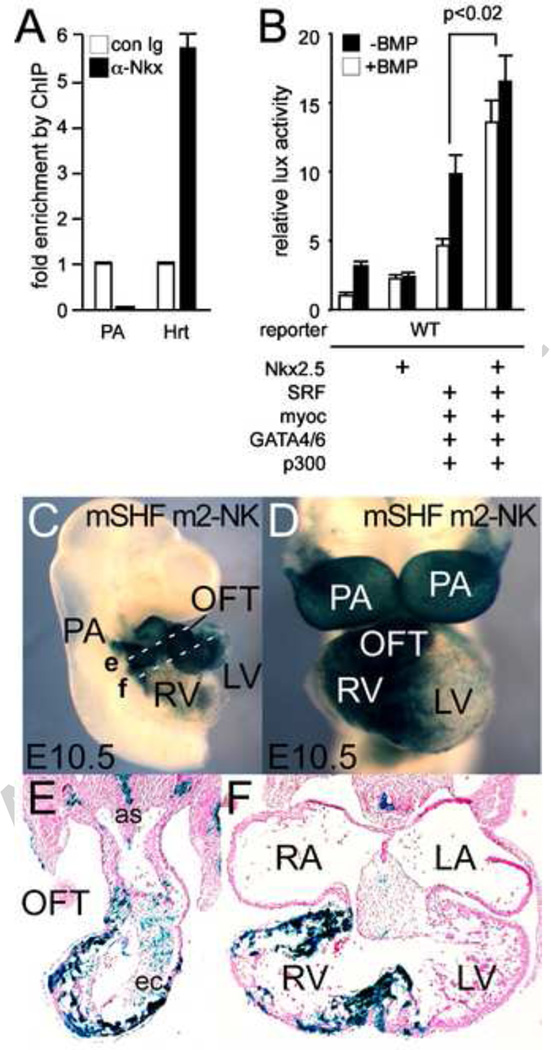
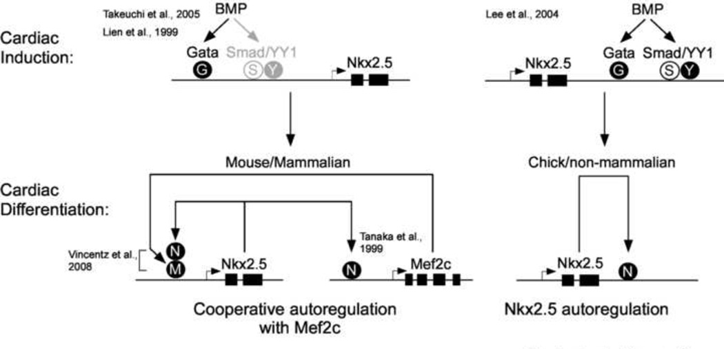
The cardiac homeobox gene Nkx2.5 plays a key and dosage-sensitive role in the differentiation of outflow tract and right ventricle from progenitors of the second heart field (SHF) and Nkx2.5 mutation is strongly associated with human outflow tract congenital heart disease (OFT CHD). Therefore defining the regulatory mechanisms controlling Nkx2.5 expression in SHF populations serves an important function in understanding the etiology of complex CHD. Through a comparative analysis of regulatory elements controlling SHF expression of Nkx2.5 in the chicken and mouse, we have found evidence that Nkx2.5 autoregulation is important for maintaining Nkx2.5 expression during SHF differentiation in both species. However the mechanism of Nkx2.5 maintenance differs between placental mammals and non-mammalian vertebrates: in chick Nkx2.5 binds directly to a genomic enhancer element that is required to maintain Nkx2.5 expression in the SHF. In addition, it is likely that this is true in other non-mammalian vertebrates given that they possess a similar genomic organization. By contrast, in placental mammals, Nkx2.5 autoregulation in the SHF functions indirectly through Mef2c. These data underscore a tight relationship in mammals between Nkx2.5 and Mef2c in SHF transcriptional regulation, and highlight the potential for evolutionary cis-regulatory analysis to identify core, conserved components of the gene networks controlling heart development.


Figures

Figure 1
Figure 1. Nkx2.5 directly regulates the CNkx2.5-SHF enhancer
A. A schematic of the 200 bp BMPRE of the chick Nkx2.5 gene depicting previously characterized Smad4 (green circles), GATA4/6 (red and orange squares) and YY1 (blue rectangle) binding elements is shown over an expanded view of nt 35–50 (Lee et al., 2004) which contains an Nkx2.5 binding consensus (yellow rectangle). The 10 bp M5 linker scanning mutation of BMPRE (nt 37–47) overlapping Nkx2.5 and Smad4 binding consensus sites is shown over the affected nucleotides in red, as is the smaller 3 bp mutation (designated Nkx2.5 mutant or nm) specifically targeting the Nkx2.5 binding element. B. BMP mediated reporter gene activation of wild-type Nkx2.5-lux-BMPRE reporter or reporter bearing the M5 or 3 bp nm mutations of the Nkx2.5 consensus site. The M5 mutation results in an approximately 50% decrease in BMP induced Nkx2.5-lux-BMPRE driven luciferase activity while the nm mutation results in a 4-fold decrease in luciferase activity. C. Data are shown for gel shifts performed with control (lanes 1 and 2), or Nkx2.5 cell extracts (lanes 3 and 4), or purified control GST (lanes 5 and 6) and GST-Smad4-MH1 domain proteins (lanes 7 and 8). Gel shifts were performed using double stranded oligonucleotides representing the wild-type BMPRE Nkx2.5 consensus (lanes 1, 3, 5, and 7) or the Nkx2.5 consensus bearing the nm mutation (lanes 2, 4, 6, 8). D. ChIP assay of Nkx2.5 binding to CNkx2.5-SHF-GFP reporter detects specific and selective enrichment (approx. 15 x) of amplicons from the BMPRE surrounding the Nkx2.5 binding consensus (Nkx site) using α-Nkx2.5 antibody vs. control IgG. No appreciable enrichment is observed of control GFP coding regions of the reporter (control). E. While addition of Nkx2.5 alone does not lead to a significant increase in CNkx2.5-SHF-lux luciferase activity in response to BMP (second column set from left), combinatorial addition of SRF, SRF cofactor Myocardin and cardiac GATAs 4 and 6 increases BMP activation of Nkx2.5-SHF-lux approximately 20-fold. The BMP response is further amplified 50–100 fold by addition of p300. F. CNkx2.5-SHF-lux response to Nkx2.5 and SRF, GATA4/6, SRF, Myocardin and p300 co-expression and BMP stimulation is reduced approximately 70% by site-specific mutation (nm) of the Nkx2.5 binding site on the CNkx2.5-SHF enhancer as compared to wild-type enhancer (WT). G. Multi-TF and BMP activation of the wild-type CNkx2.5-SHF-lux reporter is similarly reduced approximately 75% by use of a mutated non-DNA binding Nkx2.5 isoform (DN Nkx2.5) as compared to wild-type (WT Nkx2.5).
Figure 2
Figure 2. Nkx2.5 autoregulation is required for Nkx2.5-SHF enhancer expression in aortic pole myocytes but not SHF progenitors
A–F: Shown are representative lacZ expression patterns of stable lines transgenic for the CNkx2.5-SHF nm-lacZ reporter construct (nm lacZ). A–C: LacZ expression at E8.5 in PA mesoderm, ectoderm and endoderm is similar to that seen with the wild-type Nkx2.5 SHF enhancer construct (Lee et al., 2004) with staining observed in pharyngeal arch regions containing SHF progenitors, but absent in FHF-derived heart. D–F: While still expressed in PA regions, CNkx2.5-SHF nm reporter fails to maintain lacZ expression fails in differentiating OFT and RV of E10.5 transgenic embryos except in a few cells of the SHF (F, black arrowheads). G–I: Whole mount and sectioned whole mount in situ hybridization for cre mRNA expression in CNkx2.5-SHF nm-cre stable transgenic mouse line (nm cre). Cre is expressed in pharyngeal arch endoderm, mesoderm and ectoderm overlapping with SHF mesoderm (black arrowheads) at E8.5 (6 somite stage) (I) and retained primarily in pharyngeal populations at E10.5 (H). J–L: Lineage tracing using R26R ROSA lacZ reporter strain and CNkx2.5-SHF nm-cre driver at E10.5 confirms that cre expression from the CNkx2.5-SHF nm-cre transgene marks pharyngeal arch ectoderm, endoderm, SHF progenitors (black arrowheads) and OFT and right ventricular segments of the heart similar to wild-type CNkx2.5-SHF-cre (SHF-Cre) (M–O). Abbreviations: PA: pharyngeal arch; LV: left ventricle; RA: right atrium; RV: right ventricle; OFT: out flow tract; Ao: aorta; PuA: pulmonary artery; SV: sinus venosus; SHF: second heart field mesoderm; PE: pharyngeal endoderm; AS: aortic sac; EC: endocardium. Dotted lines in A, B, and I outline developing OFT/RV (A, I) and RV and LV (B) at E8.5.
Figure 3
Figure 3. Divergent conserved enhancers mediate SHF expression of Nkx2.5 in mammalian and non-mammalian vertebrates
Shown in A is the central BMP response element contained with the chicken Gallus gallus (Gg) Nkx2.5 SHF enhancer (CAR3) aligned with homologous 3’ flanking regions in the Nkx2.5 genes from the turkey Meleagris gallopavo (Mg), Budgarigar or parakeet Melopsittacus undalatus (Mu), medium ground finch Geospiza fortis (Gf), zebrafinch Taenopygia guttata (Tg), frog Xenopustropicalis (Xt) and opossum Monodelphis domestica (Md). Homologous flanking regions are at varying distances 3’ from the transcriptional start site in these species, as shown in the schematic at top. Conserved residues are shown in gray and divergent residues are highlighted in red. Consensus binding motifs are shown as colored boxes for cardiac GATA (red), Smad4 (green), YY1 (blue) and Nkx2.5 (yellow). Underlined region highlights the previously characterized chicken Nkx2.5 BMPRE. B. SHF enhancer conserved in mammalian species. Shown are aligned Nkx2.5-5’ enhancer sequences from mouse Mus musculus (Mm), rat Rattus norvegicus (Rn), human Homo sapiens (Hs), chimp Pan troglodytes (Pt), orangutan Pongo pygmaetus abelii (Pa), dog Canis familiaris (Cf), horse Equus caballus (Ec), and opossum Monodelphis domestica (Md). TF site highlighting is as in A, except that a conserved Mef2 CArG binding consensus rather than an NKE is in yellow. Note that both enhancer motifs are conserved in opossum, but lack either a strong Nkx2.5 binding motif (3’ enhancer) or Mef2 binding CArG consensus (5’ enhancer).
Figure 4
Figure 4. Mef2c interaction is required for SHF-specific expression of mNkx2.5-SHF enhancer in OFT and RV
Shown in AD are representative transient whole mount and section expression data for mNkx2.5-SHF-lacZ reporter transgenes combining the conserved 5’ enhancer from mouse Nkx2.5 and the chicken Nkx2.5 proximal promoter. Expression at E11.0 is observed in branchial arch ectoderm and mesoderm, and in the SHF derivatives of OFT and RV in the wild-type transgene mNkx2.5-SHF-lacZ (mSHF WT). Planes of section in C and D are shown in A by white dashed lines c and d. E. EMSA demonstrates preferential binding of Mef2c to the CArG consensus in the conserved 5’ enhancer of mouse Nkx2.5 over SRF (compare lane 3 to lane 4 and to binding of Mef2c to control Mef2c site in control lanes 8 and 9). Binding is lost upon mutation of the central AT residues in the 5’ mouse enhancer site (lane 4–6; 10–11). F. ChIP experiments show that Mef2c is associated with the 5’ mNkx2.5 SHF enhancer region and mNkx2.5 promoter proximal region in E10.5 heart tissue (Hrt) vs. pharyngeal arch tissue. G. While overexpression of Mef2c alone has a modest impact on mNkx2.5-SHF lux activity, much greater enhancement of basal and BMP-activated expression is observed with combined expression of Mef2c with SRF, Myocardin, p300 and cardiogenic GATAs. Increased activation is lost with Mef2c CArG box mutation (right-most columns). H. Mutation of Mef2c binding CArG box consensus results in loss of mNkx2.5-SHF enhancer activity. Representative Xgal staining pattern of E10.5 mouse embryo transgenic for mNkx2.5-SHF m2c-lacZ transgenic reporter bearing a point mutation of the consensus Mef2c binding site (mSHF m2c). LacZ expression is lost in OFT and RV while expression in PA SHF mesoderm is relatively maintained (arrow). Embryonic stages are shown in lower left. Abbreviations are as in previous figures.
Figure 5
Figure 5. Mef2c CArG and NKE consensus sites are functionally interchangeable on the mNkx2.5 SHF enhancer
A: ChIP results demonstrating Nkx2.5 binding to the native 5’ mNkx2.5-SHF enhancer region in vivo in E10.5 heart vs. PA tissue. B: mNkx2.5-lux reporter gene is not significantly activated by Nkx2.5 co-expression alone, but addition of Nkx2.5 along with Mef2c and other cardiac TFs (multi-TF: SRF, Myocardin. Gata4 and 6, p300) results in significantly increased activation. CF: Whole mount (C, D) and section (E, F) analysis of E10.5 embryo transgenic for the mNkx2.5-SHF (m2→NKE) lacZ reporter where the CArG consensus binding site has been altered to the Nkx2.5 binding NKE site from the cNkx2.5-SHF enhancer. LacZ expression is highly similar to both CNkx2.5-SHF and mNkx2.5-SHF lacZ reporters with expression largely in pharyngeal arch, OFT and RV myocardium. Planes of section in E and F are shown in C by dashed white lines e and f, respectively. Abbreviations are as in previous figures.
Figure 6
Figure 6. Summary model of direct and indirect autoregulation of Nkx2.5 SHF enhancers
Annotated model shows models of early activation of both mouse/mammalian 5’ Nkx2.5 and Chick/non-mammalian 3’ SHF enhancers by BMP via Gata, Smad and YY1 mediated mechanisms during early cardiac induction in SHF progenitors and later maintenance of expression in cardiac myocytes of the aortic pole. Mouse/mammalian enhancer expression is maintained during cardiac differentiation in part through recruitment of Nkx2.5 by protein-protein interaction with Mef2c in emerging heart cells (left). In chick and other avian or non-mammalian species SHF expression is maintained by direct recruitment of Nkx2.5 to consensus enhancer binding sites. Model incorporates both findings in this work and referenced findings from previous studies.

References

    1. Alsan BH, Schultheiss TM. Regulation of avian cardiogenesis by Fgf8 signaling. Development (Cambridge, England) 2002;129:1935–1943. - PubMed
    1. Arnold MA, Kim Y, Czubryt MP, Phan D, McAnally J, Qi X, Shelton JM, Richardson JA, Bassel-Duby R, Olson EN. MEF2C transcription factor controls chondrocyte hypertrophy and bone development. Dev Cell. 2007;12:377–389. - PubMed
    1. Barron M, Gao M, Lough J. Requirement for BMP and FGF signaling during cardiogenic induction in non-precardiac mesoderm is specific, transient, and cooperative. Dev Dyn. 2000;218:383–393. - PubMed
    1. Barth JL, Clark CD, Fresco VM, Knoll EP, Lee B, Argraves WS, Lee KH. Jarid2 is among a set of genes differentially regulated by Nkx2.5 during outflow tract morphogenesis. Dev Dyn. 2010;239:2024–2033. - PMC - PubMed
    1. Belaguli NS, Sepulveda JL, Nigam V, Charron F, Nemer M, Schwartz RJ. Cardiac tissue enriched factors serum response factor and GATA-4 are mutual coregulators. Mol. Cell Biol. 2000;20:7550–7558. - PMC - PubMed

Publication types

MeSH terms