. 2013 Jan;296(1):133-45.
doi: 10.1002/ar.22620. Epub 2012 Nov 15.

Tensor tympani motoneurons receive mostly excitatory synaptic inputs

Affiliations

Tensor tympani motoneurons receive mostly excitatory synaptic inputs

Thane E Benson et al. Anat Rec (Hoboken). 2013 Jan.

Abstract

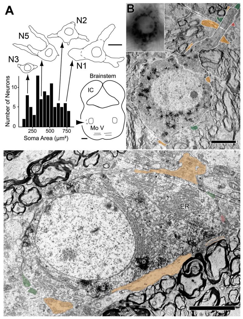
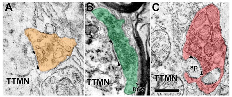
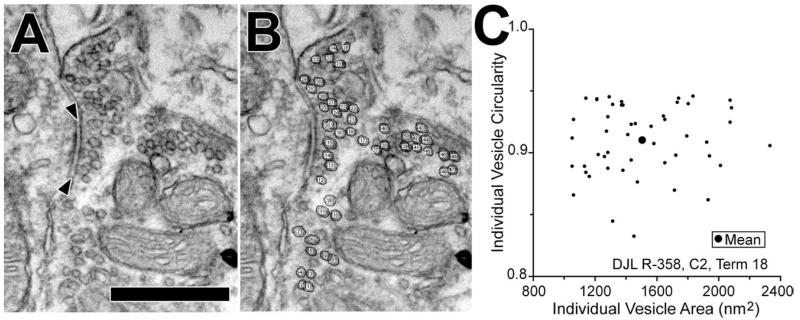
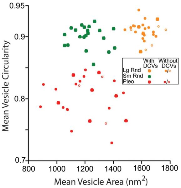
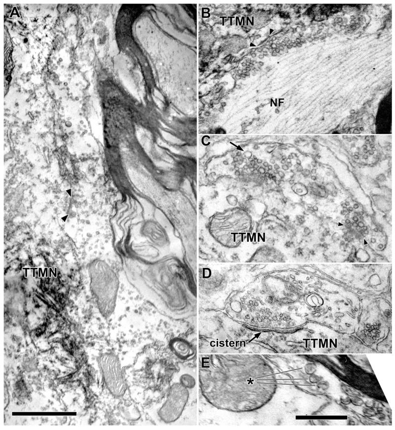
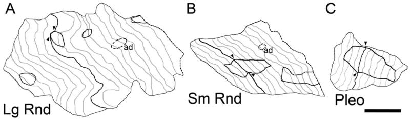
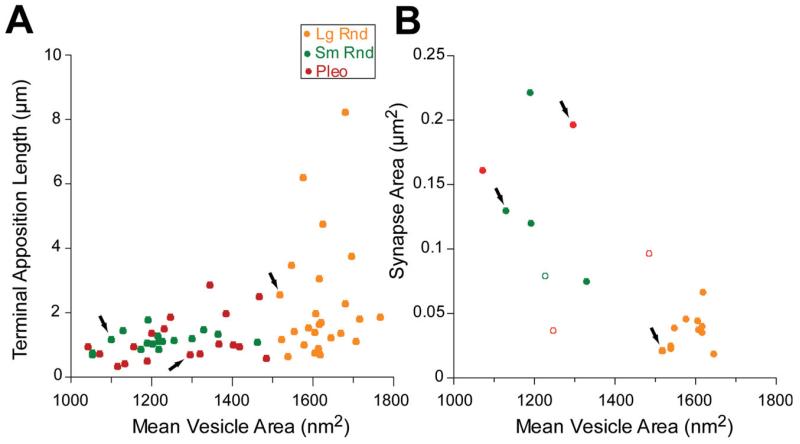
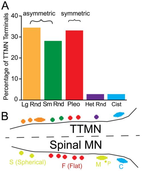
The tensor tympani is a middle ear muscle that contracts in two different situations: in response to sound or during voluntary movements. To gain insight into the inputs and neural regulation of the tensor tympani, we examined the ultrastructure of synaptic terminals on labeled tensor tympani motoneurons (TTMNs) using transmission electron microscopy. Our sample of six TTMNs received 79 synaptic terminals that formed 126 synpases. Two types of synapses are associated with round vesicles and form asymmetric junctions (excitatory morphology). One of these types has vesicles that are large and round (Lg Rnd) and the other has vesicles that are smaller and round (Sm Rnd) and also contains at least one dense core vesicle. A third synapse type has inhibitory morphology because it forms symmetric synapses with pleomorphic vesicles (Pleo). These synaptic terminals can be associated with TTMN spines. Two other types of synapse are found on TTMNs but they are uncommon. Synaptic terminals of all types form multiple synapses but those from a single terminal are always the same type. Terminals with Lg Rnd vesicles formed the most synpases per terminal (avg. 2.73). Together, the synaptic terminals with Lg Rnd and Sm Rnd vesicles account for 62% of the terminals on TTMNs, and they likely represent the pathways driving the contractions in response to sound or during voluntary movements. Having a high proportion of excitatory inputs, the TTMN innervation is like that of stapedius motoneurons but proportionately different from other types of motoneurons.


Figures

Fig. 1
Fig. 1
Example labeled TTMNs and their synaptic terminals. A: Plot of the soma areas of 92 labeled TTMNs. Above the plot are drawings of four of the TTMNs studied extensively in the electron microscope (Table 1). On the drawings, dashed lines indicate the outlines of the somata used for measurements. Arrows indicate where their soma areas fall on the plot; these areas span those seen in the general population. Scale bar: 25 μm. Brainstem outline at lower right shows the positions of the labeled neurons (arrowhead) relative to the trigeminal motor nucleus (Mo V). Scale bar: 1 mm. B: Low power electron micrograph of TTMN N2 (outlined in white) with numerous reaction product crystals in the cytoplasm. Inset at upper left is a light micrograph of the same motoneuron. Similarities of the neuron in the two micrographs include distribution of black reaction product and the position of the nucleolus. Three other dendrites (faintly illustrated in the inset) are seen extending in different directions from this neuron. Synaptic terminals (colored) are seen on the soma (three terminals) and on the proximal dendrite (seven terminals). Scale bar = 5 μm. C: Low power electron micrograph of TTMN N4, showing reaction product crystals in the cytoplasm (asterisks). One proximal dendrite, just beginning to emerge from the soma, is identified as a dendrite because of the orientation of rough endoplasmic reticulum (ER) perpendicular to the nuclear membrane. Synaptic terminals are colored. One terminal (orange) between the TTMN and the scale bar is exceptionally large and consists in this section of two separate segments – the segment at the right has orange outlines because it does not contact the TTMN until deeper sections. Scale bar = 5 μm.
Fig. 2
Fig. 2
Examples synaptic terminals on TTMNs that illustrate the synaptic vesicles. The vesicles contained in the terminal in A are large and round (Lg Rnd), those in the terminal in B are smaller and round (Sm Rnd), and those in the terminal in C are pleomorphic (Pleo). As illustrated, our criteria for synapses requires a cleft between pre- and postsynaptic membrane (arrowheads), postsynaptic dense material, and synaptic vesicles. The terminal in B has a dense core vesicle (DCV). The terminal in C is associated with a TTMN spine (sp), curved in this case, and seen in two parts as it invaginates the terminal. Scale bar = 0.5 μm.
Fig. 3
Fig. 3
Vesicle measurements at a synapse on TTMN N2. A: Electron micrograph of the synapse (arrowheads) showing the clear synaptic vesicles packed within the terminal. Scale bar = 0.5 μm. B: Same micrograph after vesicles were circled and numbered. C: Scatter plot of the circularity vs. area for 47 individual clear vesicles in B. The large dot shows the mean area and mean circularity. Mean data like these were used to compare synapses (see Fig. 4).
Fig. 4
Fig. 4
Plots of mean vesicle circularity versus mean vesicle area for the three common types of synapses on TTMNs. Each point represents measurements from all synapses of a single terminal. Symbol size distinguishes terminals with or without dense core vesicles (DCVs, see key), with open symbols representing terminals in which those vesicles were not seen but for which four or fewer sections are available. Color coding indicates cluster as identified by the kmeans algorithm. The centroids of the clusters were: Lg Rnd (1,615, 0.91), Sm Rnd (1,213, 0.90), and Pleo (1,199, 0.80). Het Rnd and Cist terminals (see Fig. 5) were not plotted.
Fig. 5
Fig. 5
Examples of less common types of terminals on and near TTMNs. A: Electron micrograph of a terminal containing Lg Rnd vesicles that apposes the TTMN for over 6 μm, one of the longest appositions in our study. A synapse is denoted by the arrowheads. Scale bar = 1 μm. B: Terminal with Lg Rnd vesicles with a high neurofilament (NF) content. This terminal appears as the exceptionally large terminal of Fig. 3 and the NF content is high throughout the terminal. C: Terminal that has round vesicles of heterogeneous size (Het Rnd). Most vesicles are comparable to Lg Rnd vesicles but a few are quite large (arrow). A synapse is indicated with arrowheads. D: A Cist terminal with a bi-laminar cistern rather than a postsynaptic density in the adjacent portion of the motoneuron. The terminal contains round vesicles. E: Unmyelinated axon containing dense core vesicles (about eight in total, asterisk points to three) that are larger than those in other terminals. This axon was not observed to contact a labeled TTMN (nearest TTMN was 1.9 μm away, out of the frame of this micrograph). Scale bar = 0.5 μm for B-E.
Fig. 6
Fig. 6
Three synaptic terminals in en face representations of their appositions with TTMNs. A: Lg Rnd; B: Sm Rnd; C: Pleo. The apposition is drawn for individual sections (in gray lines plus one dark line for the section used for apposition measurement) and each is staggered from the next by the section thickness (80 nm). Darkly outlined areas indicate locations of synapses (shown for single sections by arrowheads) and dashed areas indicate adherens junctions (ad). A smooth contour is used between adjacent sections. The number of sections available for the Lg Rnd terminal is 21, for the Sm Rnd terminal is 13, and for the Pleo terminal is 13. Dashed lines for the right-most sections of the Lg Rnd and Sm Rnd terminals indicate that the apposition appeared to extend beyond our series of sections. Scale bar: 0.5 um.
Fig. 7
Fig. 7
Plots of TTMN terminal apposition length (A) and synapse area (B). Arrows indicate the data points corresponding to the terminals in Fig. 6. Data in B are from all 19 synapses that are completely sectioned. Here, solid symbols represent synapses where the first and last parts were included in the series, whereas three open symbols denote where a section is missing at one end of the synapse (and thus represent a least estimate of their areas because the next section contains no synapse).
Fig. 8
Fig. 8
A: Percentages of each type of synaptic terminal on all six TTMNs examined in the electron microscope. Lg Rnd-Large Round; Sm Rnd-Small Round; Pleo-Pleomorphic; Het Rnd-Heterogeneous Round; Cist-Cistern. Brackets indicate the synaptic types that are asymmetric (having a prominent postsynaptic density) and those that are symmetric (having minimal postsynaptic density). B: Schematic of synaptic terminal types found on TTMNs, and comparison to types found on spinal α-motoneurons (after Conradi et al., 1979). Apparent correspondences are indicated with similar colors. S: spherical-vesicle synaptic terminals; M: large terminals containing spherical vesicles that receive input from other, presynaptic (P) terminals, or which have a synaptic complex with six or more “Taxi” bodies; F: flattened-vesicle synaptic terminals; C: cistern synaptic terminals.

Similar articles

Cited by

References

    1. Ahmari S, Buchanan J, Smith S. Assembly of presynaptic active zones from cytoplasmic transport packets. Nat Neurosci. 2000;3:445–451. - PubMed
    1. Bae YC, Tatsuzo N, Ihn HJ, Choi MH, Yoshida A, Moritani M, Shiho H, Shigenaga Y. Distribution pattern of inhibitory and excitatory synapses in the dendritic tree of single masseter alpha-motoneurons in the cat. J Comp Neurol. 1999;414:454–468. - PubMed
    1. Benson TE, Berglund AM, Brown MC. Synaptic input to cochlear-nucleus dendrites that receive medial olivocochlear synapses. J Comp Neurol. 1996;365:27–41. - PubMed
    1. Berrebi AS, Spirou GA. PEP-19 immunoreactivity in the cochlear nucleus and superior olive of the cat. Neuroscience. 1998;83:535–554. - PubMed
    1. Billig I, Yeager MS, Blikas A, Raz Y. Neurons in the cochlear nuclei controlling the tensor tympani in the rat: a study using pseudorabies virus. Brain Res. 2007;1154:124–136. - PMC - PubMed

Publication types

LinkOut - more resources