. 2013 Feb;83(2):227-34.
doi: 10.1002/cyto.a.22228. Epub 2012 Nov 16.

Real-time cell viability assays using a new anthracycline derivative DRAQ7®

Affiliations

Real-time cell viability assays using a new anthracycline derivative DRAQ7®

Jin Akagi et al. Cytometry A. 2013 Feb.

Abstract

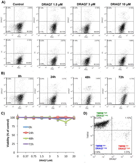
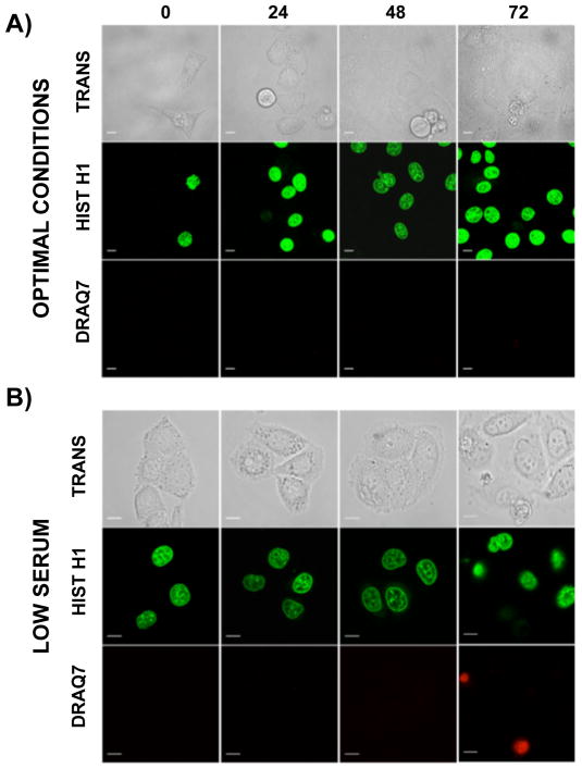
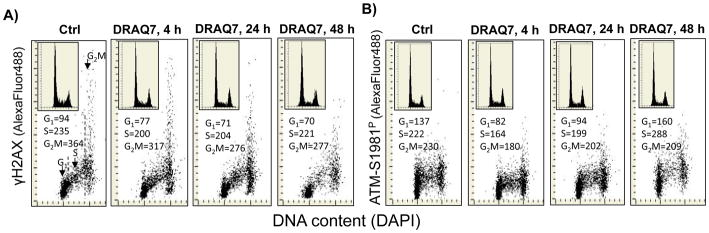
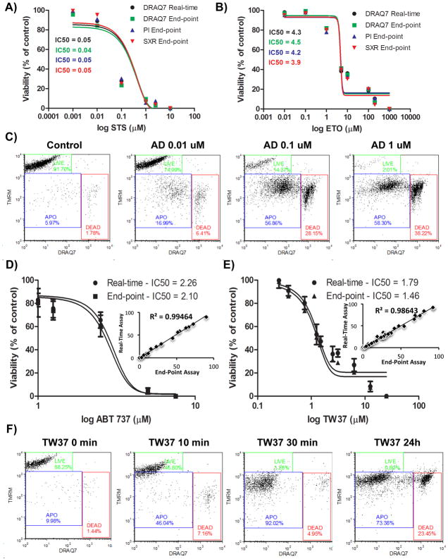
The exclusion of charged fluorescent dyes by intact cells has become a well-established assay for determining viability of cells. In search for a noninvasive fluorescent probe capable of long-term monitoring of cell death in real-time, we evaluated a new anthracycline derivative DRAQ7. The novel probe does not penetrate the plasma membrane of living cells but when the membrane integrity is compromised, it enters and binds readily to nuclear DNA to report cell death. It proved to be nontoxic to a panel of cancer cell lines grown continuously for up to 72 h and did not induce any detectable DNA damage signaling when analyzed using laser scanning microscopy and flow cytometry. The DRAQ7 provided a sensitive, real-time readout of cell death induced by a variety of stressors such as hypoxia, starvation, and drug-induced cytotoxicity. The overall responses to anticancer agents and resulting pharmacological dose-response profiles were not affected by the growth of tumor cells in the presence DRAQ7. Moreover, we for the first time introduced a near real-time microflow cytometric assay based on combination of DRAQ7 and mitochondrial inner membrane potential (ΔΨ(m) ) sensitive probe TMRM. We provide evidence that this low-dosage, real-time labeling procedure provides multiparameter and kinetic fingerprint of anticancer drug action.


Figures

Figure 1
Figure 1
DRAQ7 probe is non-toxic over a wide concentration range. A) THP1α cells were treated with 0.35 – 10 μM of DRAQ7 for up to 24 hours to assess impact of the dye upon cell viability. The fluorescence levels of cells cultured with DRAQ7 were comparable to end-point staining with 1 μg/ml of PI (bottom panel). Both U937 and THP1α cells remained viable when grown with DRAQ7 probe as evidenced by the lack of cell subpopulation exhibiting enhanced fluorescence signal intensity in the top right quadrants. B) The extreme concentration of DRAQ7 (20 μM) is non-toxic to THP1α cells for up to 72 hours of continuous exposure. C) Comparative analysis of cell viability across the broad range of doses and incubation times. Data were derived from a DRAQ7neg gate (deemed live) and cross-validated with counterstaining with a spectrally dissimilar plasma membrane permeability stain PI (1 μg/ml). D) Mitochondrial function is not affected in THP1α cells cultured for up to 72 hours with 20 μM of DRAQ7 as assessed by the multiparameter labeling with ΔΨm sensitive probe tertamethylrhodamine methyl ester (TMRM; 200 nM). Most cells featured intact and energized mitochondria indicated by TMRMhigh and DRAQ7neg (deemed live) subpopulation in the upper left quadrant.
Figure 2
Figure 2
Confocal microscopy studies of DRAQ7 reporting cell death. Transmitted light images, fluorescence images of GFP-tagged histone H1, and images of DRAQ7 in cells grown under optimal conditions (A), in medium with a low concentration (2%) of serum (B). A loss of plasma membrane integrity is reported by DRAQ7 entering and staining the nuclear DNA. Images were collected using the scan speed of 100 Hz, except for the frames presented in panel A (0h, 24h, 48h) where the scan speed was 400Hz. The brightness, contrast and γ function were adjusted in fluorescence images of DRAQ7 γ = 0.5; in images of GFP-tagged histone H1 γ = 0.75. Processing was required to visualize the weak fluorescence signals of DRAQ7. Scale bars: 10 μm.
Figure 3
Figure 3
Effects of prolonged cell culture with DRAQ7 on DNA damage responses. A) Expression of γH2AX of A549 cells, untreated (Ctrl) and exposed to 3 μM DRAQ in cultures for 4, 24 and 48 h. The mean values of γH2AX expression estimated for subpopulations of cells in G1, S and G2M phases of the cell cycle are shown in the respective panels. The insets present DNA content frequency histograms from the respective cultures. B) Expression of ATM-S1981P of A549 cells, untreated (Ctrl) and exposed to 3 μM DRAQ7 in cultures for 4, 24 and 48 h. The mean values of ATM-S1981P expression estimated for subpopulations of cells in G1, S and G2M phases of the cell cycle are shown in the respective panels. The insets present DNA content frequency histograms from the respective cultures.
Figure 4
Figure 4
Pharmacological profiling is not affected by the growth of hematopoietic cancer cells in the continuous presence of DRAQ7 probe. A) THP1α were challenged for up to 24 hours with increasing doses of staurosporine (STS) in the presence of 3 μM DRAQ7 (real-time assay). For comparison cell were also analyzed using standard (end-point) assay using 3 μM of DRAQ7, 1 μg/ml of PI and 100 nM of SYTOX Red probe as described under Materials and Methods. Curve fitting and IC50 values were calculated by plotting the data from DRAQ7neg gate (deemed live). B) THP1α were challenged for up to 24 hours with increasing doses of Etoposide (ETO) in the presence of 3 μM DRAQ7 (real-time assay). For comparison cell were also analyzed using standard (end-point) assays as described in A. Data analysis was performed analogically to conditions shown on A. C) THP1α were challenged for up to 24 hours with increasing doses of Actinomycin D (AD) in the presence of 3 μM DRAQ7 and 200 nM of (ΔΨm sensitive probe TMRM real-time assay) and immediately sampled in complete medium using the microfluidic chip-based cytometer. D) THP1α cells were challenged for up to 24 hours with increasing doses of small-molecule Bcl-2 inhibitor ABT-737 in the presence of 3 μM DRAQ7 and 200 nM of ΔΨm sensitive probe TMRM (real-time assay). Alternatively cells were labeled using an end-point static assay with identical DRAQ7 concentrations. Pharmacological dose-response curve fitting and IC50 calculations were performed using data from TMRMhigh / DRAQ7neg gate (deemed live; LIVE). Inset shows a cumulative Pearson linear correlation analysis of all data acquired from gates TMRMhigh / DRAQ7neg (deemed live; LIVE), TMRMlow / DRAQ7neg (deemed apoptotic; APO), and TMRMlow / DRAQ7high (deemed late apoptotic/necrotic; DEAD). E) THP1α cells were challenged for up to 24 hours with increasing doses of small-molecule Bcl-2 inhibitor TW-37. Condition and data analysis were identical to these described in D. For A–E note excellent agreement between results obtained with end-point vs. real-time no-wash protocols that indicates no interactions between the probe and anti-cancer drugs (Pearson linear correlation R2 = 0.98–99 at p<0.01). F) Kinetic analysis of small-molecule Bcl-2 inhibitor TW37 using real-time DRAQ7 (3 μM) / TMRM (200 nM) assay and time-lapse sampling using microfluidic chip-based cytometer. Note that even though the dissipation of ΔΨm occurs within 10–15 min following challenge with BH3 mimetic TW37, the gradual loss of plasma integrity occurs only after 8 hours of stimulation while nearly 74% of cells can sill be considered TMRMlow/DRAQ7neg after 24 hours.

Similar articles

Cited by

References

    1. Wlodkowic D, Skommer J, Pelkonen J. Multiparametric analysis of HA14-1-induced apoptosis in follicular lymphoma cells. Leuk Res. 2006;30:1187–92. - PubMed
    1. Wlodkowic D, Skommer J, Pelkonen J. Brefeldin A triggers apoptosis associated with mitochondrial breach and enhances HA14-1- and anti-Fas-mediated cell killing in follicular lymphoma cells. Leuk Res. 2007;31:1687–700. - PubMed
    1. Wlodkowic D, Skommer J, Darzynkiewicz Z. Cytometry in cell necrobiology revisited. Recent advances and new vistas. Cytometry A. 2010;77:591–606. - PMC - PubMed
    1. Darzynkiewicz Z, Juan G, Li X, Gorczyca W, Murakami T, Traganos F. Cytometry in cell necrobiology: analysis of apoptosis and accidental cell death (necrosis) Cytometry. 1997;27:1–20. - PubMed
    1. Khoshmanesh K, Akagi J, Nahavandi S, Skommer J, Baratchi S, Cooper JM, Kalantar-Zadeh K, Williams DE, Wlodkowic D. Dynamic analysis of drug-induced cytotoxicity using chip-based dielectrophoretic cell immobilization technology. Anal Chem. 2011;83:2133–44. - PubMed

Publication types

MeSH terms

LinkOut - more resources