Review
. 2012 Dec;2(12):1091-9.
doi: 10.1158/2159-8290.CD-12-0329. Epub 2012 Nov 19.

The initial hours of metastasis: the importance of cooperative host-tumor cell interactions during hematogenous dissemination

Affiliations
Review

The initial hours of metastasis: the importance of cooperative host-tumor cell interactions during hematogenous dissemination

Myriam Labelle et al. Cancer Discov. 2012 Dec.

Abstract

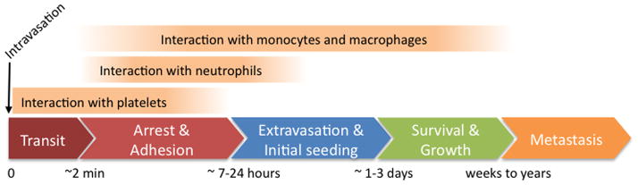
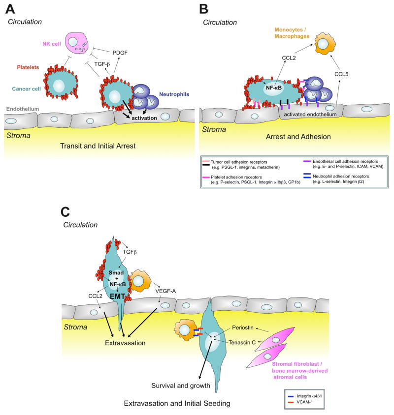
Tumor cells transit from the primary tumor via the blood circulation to form metastases in distant organs. During this process, tumor cells encounter a number of environmental challenges and stimuli that profoundly impact their metastatic potential. Here, we review the cooperative and dynamic host-tumor cell interactions that support and promote the hematogenous dissemination of cancer cells to sites of distant metastasis. In particular, we discuss what is known about the cross-talk occurring among tumor cells, platelets, leukocytes, and endothelial cells and how these cell-cell interactions are organized both temporally and spatially at sites of extravasation and in the early metastatic niche.

Significance: Metastasis is a function not only of tumor cells but also involves cooperative interactions of those cells with normal cells of the body, in particular platelets and leukocytes. These other cell types alter the behavior of the tumor cells themselves and of endothelial cells lining the vasculature and assist in tumor cell arrest and extravasation at sites of metastasis and subsequently in the establishment of tumor cells in the early metastatic niche. A better understanding of the important role that these contact and paracrine interactions play during metastasis will offer new opportunities for therapeutic intervention.


Conflict of interest statement

The authors disclose no potential conflicts of interest.

Figures

Figure 1
Figure 1. Temporal dynamics of host-tumor cell interactions during the early steps of the metastatic cascade
Tumor cells intravasate, rapidly transit through the circulation, and arrest in the vasculature of a secondary organ, generally within a few minutes. During this period, platelets form aggregates around CTCs or arrested tumor cells. Neutrophils also interact with tumor cells within the first day. Seven to 48 hours after tail-vein injection of tumor cells, monocytes/macrophages are also recruited to their vicinity. Extravasation typically takes place within the first 1–3 days after initial arrest. By that time, most tumor cells have exited the bloodstream and seeded into the stroma of the secondary site and additional myeloid cells are recruited to this initial metastatic niche. The tumor cells may reinitiate growth to form metastases within a few weeks. Alternatively, tumor cells can survive and stay dormant for a long period before reinitiating growth and thus form clinically relevant metastases only months or years later. Overall, only few cells successfully complete the metastatic cascade and give rise to overt metastases. So far, most studies of host-tumor cell interactions during metastasis have been performed at single time points arbitrarily chosen by investigators while only few studies have examined the temporal recruitment of host cells by real-time imaging or sampling at multiple time points. This scheme is thus a tentative summary of many independent studies reporting experimental observations at different time points and obtained with different model systems.
Figure 2
Figure 2. Examples of host-tumor cell interactions during the arrest and extravasation steps of the metastatic cascade
A. Upon entry into the blood circulation, tumor cells become exposed to interactions with various blood cells. Platelet-tumor cell interactions may occur soon after the entry of tumor cells into the circulation, when they are still circulating, or very early upon initial arrest. Overall, the interaction of platelets with tumor cells has various prometastatic functions during the vascular phase of the metastatic cascade. Early on, the formation of a platelet-rich thrombus around tumor cells protects tumor cells from shear stress and against lysis by NK cells. The formation of platelet microthrombi around tumor cells may also favor arrest and adhesion to the endothelium. Simultaneously, neutrophils may be recruited to the platelet-tumor cell aggregates, and participate in endothelial cell activation. B. Cytokines and chemokines secreted by platelets, the tumor cells, neutrophils and the activated endothelium promote the recruitment of monocytes, which, in concert with the other cells already present in this “niche”, further activate the endothelium and enhance tumor cell extravasation. C. Direct signaling between platelets and tumor cells also contributes to extravasation by inducing a more invasive pro-metastatic phenotype in tumor cells and probably also through effects on the endothelium. The recruited BMDCs and other host cells such as fibroblasts then contribute to the remodeling of the microenvironment to further support initial tumor cell survival and growth in the tissue parenchyma, eventually leading to the formation of overt metastases.

References

    1. Fidler IJ. Metastasis: guantitative analysis of distribution and fate of tumor embolilabeled with 125 I-5-iodo-2′-deoxyuridine. J Natl Cancer Inst. 1970;45:773–82. - PubMed
    1. Chambers AF, Groom AC, MacDonald IC. Dissemination and growth of cancer cells in metastatic sites. Nat Rev Cancer. 2002;2:563–72. - PubMed
    1. Joyce JA, Pollard JW. Microenvironmental regulation of metastasis. Nat Rev Cancer. 2009;9:239–52. - PMC - PubMed
    1. Khanna C, Hunter K. Modeling metastasis in vivo. Carcinogenesis. 2005;26:513–23. - PubMed
    1. Cristofanilli M, Budd GT, Ellis MJ, Stopeck A, Matera J, Miller MC, et al. Circulating tumor cells, disease progression, and survival in metastatic breast cancer. N Engl J Med. 2004;351:781–91. - PubMed

Publication types