

RNA interference-mediated silencing of Atp6i prevents both periapical bone erosion and inflammation in the mouse model of endodontic disease
- PMID: 23166162
- PMCID: PMC3639609
- DOI: 10.1128/IAI.00756-12
RNA interference-mediated silencing of Atp6i prevents both periapical bone erosion and inflammation in the mouse model of endodontic disease
Abstract
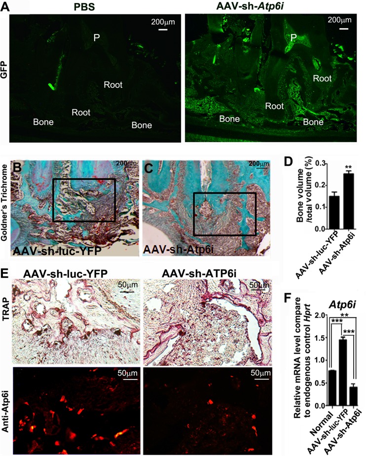
Dental caries is one of the most prevalent infectious diseases in the United States, affecting approximately 80% of children and the majority of adults. Dental caries may lead to endodontic disease, where the bacterial infection progresses to the root canal system of the tooth, leading to periapical inflammation, bone erosion, severe pain, and tooth loss. Periapical inflammation may also exacerbate inflammation in other parts of the body. Although conventional clinical therapies for this disease are successful in approximately 80% of cases, there is still an urgent need for increased efficacy of treatment. In this study, we applied a novel gene-therapeutic approach using recombinant adeno-associated virus (AAV)-mediated Atp6i RNA interference (RNAi) knockdown of Atp6i/TIRC7 gene expression to simultaneously target periapical bone resorption and periapical inflammation. We found that Atp6i inhibition impaired osteoclast function in vitro and in vivo and decreased the number of T cells in the periapical lesion. Notably, AAV-mediated Atp6i/TIRC7 knockdown gene therapy reduced bacterial infection-stimulated bone resorption by 80% in the mouse model of endodontic disease. Importantly, Atp6i(+/-) mice with haploinsufficiency of Atp6i exhibited protection similar to that in mice with bacterial infection-stimulated bone erosion and periapical inflammation, which confirms the potential therapeutic effect of AAV-small hairpin RNA (shRNA)-Atp6i/TIRC7. Our results demonstrate that AAV-mediated Atp6i/TIRC7 knockdown in periapical tissues can inhibit endodontic disease development, bone resorption, and inflammation, indicating for the first time that this potential gene therapy may significantly improve the health of those who suffer from endodontic disease.
Figures


Similar articles
-
Silencing of Ac45 Simultaneously Inhibits Osteoclast-Mediated Bone Resorption and Attenuates Dendritic Cell-Mediated Inflammation through Impairing Acidification and Cathepsin K Secretion.Infect Immun. 2020 Dec 15;89(1):e00436-20. doi: 10.1128/IAI.00436-20. Print 2020 Dec 15. Infect Immun. 2020. PMID: 33077625 Free PMC article.
-
RNAi-mediated silencing of Atp6i and Atp6i haploinsufficiency prevents both bone loss and inflammation in a mouse model of periodontal disease.PLoS One. 2013;8(4):e58599. doi: 10.1371/journal.pone.0058599. Epub 2013 Apr 5. PLoS One. 2013. PMID: 23577057 Free PMC article.
-
C1 Silencing Attenuates Inflammation and Alveolar Bone Resorption in Endodontic Disease.J Endod. 2019 Jul;45(7):898-906. doi: 10.1016/j.joen.2019.02.024. Epub 2019 May 16. J Endod. 2019. PMID: 31104818 Free PMC article.
-
Inflammatory Response Mechanisms of the Dentine-Pulp Complex and the Periapical Tissues.Int J Mol Sci. 2021 Feb 2;22(3):1480. doi: 10.3390/ijms22031480. Int J Mol Sci. 2021. PMID: 33540711 Free PMC article. Review.
-
T-cell immune response cDNA 7 in allograft rejection and inflammation.Curr Opin Investig Drugs. 2007 May;8(5):401-10. Curr Opin Investig Drugs. 2007. PMID: 17520869 Review.
Cited by
-
Calcium silicate-based cements and functional impacts of various constituents.Dent Mater J. 2017 Jan 31;36(1):8-18. doi: 10.4012/dmj.2015-425. Epub 2016 Oct 22. Dent Mater J. 2017. PMID: 27773894 Free PMC article. Review.
-
Inhibition of Rgs10 Expression Prevents Immune Cell Infiltration in Bacteria-induced Inflammatory Lesions and Osteoclast-mediated Bone Destruction.Bone Res. 2013 Sep 1;1(3):267-281. doi: 10.4248/BR201303005. Bone Res. 2013. PMID: 24761229 Free PMC article.
-
Platelet-rich plasma inhibits RANKL-induced osteoclast differentiation through activation of Wnt pathway during bone remodeling.Int J Mol Med. 2018 Feb;41(2):729-738. doi: 10.3892/ijmm.2017.3258. Epub 2017 Nov 16. Int J Mol Med. 2018. PMID: 29207140 Free PMC article.
-
Knockout and Double Knockout of Cathepsin K and Mmp9 reveals a novel function of Cathepsin K as a regulator of osteoclast gene expression and bone homeostasis.Int J Biol Sci. 2022 Aug 29;18(14):5522-5538. doi: 10.7150/ijbs.72211. eCollection 2022. Int J Biol Sci. 2022. PMID: 36147479 Free PMC article.
-
Silencing of Ac45 Simultaneously Inhibits Osteoclast-Mediated Bone Resorption and Attenuates Dendritic Cell-Mediated Inflammation through Impairing Acidification and Cathepsin K Secretion.Infect Immun. 2020 Dec 15;89(1):e00436-20. doi: 10.1128/IAI.00436-20. Print 2020 Dec 15. Infect Immun. 2020. PMID: 33077625 Free PMC article.
References
-
- World Health Organization 12 September 2012, date accessed Oral health. World Health Organization, Geneva, Switzerland: http://www.who.int/oral_health/disease_burden/global/en/
-
- Nair PN. 1997. Apical periodontitis: a dynamic encounter between root canal infection and host response. Periodontol. 2000 13:121–148 - PubMed
-
- Stashenko P. 1990. Role of immune cytokines in the pathogenesis of periapical lesions. Endod. Dent. Traumatol. 6:89–96 - PubMed
-
- Kakehashi S, Stanley HR, Fitzgerald RJ. 1965. The effects of surgical exposures of dental pulps in germ-free and conventional laboratory rats. Oral Surg. Oral Med. Oral Pathol. 20:340–349 - PubMed
-
- Nair PN. 2004. Pathogenesis of apical periodontitis and the causes of endodontic failures. Crit. Rev. Oral Biol. Med. 15:348–381 - PubMed
Publication types
MeSH terms
Substances
Grants and funding
LinkOut - more resources
Full Text Sources
Other Literature Sources
Molecular Biology Databases
