. 2013 Feb;33(3):557-70.
doi: 10.1128/MCB.01016-12. Epub 2012 Nov 19.

TCPTP regulates SFK and STAT3 signaling and is lost in triple-negative breast cancers

Affiliations

TCPTP regulates SFK and STAT3 signaling and is lost in triple-negative breast cancers

Benjamin J Shields et al. Mol Cell Biol. 2013 Feb.

Erratum in

  • Correction.
    [No authors listed] [No authors listed] Mol Cell Biol. 2023;43(11):629. doi: 10.1080/10985549.2023.2277100. Epub 2023 Nov 17. Mol Cell Biol. 2023. PMID: 37955527 Free PMC article. No abstract available.

Abstract

Tyrosine phosphorylation-dependent signaling, as mediated by members of the epidermal growth factor receptor (EGFR) family (ErbB1 to -4) of protein tyrosine kinases (PTKs), Src family PTKs (SFKs), and cytokines such as interleukin-6 (IL-6) that signal via signal transducer and activator of transcription 3 (STAT3), is critical to the development and progression of many human breast cancers. EGFR, SFKs, and STAT3 can serve as substrates for the protein tyrosine phosphatase TCPTP (PTPN2). Here we report that TCPTP protein levels are decreased in a subset of breast cancer cell lines in vitro and that TCPTP protein is absent in a large proportion of "triple-negative" primary human breast cancers. Homozygous TCPTP deficiency in murine mammary fat pads in vivo is associated with elevated SFK and STAT3 signaling, whereas TCPTP deficiency in human breast cancer cell lines enhances SFK and STAT3 signaling. On the other hand, TCPTP reconstitution in human breast cancer cell lines severely impaired cell proliferation and suppressed anchorage-independent growth in vitro and xenograft growth in vivo. These studies establish TCPTP's potential to serve as a tumor suppressor in human breast cancer.


Figures

Fig 1
Fig 1
TCPTP expression in human breast cancer cell lines. Cell lysates from the indicated asynchronous human breast cancer cell lines and immortalized normal breast epithelial cell lines were resolved by SDS-PAGE and immunoblotted for TCPTP (CF4) and tubulin (A) or PTP1B, SHP-2, and actin (B). The TCPTP variants, TC48 and TC45, are indicated, and the TC48/TC45 ratios shown. Cells with a low TC45 (TC48/TC45 ratios ≥ 2) or TCPTP (TC48 and TC45) level are indicated by one star or two stars, respectively.
Fig 2
Fig 2
TC48 and TC45 differentially contribute to PTK signaling and anchorage-independent growth. (A) HeLa cells stably expressing control, TCPTP (TC48 and TC45), or TC48 shRNAs were processed for immunoblot analysis with antibodies to TCPTP and actin. (B to G) HeLa cells stably expressing control, TCPTP, or TC48 shRNAs (B to G) or transiently transfected with control (GFP) or TC45-specific siRNAs (F and G) were serum starved and left unstimulated or stimulated with 10 ng/ml EGF, pulsed with 10 ng/ml IL-6 for 10 min and then chased, or detached for 30 min and stimulated with plate-bound collagen. Lysates were then collected at the indicated times and resolved by SDS-PAGE and immunoblotted with antibodies to phospho-(Y705)-STAT3 (p-STAT3), phospho-(Y418)-SFK (p-SFK), phospho-(Y1022/Y1023)-JAK1 (p-JAK1), phospho-(S473)-Akt (p-Akt), or phospho-ERK1/2 (p-ERK1/2) and the corresponding indicated proteins and loading controls. (E) HeLa cells stably expressing control, TCPTP, or TC48 shRNAs were grown in soft agar for 4 to 5 weeks, and colonies were stained with crystal violet and counted. Representative images and data (means ± standard errors of the means [SEM]) from triplicate determinations from three independent experiments are shown; significance was calculated using a two-tailed Student's t test (*, P < 0.05).
Fig 3
Fig 3
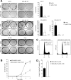
TC45 reconstitution suppresses breast cancer cell growth. (A) The indicated breast cancer cell lines and HeLa cells were transduced with pWZL-hygro or TC45-pWZL-hygro retroviruses and selected with 100 μg/ml hygromycin for 1 to 5 weeks for the formation of foci/colonies. Representative images from three independent experiments are shown. MCF-7, MDA-MB-175, and MDA-MB-231 colonies/well from three independent experiments were counted; significance was calculated using a two-tailed Student's t test (*, P < 0.05; **, P < 0.01). (B to D) MDA-MB-231 cells were transduced with pWZL-hygro and TC45-pWZL-hygro retroviruses for 24 h, selected with 100 μg/ml hygromycin for 7 days, and then subjected to passage into 24-well plates (2 × 10⁴ cells/well) followed by the counting of cells at the indicated times using a hemocytometer (B), fixed in 95% (vol/vol) ethanol, stained with propidium iodide, and processed for flow cytometry (2c and 4c cells are indicated) (C), or stained with annexin V-fluorescein isothiocyanate and propidium iodide and analyzed by flow cytometry (annexin V-positive, propidium iodide [PI]-negative cells were defined as apoptotic) (D). Data shown represent means ± standard deviations (SD) of the results of triplicate determinations and are representative of results from two independent retroviral transductions. In panel C, values correspond to the percentages of sub-G1, G0/G1, S, and G2/M cells.
Fig 4
Fig 4
TC45 overexpression attenuates MDA-MB-231 cell PTK signaling. MDA-MB-231 cells were transfected transiently with pCG control or pCG-TC45 and then left untreated (A) or serum starved and stimulated with either 1 ng/ml IL-6 or 10 ng/ml EGF or detached for 30 min and stimulated with plate-bound collagen for the indicated times (B to D), and lysates were processed for immunoblot analysis with antibodies to phosphotyrosine (pTyr), p-STAT3, p-SFK, or p-EGFR 1068, TCPTP, and the indicated loading controls. Results shown in panels A to C are representative of three independent experiments, and those in panel D are representative of two independent experiments. In panels B and C, IL-6-induced pSTAT3 and EGF-induced p-EGFR Y1068 and the p-SFK species indicated by arrows were quantified from 3 experiments and normalized as indicated. AU, arbitrary units. Statistical significance was calculated using a two-tailed Student's t test (*, P < 0.05; **, P < 0.01).
Fig 5
Fig 5
TCPTP knockdown enhances PTK signaling in SK-BR-3 and T47-D breast cancer cells. SK-BR-3 (A and B), HCC-1954 (C), and T47-D (D and E) cells stably expressing control or TCPTP shRNAs were serum starved and left unstimulated or stimulated with 10 ng/ml EGF, pulsed with the indicated concentrations of IL-6, and chased, or stimulated with 100 ng/ml prolactin, and lysates were collected at the times indicated, resolved by SDS-PAGE, and processed for immunoblot analysis with antibodies to p-EGFR Y1173, p-EGFR Y1068, p-STAT3, p-SFK, p-ERK1/2, p-Akt, p-STAT5, and TCPTP and the indicated loading controls. In panels B and D, IL-6-induced pSTAT3 from 3 experiments was quantified and normalized to STAT3. AU, arbitrary units. Statistical significance was calculated using a two-tailed Student's t test (*, P < 0.05).
Fig 6
Fig 6
TC45 attenuates MDA-MB-231 cell tumorigenicity. MDA-MB-231-TC45 cells were cultured for 3 days in the absence or presence of 2 μg/ml doxycycline (DOX) for the induction of TC45 expression and then either grown in soft agar with or without DOX (A) or injected into the flanks of BALB/c nu/nu mice and tumor volumes were measured every 2 days using calipers (B). Tumors were extracted after 16 days and homogenates were processed for immunoblot analysis to assess TC45 protein levels. Results in panel A represent the means ± SEM of the results from triplicate determinations from three independent experiments; significance was calculated using a two-tailed Student's t test (*, P < 0.05). Results in panel B represent the means ± SEM of the tumor volumes of 10 mice per group and are representative of two independent experiments; significance was determined using a two-tailed Mann-Whitney U test (*, P < 0.05).
Fig 7
Fig 7
CMP6 and SU6656 suppress MDA-MB-231 focus formation and anchorage-independent growth. (A) MDA-MB-231 cells were seeded at a density of 2 × 10³ cells/well in a 6-well dish and cultured in the absence or presence of 2 μM CMP6 or 5 μM SU6656 for 7 days and then fixed with 3.2% paraformaldehyde and stained with 0.05% crystal violet. (B) MDA-MB-231 cells were seeded at a density of 2 × 10³ cells/well in a 6-well dish and cultured in the absence or presence of 10 μM CMP6 or 10 μM SU6656 for 60 min and processed for immunoblotting as indicated. (C) MDA-MB-231 cells were cultured in 0.3% (wt/vol) soft agar in the absence or presence of 10 μM CMP6 or 10 μM SU6656 for 10 to 14 days and colonies stained with 0.05% crystal violet and counted using a dissecting microscope. Results in panels A and B are representative of two independent experiments performed in triplicate. Results in panel C represent the means ± SEM of triplicate determinations from three independent experiments; significance was calculated using a two-tailed Student's t test (**, P < 0.01).
Fig 8
Fig 8
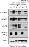
TCPTP deficiency in mice enhances STAT3 Y705 and SFK Y418 phosphorylation in mammary fat pads. The indicated tissues were extracted from 5- to 6-week-old Ptpn2ex2/ex2 (C57BL/6) TCPTP-deficient mice and wild-type littermates and homogenates processed for immunoblot analysis with the indicated antibodies.
Fig 9
Fig 9
TCPTP expression in human breast cancer. (A) ER+ ErbB2, ER ErbB2+, and ER ErbB2 primary human breast cancers were homogenized, and clarified extracts were processed for immunoblot analysis with antibodies to TCPTP (CF4) and actin. The TCPTP variants, TC48 and TC45, are indicated, and the TC48/TC45 ratios are shown. Cells with a low TC45 (TC48/TC45 ratios ≥ 2) or TCPTP (TC48 and TC45) level are indicated by one star or two stars, respectively. (B) HeLa cells expressing control or TCPTP-specific shRNAs were injected into the flanks of BALB/c nu/nu mice, and tumors were extracted after 7 weeks and processed for immunoblot analysis or fixed in formalin and processed for immunohistochemistry; TCPTP was detected with the CF4 monoclonal, HRP-conjugated secondary antibodies and DAB (staining TCPTP-positive cells brown). (C) Formalin-fixed and paraffin-embedded breast cancers were deparaffinized and rehydrated, and sections were processed for immunohistochemistry, stained for TCPTP (CF4), and counterstained with hematoxylin. Representative images (×400) from TCPTP-positive and -negative tumors (arrows indicate TCPTP-positive lymphocytes) and surrounding normal tissue (showing TCPTP expression in a normal terminal duct lobular unit) are shown. Scale bar, 50 μm.

Comment in

Similar articles

Cited by

References

    1. Reddy KB. 2011. Triple-negative breast cancers: an updated review on treatment options. Curr. Oncol. 18:e173–e179 - PMC - PubMed
    1. Yarden Y. 2001. Biology of HER2 and its importance in breast cancer. Oncology 61(Suppl 2):1–13 - PubMed
    1. Nielsen TO, Hsu FD, Jensen K, Cheang M, Karaca G, Hu Z, Hernandez-Boussard T, Livasy C, Cowan D, Dressler L, Akslen LA, Ragaz J, Gown AM, Gilks CB, van de Rijn M, Perou CM. 2004. Immunohistochemical and clinical characterization of the basal-like subtype of invasive breast carcinoma. Clin. Cancer Res. 10:5367–5374 - PubMed
    1. Muthuswamy SK, Li D, Lelievre S, Bissell MJ, Brugge JS. 2001. ErbB2, but not ErbB1, reinitiates proliferation and induces luminal repopulation in epithelial acini. Nat. Cell Biol. 3:785–792 - PMC - PubMed
    1. Biscardi JS, Belsches AP, Parsons SJ. 1998. Characterization of human epidermal growth factor receptor and c-Src interactions in human breast tumor cells. Mol. Carcinog. 21:261–272 - PubMed

Publication types

MeSH terms

Substances