. 2013 Feb;33(3):582-95.
doi: 10.1128/MCB.01225-12. Epub 2012 Nov 19.

TAK1 is essential for osteoclast differentiation and is an important modulator of cell death by apoptosis and necroptosis

Affiliations

TAK1 is essential for osteoclast differentiation and is an important modulator of cell death by apoptosis and necroptosis

Betty Lamothe et al. Mol Cell Biol. 2013 Feb.

Abstract

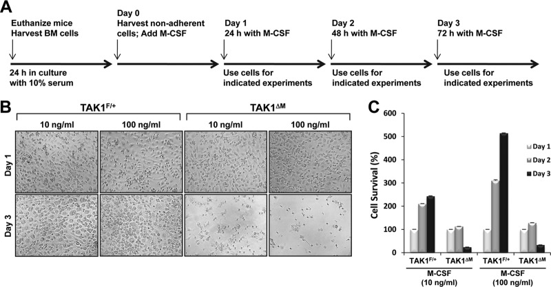
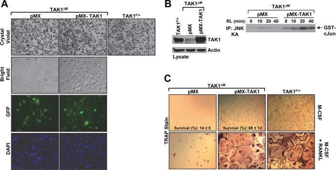
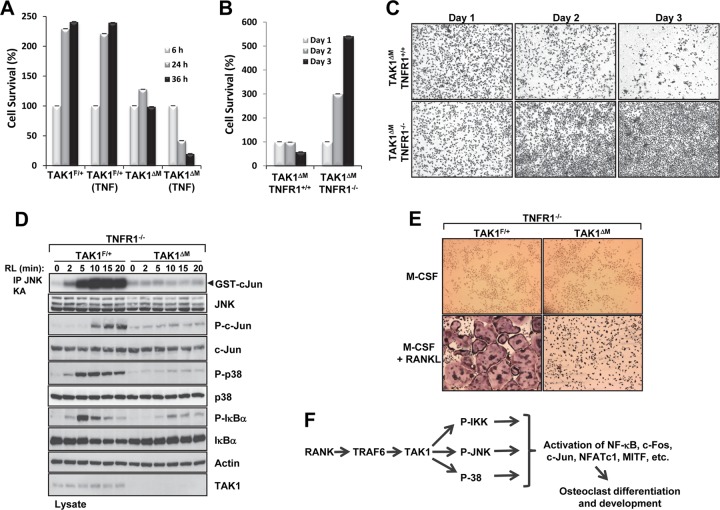
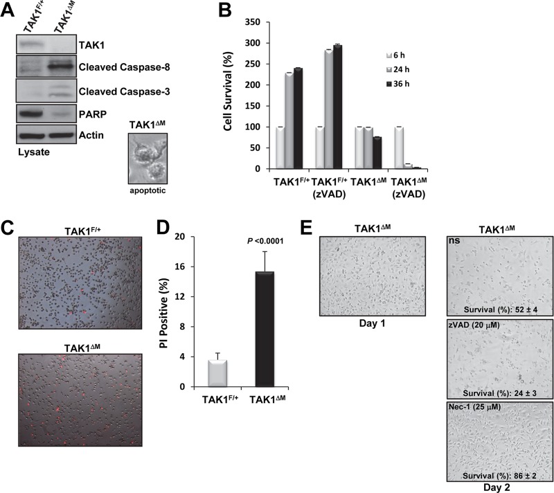
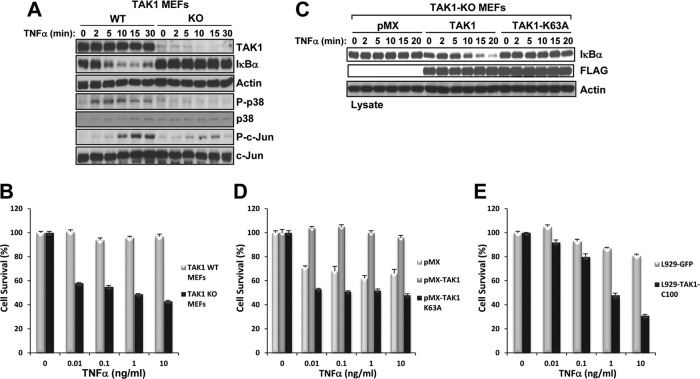
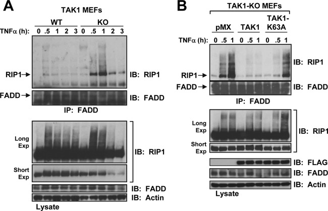
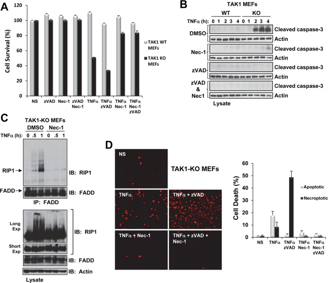
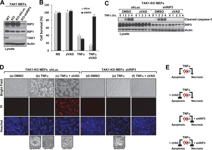
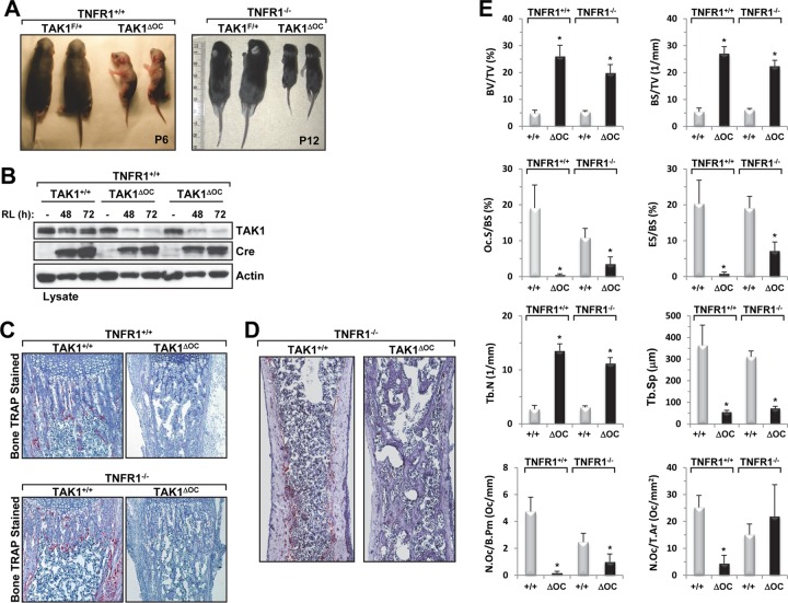
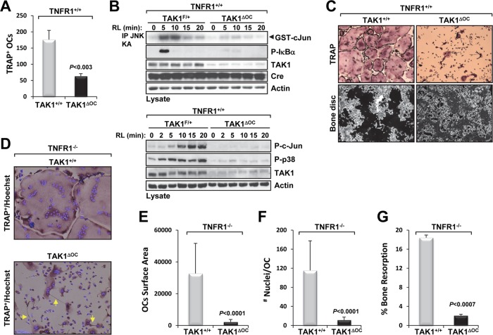
Transforming growth factor β (TGF-β)-activated kinase 1 (TAK1), a mitogen-activated protein 3 (MAP3) kinase, plays an essential role in inflammation by activating the IκB kinase (IKK)/nuclear factor κB (NF-κB) and stress kinase (p38 and c-Jun N-terminal kinase [JNK]) pathways in response to many stimuli. The tumor necrosis factor (TNF) superfamily member receptor activator of NF-κB ligand (RANKL) regulates osteoclastogenesis through its receptor, RANK, and the signaling adaptor TRAF6. Because TAK1 activation is mediated through TRAF6 in the interleukin 1 receptor (IL-1R) and toll-like receptor (TLR) pathways, we sought to investigate the consequence of TAK1 deletion in RANKL-mediated osteoclastogenesis. We generated macrophage colony-stimulating factor (M-CSF)-derived monocytes from the bone marrow of mice with TAK1 deletion in the myeloid lineage. Unexpectedly, TAK1-deficient monocytes in culture died rapidly but could be rescued by retroviral expression of TAK1, inhibition of receptor-interacting protein 1 (RIP1) kinase activity with necrostatin-1, or simultaneous genetic deletion of TNF receptor 1 (TNFR1). Further investigation using TAK1-deficient mouse embryonic fibroblasts revealed that TNF-α-induced cell death was abrogated by the simultaneous inhibition of caspases and knockdown of RIP3, suggesting that TAK1 is an important modulator of both apoptosis and necroptosis. Moreover, TAK1-deficient monocytes rescued from programmed cell death did not form mature osteoclasts in response to RANKL, indicating that TAK1 is indispensable to RANKL-induced osteoclastogenesis. To our knowledge, we are the first to report that mice in which TAK1 has been conditionally deleted in osteoclasts develop osteopetrosis.


Figures

Fig 1
Fig 1
TAK1-deficient BMMs undergo spontaneous cell death in culture. (A) Timeline of the different experimental procedures. (B and C) High-dose M-CSF does not rescue TAK1ΔM monocytes from cell death. BM cells were treated with M-CSF (10 or 100 ng/ml), and on the morning of day 1, the cells were lifted from the plate, counted, and replated in the presence of M-CSF (10 or 100 ng/ml). Representative phase-contrast images were taken the evening of day 1 and the evening of day 3 (B). Cell survival was measured by using the XTT assay (C).
Fig 2
Fig 2
Retroviral delivery of TAK1 rescues survival of TAK1-deficient monocytes and osteoclast differentiation. (A to C) TAK1ΔM BMMs were infected with retroviral supernatant expressing empty vector (pMX) or TAK1 (pMX-TAK1). After 2 days, phase-contrast and fluorescence microscopy images of the infected TAK1ΔM BMMs and control TAK1F/+ BMMs were obtained. The cells were then fixed and stained with crystal violet (A). The cells were lysed and immunoblotted as indicated (B, left panel). Infected TAK1ΔM BMMs were stimulated with RANKL (RL,100 ng/ml) as indicated, and JNK activity was measured using an in vitro kinase assay (B, right panel). The remaining cells were treated with M-CSF (10 ng/ml) in the absence or presence of RANKL (100 ng/ml) for 4 days and then stained for TRAP (C). Percent cell survival was estimated by counting the relative number of cells in 15 independent fields (C, top panels). IP, immunoprecipitation; DAPI, 4′,6-diamidino-2-phenylindole.
Fig 3
Fig 3
Survival of TAK1-deficient BMMs is rescued by deletion of TNFR1, and TAK1 is indispensable to osteoclast formation. (A) TAK1ΔM BMMs are sensitive to TNF-α-induced cell death. TAK1F/+ and TAK1ΔM BMMs were treated with M-CSF (10 ng/ml) in the absence or presence of TNF-α (10 ng/ml), and cell survival was measured using the XTT assay. (B and C) TAK1ΔM BMMs with or without TNFR1 deletion were lifted from the plate on the morning of day 1, counted, and replated at low density. Cell survival was measured using the XTT assay (B), and the cells were fixed and stained with crystal violet (C). (D) TAK1-deficient monocytes on a TNFR1-null background did not respond to RANKL (RL) treatment. BMMs from TAK1ΔM-TNFR1−/− or TAK1F/+-TNFR1−/− mice were stimulated with RANKL (100 ng/ml), cell lysates were immunoblotted as indicated, and an in vitro kinase assay for JNK activity was performed. (E) BMMs derived from TAK1ΔM-TNFR1−/− or TAK1F/+-TNFR1−/− mice were cultured in the presence of M-CSF (10 ng/ml) with or without RANKL (100 ng/ml) for 4 days, fixed, and stained for TRAP. (F) Schematic of the role of TAK1 in regulating RANKL signaling and osteoclast differentiation.
Fig 4
Fig 4
TAK1-deficient BMMs die by apoptosis and necroptosis. (A) Active caspases in TAK1-deficient monocytes. TAK1F/+ and TAK1ΔM BMMs were harvested on the morning of day 2, and protein lysates were immunoblotted as indicated. A representative phase-contrast image of an apoptotic cell is shown (inset). (B) zVAD-fmk potentiates the cell death of TAK1-deficient monocytes. On the morning of day 1, TAK1F/+ and TAK1ΔM BMMs were lifted from the plate, counted, and replated at high density in the absence or presence of zVAD-fmk (10 μM). Cell survival was measured using the XTT assay. (C and D) On the morning of day 2, TAK1F/+ and TAK1ΔM BMMs were stained with PI (C), and the percentage of PI-positive cells was measured (D). (E) Nec-1 treatment protects TAK1-deficient monocytes from cell death. Representative phase-contrast images and percent survival of TAK1ΔM BMMs after 36 h of treatment with M-CSF (10 ng/ml) in the presence of either zVAD-fmk or Nec-1 are shown.
Fig 5
Fig 5
TAK1-KO MEFs have defective TNF-α signaling and are hypersensitive to TNF-α-induced cell death. (A) WT and TAK1-KO MEFs were stimulated with TNF-α (10 ng/ml), and the cell lysates were immunoblotted with the indicated antibodies. (B) WT and TAK1-KO MEFs plated in 96-well plates were treated with TNF-α for 9 h, and cell viability was assessed using crystal violet staining. (C and D) TAK1-KO MEFs expressing empty vector (pMX), TAK1, or TAK1-K63A were stimulated with TNF-α (10 ng/ml) as indicated, and the cell lysates were immunoblotted with the indicated antibodies (C). The indicated cells were plated in 96-well plates and treated with TNF-α for 9 h, and cell viability was assessed using crystal violet staining (D). (E) L929 cells expressing GFP-TAK1-C100 are hypersensitive to TNF-α-induced cell death. L929 cells expressing GFP or GFP-TAK1-C100 were plated in a 96-well plate and treated with TNF-α for 9 h, and cell viability was assessed using crystal violet staining.
Fig 6
Fig 6
RIP1 forms a complex with FADD in TAK1-KO MEFs in response to TNF-α. (A) WT and TAK1-KO MEFs were stimulated with TNF-α (10 ng/ml), and the cell lysates were immunoprecipitated with an anti-FADD antibody and immunoblotted with anti-RIP1 (top) or anti-FADD (bottom). The cell lysates were immunoblotted with the indicated antibodies (Long Exp, long exposure; Short Exp, short exposure). (B) Reconstitution of TAK1-KO MEFs with TAK1 but not TAK1-K63A blocks FADD and RIP1 interaction in response to TNF-α. TAK1-KO MEFs expressing empty vector (pMX), TAK1, or TAK1-K63A were processed as described for panel A.
Fig 7
Fig 7
Nec-1 prevents TNF-α-induced cell death of TAK1-KO MEFs. (A) WT and TAK1-KO MEFs were pretreated for 30 min with zVAD-fmk (20 μM) and/or Nec-1 (50 μM) and then treated with TNF-α (1 ng/ml) for 9 h. Cell viability was assessed using crystal violet staining. NS, nonstimulated. (B) WT and TAK1-KO MEFs were pretreated for 30 min with combinations of dimethyl sulfoxide (DMSO) (1%), zVAD-fmk (20 μM), or Nec-1 (50 μM) as indicated and then treated with TNF-α (1 ng/ml). The cell lysates were immunoblotted with the indicated antibodies. (C) TAK1-KO MEFs were pretreated for 30 min with DMSO (1%) or Nec-1 (50 μM) and then treated with TNF-α (10 ng/ml). The cell lysates were processed as described in the Fig. 6A legend. (D) TAK1-KO MEFs were pretreated as described for panel A, treated with TNF-α (1 ng/ml) for 1 h 45 min, and then stained with PI. Representative images of fluorescent PI-positive TAK1-KO MEFs under the indicated treatment conditions (left) and the percentage of cell death attributed to apoptosis (chromatin condensation) and necroptosis (PI positive) (graph, right) are shown.
Fig 8
Fig 8
Stable knockdown of RIP3 combined with caspase inhibition protects TAK1-KO MEFs from TNF-α-induced cell death. (A) The indicated cell lysates were immunoblotted with the indicated antibodies. (B and C) TAK1-KO MEFs infected with shRNA-luciferase (shLuc) or shRNA-RIP3 (shRIP3) were pretreated for 30 min with dimethyl sulfoxide (DMSO) (1%) or zVAD-fmk (20 μM) and then treated with TNF-α (1 ng/ml) for 9 h. Cell viability was measured using the CellTiter-Glo assay (B). TAK1-KO MEFs expressing shLuc or shRIP3 were pretreated as described for panel B and treated with TNF-α (1 ng/ml) for the indicated times, and the cell lysates were immunoblotted with the indicated antibodies (C). (D) TAK1-KO MEFs expressing shLuc or shRIP3 were pretreated with DMSO (1%; columns a, b, d, and e) or zVAD-fmk (20 μM; columns c and f) for 30 min, stimulated with TNF-α (1 ng/ml; columns b, c, e, and f) for 1 h 45 min, and then stained with PI (red) and Hoechst (blue). Representative phase-contrast and fluorescence microscopy images (magnification, ×10) of the cells under the indicated treatment conditions are shown. The bottom panels in columns b, c, and e show images (magnification, ×20) of cells dying by apoptosis (as indicated by membrane blebs) or by necroptosis (as indicated by rounded, necrosis-like morphology). (E) Schematic of TNF-α-induced cell death in TAK1-deficient MEFs.
Fig 9
Fig 9
Conditional deletion of TAK1 in osteoclasts results in growth retardation, postnatal lethality, and severe osteopetrosis in TAK1ΔOC mice. (A) Growth retardation of TAK1ΔOC pups. Photographs of 6-day-old TAK1ΔOC mice and 12-day-old TAK1ΔOC-TNFR1−/− mice with their respective control littermates. (B) RANKL-induced loss of TAK1 expression in TAK1ΔOC differentiating osteoclasts. TAK1+/+ and TAK1ΔOC spleen-derived monocytes were cultured with M-CSF (10 ng/ml) in the absence or presence of RANKL (RL, 100 ng/ml), and the cell lysates were immunoblotted with the indicated antibodies. (C) Representative images of TRAP-stained osteoclasts (red) in the femurs of the indicated mice (magnification, ×40). (D) Representative images of TRAP-stained sections in the femurs of the indicated mice. (E) Results of the histomorphometric analysis of femurs from 6-day-old male TAK1+/+-TNFR1+/+ mice (n = 4), 6-day-old male TAK1ΔOC-TNFR1+/+ mice (n = 5), 10-day-old male TAK1+/+-TNFR1−/− mice (n = 5), and 10-day-old TAK1ΔOC-TNFR1−/− mice (n = 5). BV/TV, bone volume per total volume; BS/TV, bone surface per total volume; Tb.N, trabecular number; Tb.Sp, trabecular spacing; Oc.S/BS, percentage of bone surface covered with osteoclasts; ES/BS, percentage of bone surface eroded by osteoclasts; N.Oc/B.Pm, number of osteoclasts per bone perimeter; N.Oc/T.Ar, number of osteoclasts per tissue area of interest. *, P < 0.001.
Fig 10
Fig 10
Ex vivo analysis of TAK1ΔOC osteoclast formation and resorption activity. (A) TAK1ΔOC osteoclast development is defective. Spleen-derived monocytes from TAK1+/+ and TAK1ΔOC mice were treated with M-CSF (10 ng/ml) and RANKL (100 ng/ml) for 72 h on plastic dishes or bone discs. Cells were stained for TRAP, and multinucleated TRAP+ cells were quantified. (B) TAK1ΔOC osteoclasts did not respond to RANKL treatment. Spleen-derived monocytes from the indicated mice were treated as described for panel A, starved for 2 h followed by RANKL (RL, 100 ng/ml) treatment for the indicated times, and processed as described in the legend to Fig. 3D. (C) Representative images of TRAP staining (top panels) and bone resorption (black areas, bottom panels) (magnification, ×10) are shown. (D to G) TAK1ΔOC-TNFR1−/− differentiating osteoclasts have defects similar to those of TAK1ΔOC-TNFR1+/+osteoclasts. Spleen-derived monocytes were treated as described for panel A on plastic dishes (D to F) or bone discs (G). Representative images of TRAP and Hoechst staining (magnification, ×20) are shown with yellow arrows pointing at spikelike projections (D). Graphic representations of the area occupied by osteoclasts (E), the number of nuclei per osteoclast (F), and the percent bone resorption (G) are shown.

Similar articles

Cited by

References

    1. Dougall WC, Glaccum M, Charrier K, Rohrbach K, Brasel K, De Smedt T, Daro E, Smith J, Tometsko ME, Maliszewski CR, Armstrong A, Shen V, Bain S, Cosman D, Anderson D, Morrissey PJ, Peschon JJ, Schuh J. 1999. RANK is essential for osteoclast and lymph node development. Genes Dev. 13:2412–2424 - PMC - PubMed
    1. Kong YY, Yoshida H, Sarosi I, Tan HL, Timms E, Capparelli C, Morony S, Oliveira-dos Santos AJ, Van G, Itie A, Khoo W, Wakeham A, Dunstan CR, Lacey DL, Mak TW, Boyle WJ, Penninger JM. 1999. OPGL is a key regulator of osteoclastogenesis, lymphocyte development and lymph-node organogenesis. Nature 397:315–323 - PubMed
    1. Darnay BG, Besse A, Poblenz AT, Lamothe B, Jacoby JJ. 2007. TRAFs in RANK signaling. Adv. Exp. Med. Biol. 597:152–159 - PubMed
    1. Teitelbaum SL. 2007. Osteoclasts: what do they do and how do they do it? Am. J. Pathol. 170:427–435 - PMC - PubMed
    1. Lomaga MA, Yeh WC, Sarosi I, Duncan GS, Furlonger C, Ho A, Morony S, Capparelli C, Van G, Kaufman S, van der Heiden A, Itie A, Wakeham A, Khoo W, Sasaki T, Cao Z, Penninger JM, Paige CJ, Lacey DL, Dunstan CR, Boyle WJ, Goeddel DV, Mak TW. 1999. TRAF6 deficiency results in osteopetrosis and defective interleukin-1, CD40, and LPS signaling. Genes Dev. 13:1015–1024 - PMC - PubMed

Publication types

MeSH terms

Substances

LinkOut - more resources