. 2013 Jan 4;288(1):442-54.
doi: 10.1074/jbc.M112.413922. Epub 2012 Nov 19.

The role of UNC93B1 protein in surface localization of TLR3 receptor and in cell priming to nucleic acid agonists

Affiliations

The role of UNC93B1 protein in surface localization of TLR3 receptor and in cell priming to nucleic acid agonists

Jelka Pohar et al. J Biol Chem. .

Abstract

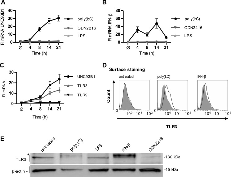
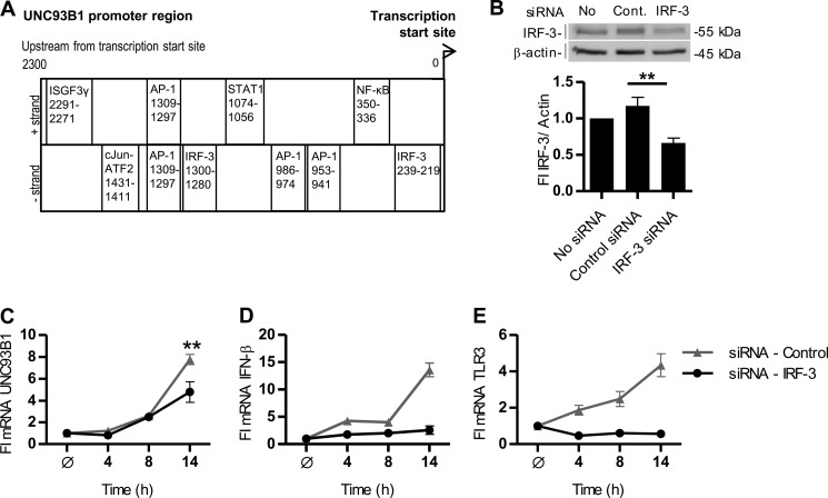
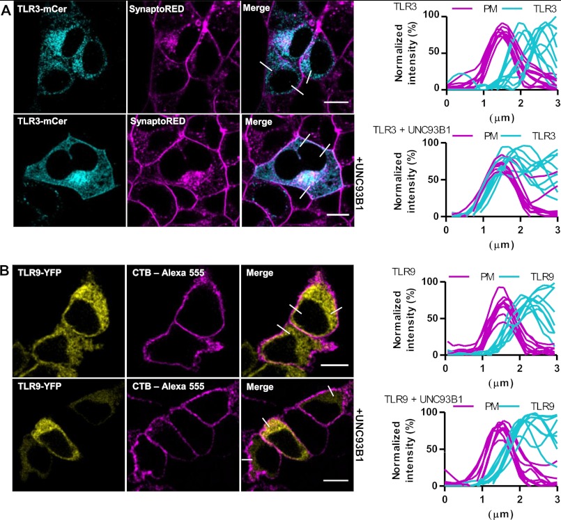
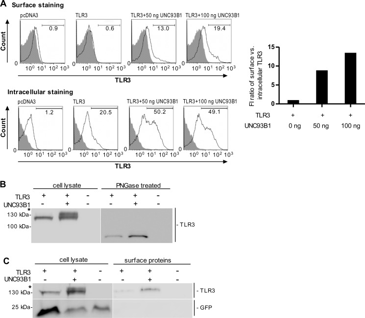
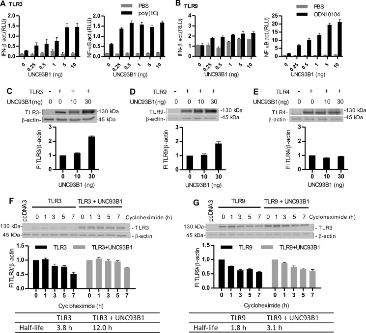
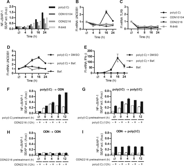
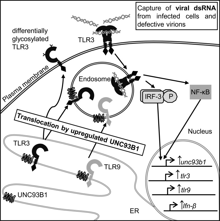
Translocation of nucleic acid-sensing (NAS) Toll-like receptors (TLRs) to endosomes is essential for response to microbial nucleic acids as well as for prevention of the autoimmune response. The accessory protein UNC93B1 is indispensable for activation of NAS TLRs because it regulates their response through trafficking to endosomes. We observed that poly(I:C) up-regulates transcription of UNC93B1 and promotes trafficking of TLR3 to the plasma membrane in human epithelial cell line. Up-regulation of UNC93B1 is triggered through TLR3 activation by poly(I:C). Further studies revealed that expression of UNC93B1 promotes trafficking of differentially glycosylated TLR3, but not other NAS TLRs, to the plasma membrane. UNC93B1 promoter region contains binding sites for poly(I:C)- and type I interferon-inducible regulatory elements. UNC93B1 also increases the protein lifetime of TLR3 and TLR9 and augments signaling of all NAS TLRs. Furthermore, we discovered that poly(I:C) pretreatment primes B-cells to the activation by ssDNA via up-regulation of UNC93B1. Our findings identified TLR3 as the important regulator of UNC93B1 that in turn governs the responsiveness of all NAS TLRs.


Figures

FIGURE 1.
FIGURE 1.
Activation of human umbilical vein endothelial cells by poly(I:C) up-regulates transcription of UNC93B1 and surface localization of TLR3. A and B, HUVEC cells were incubated with poly(I:C) (25 μg/ml), ODN2216 (5 μm), or LPS (25 ng/ml) for the indicated times. UNC93B1 (A) and IFN-β (B) mRNA transcripts were determined by real-time PCR. C, HUVEC cells were incubated with IFN-β (1 nm) for the indicated times. Induction of UNC93B1, TLR3, and TLR9 mRNA transcripts was measured by real-time PCR. A–C, the results are represented by mean values with S.D. from triplicate wells. The representative data from three experiments are shown. D, expression of cell surface-associated TLR3 is shown. HUVEC cells were either untreated or stimulated with poly(I:C) (50 μg/ml) or IFN-β (1 nm) for 24 h. Histograms of unstained cells (filled areas) or cells stained with anti-TLR3 antibodies (solid lines) are shown. Data are representative of two experiments. E, HUVEC cells were stimulated with poly(I:C) (50 μg/ml), LPS (50 ng/ml), IFN-β (1 nm), or ODN2216 (5 μm) for 24 h, and cell lysates were separated by 5% SDS-PAGE. A Western blot was performed with anti-TLR3 antibody and anti-β-actin antibodies. * indicates the differentially glycosylated form of TLR3. The representative image from two experiments is shown.
FIGURE 2.
FIGURE 2.
Transcriptional regulation of UNC93B1. A, shown is a schematic map of the 2300 bp of the human UNC93B1 promoter region. TFBS for transcriptional factors promoting antiviral response are distributed according to the distance from the transcription start site. B–E, HUVEC cells were transfected with an siRNA pool specific to human IRF-3 or negative control siRNA non-target pool. B, HUVEC cells were lysed and subjected to Western blot. IRF-3 was detected with anti-IRF-3 antibodies. Anti-β-actin antibodies were used as a loading control. IRF-3 expression was normalized to the loading control and calculated as a percentage of the intensity of the untreated control. Densitometric analysis is shown below the blot. This experiment was repeated twice, and the representative data are shown. Statistical significance is indicated by **, p ≤ 0.05. C–E, the effect of IRF-3 knockdown on transcription of UNC93B1, IFN-β, and TLR3 is shown. 48 h after siRNA transfection cells were treated with poly(I:C) (25 μg/ml) for 4–21 h or left untreated. Induction of UNC93B1 (C), IFN-β (D), and TLR3 (E) mRNA was subsequently measured by real-time PCR. The results are represented by mean values with S.D. from the triplicate wells showing the representative data from three experiments. Statistical significance is indicated by **, p ≤ 0.05.
FIGURE 3.
FIGURE 3.
UNC93B1 augments surface localization of TLR3 in HEK293T cells. A and B, HEK293T cells were transfected with a TLR3-mCer (cyan) (A) or TLR9-YFP (yellow) (B) alone (top) or with UNC93B1 encoding plasmid (bottom). Plasma membrane markers SynaptoRed (A) and cholera toxin subunit B Alexa Fluor 555 (CTB-Alexa) (B) are shown in magenta. A and B, TLR membrane localization was evaluated from plots of normalized fluorescence intensities of TLR and plasma membrane (PM) within 3-μm line profiles (n = 9). Three representative lines are marked on merged images. Images are selected from five independent experiments. Scale bars, 10 μm.
FIGURE 4.
FIGURE 4.
UNC93B1 translocates the differentially glycosylated TLR3 to the plasma membrane. A, HEK293T cells were transfected with TLR3 alone or with UNC93B1. Cell surface and intracellular expression of TLR3 was determined using flow cytometry. Histograms of mock-transfected cells (pcDNA3, filled areas) or cells transfected with TLR3 (solid lines) are shown. The representative data from three experiments are shown. Increase of cell population expressing plasma membrane-localized TLR3 normalized by the number of cells expressing intracellular TLR3 is shown on the right. B, HEK293T cells were transiently transfected with plasmid encoding TLR3 alone or with UNC93B1. 48 h post-transfection cell lysates were loaded onto a 5% SDS-PAGE gel. TLR3 was detected on a Western blot using anti-TLR3 antibody (left). * indicates the differentially glycosylated form of TLR3. Cell lysates were treated with peptide N-glycosidase F (PNGase; right). The representative image from three experiments is shown. C, HEK293T cells were transiently transfected with TLR3 alone or with UNC93B1. 48 h post-transfection surface proteins were biotinylated. Cells were lysed, and the biotinylated proteins were isolated, blotted, and detected using anti-TLR3 antibody. Cytoplasmic mCerulean was detected using anti-GFP antibody. * indicates differentially glycosylated form of TLR3. The representative image from three experiments is shown.
FIGURE 5.
FIGURE 5.
UNC93B1 augments signaling and protein lifetime of TLR3 and TLR9. A and B, HEK293 cells were transfected with plasmid encoding TLR3 (A) or TLR9 (B) with increasing amounts of UNC93B1. Cells were cotransfected with IFN-β- or NF-κB-responsive reporter plasmids and Renilla normalization reporter plasmid. 18 h after stimulation with poly(I:C) (10 μg/ml) (A) or ODN10104 (10 μg/ml) (B), luciferase activity (relative luciferase units (RLU)) was measured in the cell lysates. The results are represented by mean values with S.D. from the triplicate wells. The representative data from three experiments are shown. C–E, HEK293T cells were transfected with TLR3 (C), TLR9 (D), and TLR4 (E) alone or with UNC93B1. A Western blot was performed using anti-TLR3 (C), anti-HA (D), or anti-Myc (E) antibodies. Anti-β-actin antibodies were used as a loading control. TLR3, TLR9, or TLR4 protein expression was normalized to the loading control and calculated as a percentage of the intensity of the proteins in cells not overexpressing UNC93B1. Densitometric analyses are shown below each blot. The representative data from three experiments are shown. F and G, HEK293T cells were transfected with TLR3 (F) or TLR9 (G) alone or with UNC93B1. 48 h post-transfection, the cells were incubated with cycloheximide for the indicated times. Cell lysates were prepared, and a Western blot was performed using anti-TLR3 (F) or anti-HA (G) antibodies. Anti-β-actin antibodies were used as a loading control. TLR3 and TLR9 protein expression was normalized to the loading control and calculated as a percentage of the intensity of the proteins in the untreated cells. Densitometric analyses and calculated half-lives of proteins are shown below each blot. The representative data from three experiments are shown.
FIGURE 6.
FIGURE 6.
Poly(I:C) pretreatment primes B-cells to the activation by ssDNA via up-regulation of UNC93B1. A–C, Ramos-Blue cells were stimulated with poly(I:C) (20 μg/ml), ODN2216 (20 μg/ml), ODN10104 (20 μg/ml), and R-848 (20 μg/ml) for the indicated times. A, NF-κB/AP-1-dependent secreted embryonic alkaline phosphatase (SEAP) activity (A630) was measured in supernatants. UNC93B1 (B) and TLR9 (C) mRNA were subsequently measured by real-time PCR. D and E, cells were stimulated with poly(I:C) (20 μg/ml) + DMSO alone or pretreated with bafilomycin A (Baf.) for the indicated times. UNC93B1 (D) and IFN-β (E) mRNA were measured by real-time PCR. The results are represented by mean values with S.D. from triplicate wells. The representative data from three experiments are shown. F–I, Ramos-Blue B-cells were pretreated with medium (Ø) or first agonist for 0–12 h and subsequently treated with second agonist for additional 12 h. Agonist used were poly(I:C) (0.5 μg/ml) and ODN2216 (0.5 μg/ml). F, the first agonist was poly(I:C), and the second agonist was ODN2216. G, the first agonist was poly(I:C), and the second agonist was also poly(I:C). H, the first agonist was ODN2216, and the second agonist was also ODN2216. I, the first agonist was ODN2216, and the second agonist was poly(I:C). Results shown in F–I were all preformed in one experiment setting. The results are represented by mean values with S.D. from triplicate wells. The representative data from three experiments are shown.
FIGURE 7.
FIGURE 7.
Model of the TLR3-responsive positive feedback loop mediating the response to nucleic acids through UNC93B1 up-regulation. Activation of TLR3 increases the transcription of UNC93B1 mRNA. UNC93B1 interacts with TLR3 and other NAS TLRs in the ER. UNC93B1 induces differential glycosylation and translocation of TLR3 to the plasma membrane where it can be activated by viral dsRNA from infected cells and defective virions and primes the responsiveness of TLR9.

Similar articles

Cited by

References

    1. Akira S., Takeda K. (2004) Toll-like receptor signalling. Nat. Rev. Immunol. 4, 499–511 - PubMed
    1. Beutler B. (2004) Innate immunity. An overview. Mol. Immunol. 40, 845–859 - PubMed
    1. McGettrick A. F., O'Neill L. A. (2010) Localisation and trafficking of Toll-like receptors. An important mode of regulation. Curr. Opin. Immunol. 22, 20–27 - PubMed
    1. Choe J., Kelker M. S., Wilson I. A. (2005) Crystal structure of human toll-like receptor 3 (TLR3) ectodomain. Science 309, 581–585 - PubMed
    1. Bell J. K., Botos I., Hall P. R., Askins J., Shiloach J., Segal D. M., Davies D. R. (2005) The molecular structure of the Toll-like receptor 3 ligand-binding domain. Proc. Natl. Acad. Sci. U.S.A. 102, 10976–10980 - PMC - PubMed

Publication types

LinkOut - more resources