. 2013 Jan 11;288(2):1150-61.
doi: 10.1074/jbc.M112.400077. Epub 2012 Nov 19.

Molecular interaction studies of HIV-1 matrix protein p17 and heparin: identification of the heparin-binding motif of p17 as a target for the development of multitarget antagonists

Affiliations

Molecular interaction studies of HIV-1 matrix protein p17 and heparin: identification of the heparin-binding motif of p17 as a target for the development of multitarget antagonists

Antonella Bugatti et al. J Biol Chem. .

Abstract

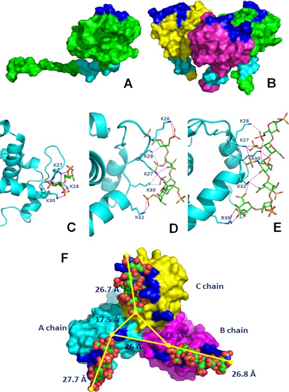
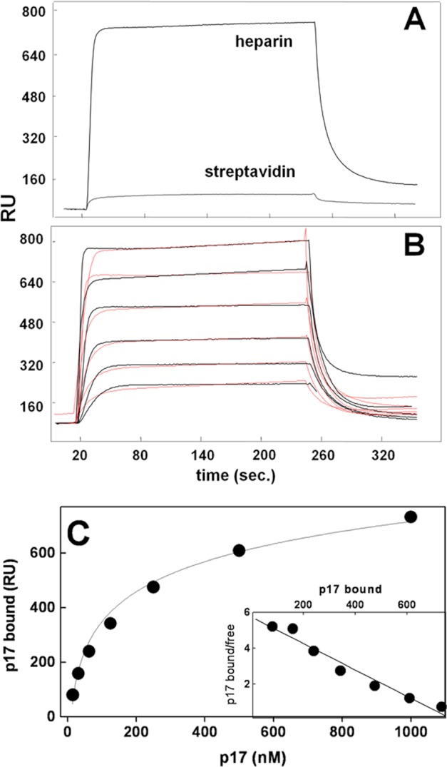
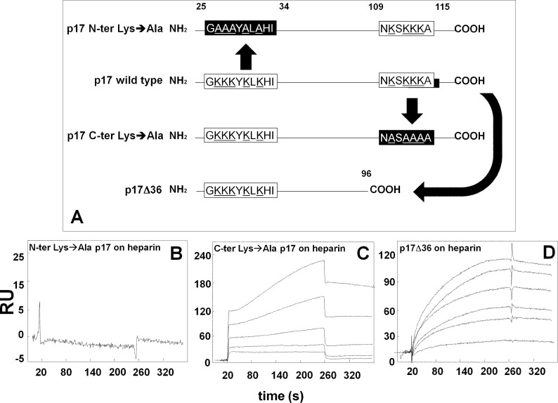
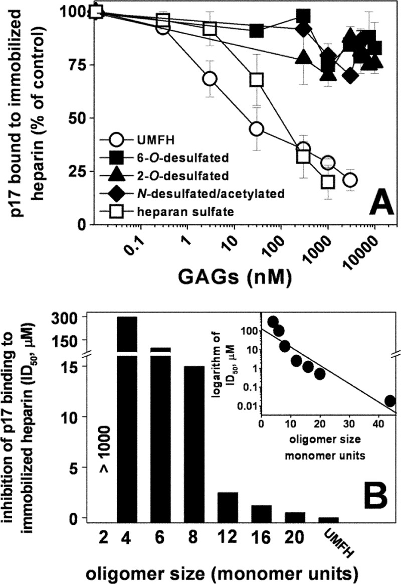
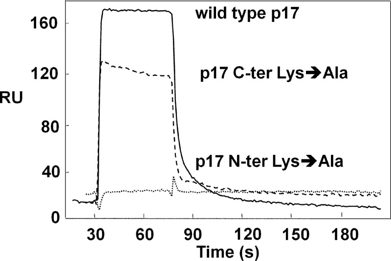
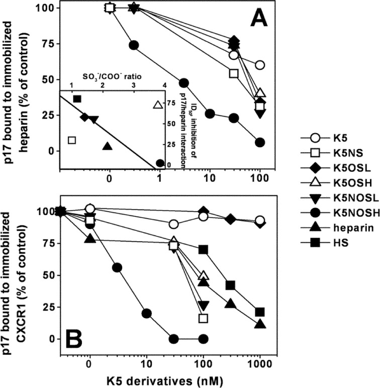
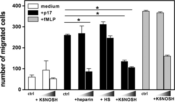
Once released by HIV(+) cells, p17 binds heparan sulfate proteoglycans (HSPGs) and CXCR1 on leukocytes causing their dysfunction. By exploiting an approach integrating computational modeling, site-directed mutagenesis of p17, chemical desulfation of heparin, and surface plasmon resonance, we characterized the interaction of p17 with heparin, a HSPG structural analog, and CXCR1. p17 binds to heparin with an affinity (K(d) = 190 nm) that is similar to those of other heparin-binding viral proteins. Two stretches of basic amino acids (basic motifs) are present in p17 N and C termini. Neutralization (Arg→Ala substitution) of the N-terminal, but not of the C-terminal basic motif, causes the loss of p17 heparin-binding capacity. The N-terminal heparin-binding motif of p17 partially overlaps the CXCR1-binding domain. Accordingly, its neutralization prevents also p17 binding to the chemochine receptor. Competition experiments demonstrated that free heparin and heparan sulfate (HS), but not selectively 2-O-, 6-O-, and N-O desulfated heparins, prevent p17 binding to substrate-immobilized heparin, indicating that the sulfate groups of the glycosaminoglycan mediate p17 interaction. Evaluation of the p17 antagonist activity of a panel of biotechnological heparins derived by chemical sulfation of the Escherichia coli K5 polysaccharide revealed that the highly N,O-sulfated derivative prevents the binding of p17 to both heparin and CXCR1, thus inhibiting p17-driven chemotactic migration of human monocytes with an efficiency that is higher than those of heparin and HS. Here, we characterized at a molecular level the interaction of p17 with its cellular receptors, laying the basis for the development of heparin-mimicking p17 antagonists.


Figures

FIGURE 1.
FIGURE 1.
Shown is the localization of basic motifs in monomeric (A) and trimeric (B) p17. The proteins are shown as Connolly surface. The chains are colored in green, yellow, and magenta. The N-terminal and C-terminal basic motifs are labeled in blue and cyan, respectively. Predicted binding modes between the N-terminal basic motif of p17 and heparin di- (C), tetra- (D), and hexasaccharide (E) are shown. Shown are stick oligosaccharides (green) and the side chains of p17 that interact with them (cyan). Protein is shown as a ribbon, and the hydrogen bonds are represented by magenta dotted lines. F, prediction of the binding mode of the heparin hexasaccharide at the three N-terminal sites. The modeled trimeric form of p17 is shown as Connolly surface, A chain, B chain, and C chain are colored in cyan, magenta, and yellow, respectively. The N-terminal basic motifs of each chain are colored in blue. The ligands are shown in the Corey, Pauling, and Koltun space filling-model.
FIGURE 2.
FIGURE 2.
SPR analysis of p17-heparin interaction. A, sensorgrams showing the binding of native p17 (500 nm) to a heparin- or streptavidin-coated sensorchip. B, blank-subtracted sensorgrams overlay showing the binding of increasing concentrations of native p17 (1000, 500, 250, 125, 62.5, and 31.25 nm) to a heparin-coated sensorchip. Black lines represent the experimental data. Red lines represent the fits. In A and B, the response (in RU) was recorded as a function of time. C, saturation curve obtained using the values of RU bound at equilibrium from injection of increasing concentrations of p17 onto a heparin-coated sensorchip. C, inset: Scatchard plot analysis of the equilibrium binding data shown in C. The correlation coefficient of the linear regression was equal to −0.96.
FIGURE 3.
FIGURE 3.
SPR analysis of the interaction of p17 mutants with heparin. A, schematic representation of the p17 mutants used in the present work. The N- and C-terminal basic motifs of p17 (white boxes) were deleted or neutralized (black boxes) by substituting positively charged lysine with alanine (underlined). Blank-subtracted sensorgrams showing the binding of p17 N-terminal (N-ter) Lys→Ala (500 nm) (B), p17 C-terminal (C-ter) Lys→Ala (500, 250, 125, 62.5, and 31.25 nm) (C) and p17Δ36 (950, 900, 800, 500, 250, and 100 nm) (D) to a heparin-coated sensorchip. The response (in RU) was recorded as a function of time.
FIGURE 4.
FIGURE 4.
Effect of chemically modified heparins or size-defined oligosaccharides on the interaction of p17 to immobilized heparin. Selectively desulfated heparins (A) or size-defined heparin oligosaccharides (B) were evaluated for their capacity to inhibit the interaction of p17 with sensorchip-immobilized heparin. In A, the responses were plotted as percentage of the binding of p17 measured in the absence of free GAGs. Each point is the mean ± S.E. of three separate determinations. In B, the responses were plotted as ID50 of inhibition. The experiment shown is representative of another one that gave similar results. UMFH, unmodified heparin. B, inset, the logarithm of the potency (ID50) of the various size-defined heparin oligosaccharides in inhibiting the binding of p17 to immobilized heparin were plotted against their length (monomer units). The correlation coefficient of the linear regression was equal to −0.93.
FIGURE 5.
FIGURE 5.
SPR analysis of the interaction of p17 mutants with CXCR1. Blank-subtracted sensorgrams showing the binding of wild type p17 (continuous line), p17 C-terminal (C-ter) Lys→Ala (dashed line), and p17 N-terminal (N-ter) Lys→Ala (dotted line) (all at 2 μm) to a CXCR1-coated sensorchip. The response in RU was recorded as a function of time.
FIGURE 6.
FIGURE 6.
Effect of biotechnological heparins on the interaction of p17 to its receptors. K5 derivatives were evaluated for their capacity to inhibit the interaction of p17 with heparin (A) or CXCR1 (B) immobilized to a sensorchip. The responses were plotted as percentage of the binding of p17 measured in the absence of any free GAG. Each point is the mean ± S.E. of three separate determinations. A, inset, the potency (ID50) of the various GAGs in inhibiting the binding of p17 to immobilized heparin were plotted against their SO3/COO ratio. The correlation coefficient of the linear regression was equal to −0.96.
FIGURE 7.
FIGURE 7.
Effect of heparin, HS and K5NOSH on monocyte migration induced by p17. Monocytes in the absence (control, ctrl) or in the presence of heparin, HS and K5NOSH (all at 1 and 10 μg/ml) were added to the top well, whereas medium alone or containing p17 (59 nm) or fMLP (10 nm) was added to the bottom well. Bars represent the mean ± S.D. of three independent experiments performed in duplicate. Statistical analysis was performed using GraphPad Prism software (version 5, GraphPad Software, Inc.) and one-way analysis of varaince and Bonferroni's post test was used to compare data. *, p < 0.01 statistically different compared with p17-stimulated cells. NT, not treated cells.

Similar articles

Cited by

References

    1. Fiorentini S., Riboldi E., Facchetti F., Avolio M., Fabbri M., Tosti G., Becker P. D., Guzman C. A., Sozzani S., Caruso A. (2008) HIV-1 matrix protein p17 induces human plasmacytoid dendritic cells to acquire a migratory immature cell phenotype. Proc. Natl. Acad. Sci. U.S.A. 105, 3867–3872 - PMC - PubMed
    1. Bryant M., Ratner L. (1990) Myristoylation-dependent replication and assembly of human immunodeficiency virus 1. Proc. Natl. Acad. Sci. U.S.A. 87, 523–527 - PMC - PubMed
    1. Cannon P. M., Matthews S., Clark N., Byles E. D., Iourin O., Hockley D. J., Kingsman S. M., Kingsman A. J. (1997) Structure-function studies of the human immunodeficiency virus type 1 matrix protein, p17. J. Virol. 71, 3474–3483 - PMC - PubMed
    1. Budka H. (1990) Human immunodeficiency virus (HIV) envelope and core proteins in CNS tissues of patients with the acquired immune deficiency syndrome (AIDS). Acta Neuropathol. 79, 611–619 - PubMed
    1. Popovic M., Tenner-Racz K., Pelser C., Stellbrink H. J., van Lunzen J., Lewis G., Kalyanaraman V. S., Gallo R. C., Racz P. (2005) Persistence of HIV-1 structural proteins and glycoproteins in lymph nodes of patients under highly active antiretroviral therapy. Proc. Natl. Acad. Sci. U.S.A. 102, 14807–14812 - PMC - PubMed

Publication types

MeSH terms

Substances