. 2013 Jan 11;288(2):1317-28.
doi: 10.1074/jbc.M112.419929. Epub 2012 Nov 19.

Interaction between the elastin peptide VGVAPG and human elastin binding protein

Affiliations

Interaction between the elastin peptide VGVAPG and human elastin binding protein

Charlotte Blanchevoye et al. J Biol Chem. .

Abstract

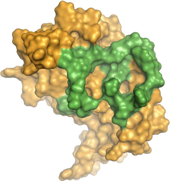
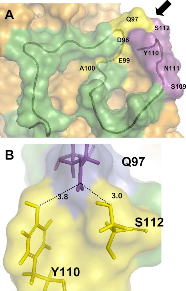
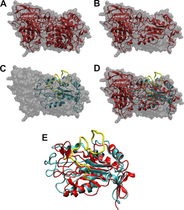
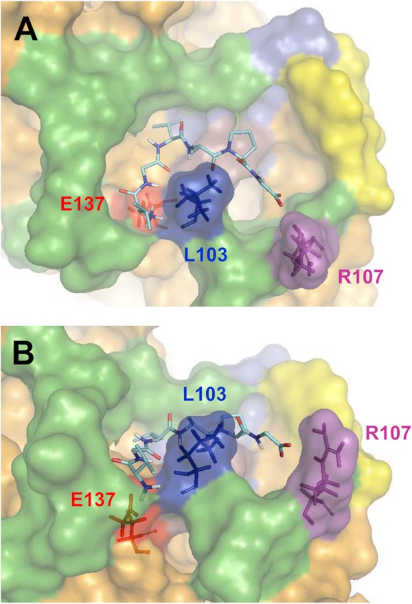
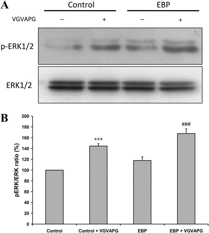
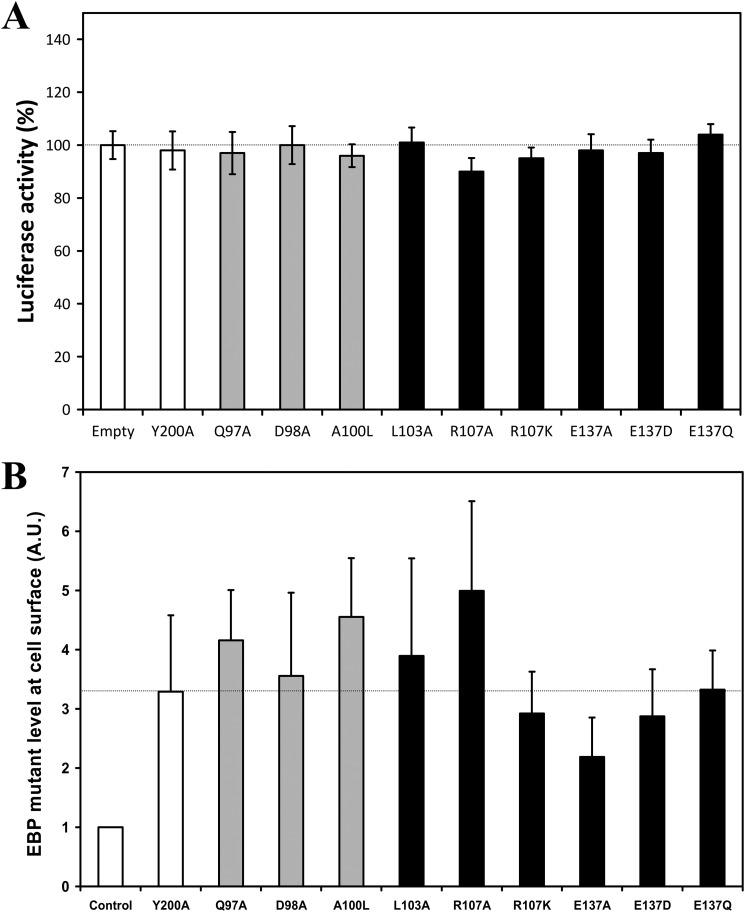
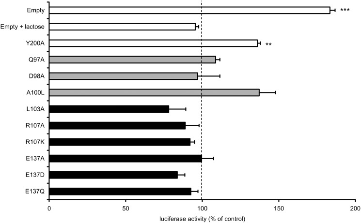
The elastin binding protein (EBP), a spliced variant of lysosomal β-galactosidase, is the primary receptor of elastin peptides that have been linked to emphysema, aneurysm and cancer progression. The sequences recognized by EBP share the XGXXPG consensus pattern found in numerous matrix proteins, notably in elastin where the VGVAPG motif is repeated. To delineate the elastin binding site of human EBP, we built a homology model of this protein and docked VGVAPG on its surface. Analysis of this model suggested that Gln-97 and Asp-98 were required for interaction with VGVAPG because they contribute to the definition of a pocket thought to represent the elastin binding site of EBP. Additionally, we proposed that Leu-103, Arg-107, and Glu-137 were essential residues because they could interact with VGVAPG itself. Site-directed mutagenesis experiments at these key positions validated our model. This work therefore provides the first structural data concerning the interaction of the VGVAPG with its cognate receptor. The present structural data should now allow the development of EBP-specific antagonists.


Figures

FIGURE 1.
FIGURE 1.
Homology model of EBP. This model was built using the MODELLER package as described under “Experimental Procedures.” The surface of EBP is colored orange. The surface corresponding to V32 is colored green.
FIGURE 2.
FIGURE 2.
Analysis of the elastin binding site. A, conformation of the V32 sequence in EBP (green tube). The surface defined by the QDEA (yellow) and SYNS (magenta) strings are labeled. The arrow indicates the orientation permitting to observe B. B, stick representation of Gln-97 (magenta) and the SYNS string (yellow) local organization. The thick sticks correspond to residues (Gln-97 (Q97), Tyr-110 (Y110), Ser-112 (S112)) orienting groups in a local geometry favorable to H-bonding. The distance between these groups are reported in angstroms.
FIGURE 3.
FIGURE 3.
Comparison of the homology model of EBP with the crystal structure of human β-galactosidase. A, crystal structure of human β-gal (Protein Data Bank code 3THC). The surface of the molecule is colored gray. The secondary structure of the enzyme is represented as a red schematic. The catalytic TIM barrel is on the right side. B, same representation than A in which the sequences spliced in EBP have been removed. C, molecular model of EBP overlaid on the surface envelope of human β-gal. The molecule is represented as a cyan schematic. The yellow line corresponds to the backbone trace of V32. D, superposition of B and C. E, overlay of β-gal TIM barrel after splicing (from B) and our EBP model.
FIGURE 4.
FIGURE 4.
Model of VGVAPG docked on EBP. A, the VGVAPG peptide, which is located in the pocket defined by the V32 sequence (green), is represented as sticks. The three residues stabilizing the interaction are colored in red (Glu-137), dark blue (Leu-103), and violet (Arg-107). The yellow surface corresponds to QDEA, the magenta surface corresponds to SYNS. B, other view of the docked peptide permitting to better appreciate the position of Glu-137, Leu-103, and Arg-107.
FIGURE 5.
FIGURE 5.
VGVAPG activates the ERK pathway in transfected COS-7 cells. A, Western blots of pERK and ERK levels in COS-7 transfected with an empty vector or with WT-EBP, after or without VGVAPG treatment. Cells were incubated for 24 h in 200 μl of serum-free DMEM with a mixture of Lipofectamine 2000 and DNA plasmids for 4 h. Cells were allowed to rest for 24 h in a serum-free medium before a 30-min VGVAPG stimulation (150 μg/ml) at 37 °C. B, corresponding densitometric analysis. The pERK/ERK ratio is compared with the corresponding basal situation in the absence of VGVAPG treatment. The excitability of the system was evaluated using a two-tailed Student's t test. Control COS-7 cells versus VGVAPG-treated COS-7 cells (***, p < 0.001); EBP-transfected cells versus VGVAPG-stimulated EBP-transfected cells (###, p < 0.001).
FIGURE 6.
FIGURE 6.
Effect of EBP mutant transfection on basal luciferase activity and EBP mutant levels at cell surface in transfected COS-7 cells. A, COS-7 cells were co-transfected with constructs encoding luciferase under the control of MMP-1 promoter, the indicated mutant EBP constructs, or with an empty vector. Cells were then stimulated with the VGVAPG peptide (150 μg/ml). Basal luciferase activity is expressed as a percentage of the control condition (empty vector). B, COS-7 cells were transiently transfected by 2 μg of EBP-FLAG mutant cDNA for 24 h. Cells were further biotinylated with 500 μg/ml of biotin, washed three times, and sonicated. Human EBP-FLAG mutants were then solubilized for 1 h at 4 °C and centrifuged at 20,000 × g for 30 min at 4 °C. The biotinylated proteins were precipitated for 45 min at 4 °C with 20 μl of streptavidin beads. After elution, retained proteins were analyzed by Western blots using an anti-FLAG antibody. The presented results correspond to the densitometric analysis of three experiments after normalization. Control, COS-7 cells transfected with the Y200A EBP-FLAG construct without biotinylation. Gray bars, mutations in the QDEA string; black bars, mutations of Leu-103, Arg-107, and Glu-137. No statistical difference was observed between the estimated EBP-FLAG levels in transfected cells as compared with the Y200A level (two-tailed Student's t test). A.U., arbitrary units.
FIGURE 7.
FIGURE 7.
Site-directed mutagenesis exploration of VGVAPG binding site. COS-7 cells were co-transfected with constructs encoding luciferase under the control of MMP-1 promoter, the indicated mutant EBP constructs, or with an empty vector. Cells were then stimulated with the VGVAPG peptide (150 μg/ml) in the absence or presence of 1 mm lactose (EBP antagonist). The results correspond to the luciferase activity measured after VGVAPG treatment. They are expressed as a percentage of luciferase activity observed for the same situation without treatment. This control value was set to 100% for all experiments (dashed line). White bars, control experiments; gray bars, mutations in the QDEA string; black bars, mutations of Leu-103, Arg-107, and Glu-137. The reported significance compares the treated versus untreated cells (one-tailed Student's t test). **, p < 0.01; ***, p < 0.001.
FIGURE 8.
FIGURE 8.
Possible role of Glu-137 in the interplay between the elastin and galactolectin sites. A, sequence alignment showing the strict conservation of Glu-137 in β-galactosidases. Alignment ruler, *, identity' :, homology; ., similarity. B, after alignment of the protein backbone of their TIM barrels (human β-Gal and human EBP), the relative positions of Glu-268 interacting with galactose (in red, from Protein Data Bank code 3THC) and Glu-137 interacting with VGVAPG (from our model) suggest a possible competition between galactose and VGVAPG for Glu-137 binding. This competition at a common site could explain why EBP galactolectin occupancy blocks elastin peptide effects.

Similar articles

Cited by

References

    1. Kielty C. M. (2006) Elastic fibres in health and disease. Expert Rev. Mol. Med. 8, 1–23 - PubMed
    1. Vrhovski B., Weiss A. S. (1998) Biochemistry of tropoelastin. Eur. J. Biochem. 258, 1–18 - PubMed
    1. Shapiro S. D., Endicott S. K., Province M. A., Pierce J. A., Campbell E. J. (1991) Marked longevity of human lung parenchymal elastic fibers deduced from prevalence of d-aspartate and nuclear weapons-related radiocarbon. J. Clin. Invest. 87, 1828–1834 - PMC - PubMed
    1. Hinek A. (1994) Nature and the multiple functions of the 67-kD elastin-/laminin binding protein. Cell Adhes. Commun. 2, 185–193 - PubMed
    1. Hinek A., Rabinovitch M., Keeley F., Okamura-Oho Y., Callahan J. (1993) The 67-kD elastin/laminin-binding protein is related to an enzymatically inactive, alternatively spliced form of β-galactosidase. J. Clin. Invest. 91, 1198–1205 - PMC - PubMed

Publication types