. 2013 Jan 4;288(1):624-32.
doi: 10.1074/jbc.M112.340455. Epub 2012 Nov 19.

Nuclear factor of activated T-cells (NFAT)C2 inhibits Notch receptor signaling in osteoblasts

Affiliations

Nuclear factor of activated T-cells (NFAT)C2 inhibits Notch receptor signaling in osteoblasts

Stefano Zanotti et al. J Biol Chem. .

Abstract

Notch receptors regulate osteoblastogenesis, and Notch activation induces cleavage and nuclear translocation of the Notch intracellular domain (NICD), which associates with Epstein-Barr virus latency C-promoter binding factor-1/suppressor of hairless/lag-1 (CSL) and induces transcription of Notch target genes, such as hairy enhancer of split-related with YRPW motif (Hey)1 and Hey2. Nuclear factors of activated T-cells (NFAT) are transcription factors that regulate osteoclastogenesis, but their function in osteoblasts is not clear. Notch inhibits NFATc1 transcription, but interactions between Notch and NFAT are understood poorly. To determine the regulation of NFAT expression by Notch, osteoblasts from Rosa(Notch) mice, where NICD is transcribed following excision of a loxP flanked STOP cassette, were used. Alternatively, wild-type C57BL/6 osteoblasts were exposed to the Notch ligand Delta-like (Dll)1 to induce Notch signaling or to bovine serum albumin as control. In Rosa(Notch) osteoblasts, Notch suppressed NFATc1 expression, increased Nfatc2 mRNA by post-transcriptional mechanisms, and had no effect on NFATc3 and NFATc4 transcripts. Induction of Nfatc2 transcripts by Notch was confirmed in C57BL/6 osteoblasts exposed to Dll1. To investigate NFATc2 function in osteoblasts, constitutively active NFATc2 was overexpressed in Rosa(Notch) osteoblasts. NFATc2 suppressed Notch transactivation and expression of Hey genes. Electrophoretic mobility shift assays revealed that NFATc2 and CSL bind to similar DNA sequences, and chromatin immunoprecipitation indicated that NFATc2 displaced CSL from the Hey2 promoter. The effects of NICD and NFATc2 in Rosa(Notch) osteoblasts were assessed, and both proteins inhibited osteoblast function. In conclusion, Notch stabilizes Nfatc2 transcripts, NFATc2 suppresses Notch signaling, and both proteins inhibit osteoblast function.


Figures

FIGURE 1.
FIGURE 1.
NICD induces NFATc2 expression in osteoblasts by post-transcriptional mechanisms. RosaNotch calvarial osteoblasts were infected with Ad-CMV-CRE (CRE, black bars or full circles) or control Ad-CMV-GFP (GFP, white bars or open circles). Wild-type C57BL/6 osteoblasts were exposed to immobilized Dll1 (Dll1, black bars), or control BSA (BSA, white bars). In A and C, RosaNotch osteoblasts were cultured for 3 days (A) or 1 day (C) after confluence, and total RNA was extracted, reverse-transcribed, and amplified by qRT-PCR. Data are expressed as ratio of Nfatc1, Nfatc2, Nfatc3, Nfatc4 (A), and NFATc2 hnRNA (C) copy number corrected for Rpl38 expression, relative to corrected expression in GFP. Values are means ± S.E., n = 4. *, significantly different between CRE and GFP, p < 0.05. Data are from representative experiments performed in duplicate. In B, total cellular extracts from RosaNotch osteoblasts were fractioned by gel electrophoresis and transferred to an Immobilon-P membrane, which was incubated with an antibody against NFATc2, stripped, and reprobed with an antibody against actin. Digital images were acquired, and signal from NFATc2 and actin antibodies was quantified by densitometry to estimate protein expression. Data are expressed as ratio of NFATc2 levels corrected for actin expression relative to corrected NFATc2 levels in GFP. Values are means of four independent experiments ± S.E. *, significantly different between CRE and GFP, p < 0.05. In D, osteoblasts from wild-type C57BL/6 were cultured for 3 days after confluence, and total RNA was extracted, reverse-transcribed, and amplified by qRT-PCR. Data are expressed as ratio of Hey2 or Nfatc2 copy number corrected for Rpl38 expression, relative to corrected expression in BSA. Values are means ± S.E., n = 4. *, significantly different between Dll1 and BSA, p < 0.05. In E, RosaNotch osteoblasts were transcriptionally arrested with DRB (time 0) and harvested at the indicated times. Total RNA was extracted and amplified by qRT-PCR, and data are expressed as the percentage of Nfatc2 mRNA copy number corrected for Rpl38 expression, relative to the corrected Nfatc2 mRNA expression before DRB treatment, plotted versus time. Data are means ± S.E., n = 4. Nfatc2 decay slopes for CRE and GFP are statistically different, p < 0.05. Data are from representative experiments performed in triplicate.
FIGURE 2.
FIGURE 2.
NFATc2 is expressed and activated in osteoblasts. RosaNotch or wild-type C57BL/6 calvarial osteoblasts were transduced with Ad-CMV-caNFATc2 tagged with HA (NFATc2, black bars) or control Ad-CMV-GFP (Control, white bars). RosaNotch osteoblasts were co-transduced with Ad-CMV-CRE (CRE) to induce Notch, or control Ad-CMV-GFP (GFP), and cultured for 72 h. In A, total cellular extracts from RosaNotch osteoblasts were fractioned by gel electrophoresis and transferred to an Immobilon-P membrane, which was incubated with an antibody against NFATc2. Membranes were stripped and reprobed with an antibody against actin. In B and D, total RNA was extracted from RosaNotch osteoblasts and amplified by qRT-PCR. Data are expressed as ratio of Nfatc2 and Rcan1.4 copy number corrected for Rpl38 expression, relative to corrected expression in control cells co-transduced with Ad-CMV-GFP. Values are means ± S.E., n = 4. *, significantly different between NFATc2 and control, p < 0.05. +, significantly different between CRE and GFP, p < 0.05. In C, wild-type C57BL/6 osteoblasts were cultured to confluence, and DNA was cross-linked to associated proteins and fragmented by sonication. DNA-protein complexes were obtained by immunoprecipitation with an HA antibody or control IgG, and DNA was purified following reversal of the cross-linking reaction. Immunoprecipitated DNA and fragmented input DNA were amplified by qPCR with primers flanking Nfatc consensus sequences required for the activity of the Rcan1.4 promoter. Data are expressed as enrichment of DNA by immunoprecipitation with the HA antibody relative to control IgG, corrected for input DNA levels. Values are means of three qPCR reactions. Data are from representative experiments performed in duplicate.
FIGURE 3.
FIGURE 3.
NFATc2 suppresses Notch canonical signaling in osteoblasts. RosaNotch calvarial osteoblasts were transduced with Ad-CMV-caNFATc2 (NFATc2, black bars) or control Ad-CMV-GFP (Control, white bars) and co-transduced with Ad-CMV-CRE (CRE) to induce Notch or control Ad-CMV-GFP (GFP). In A, osteoblasts were cultured to subconfluence and transiently transfected with the 12×CSL-Luc reporter or with the Hey1-Luc or Hey2-Luc promoter constructs, co-transfected with a CMV/β-galactosidase expression vector, and harvested after 48 h. Data shown represent luciferase/β-galactosidase activity. Values are means ± S.E., n = 6. In B, osteoblasts were cultured for 72 h after confluence, and total RNA was extracted and amplified by qRT-PCR. Data are expressed as ratio of Hey1 or Hey2 copy number, corrected for Rpl38 expression, relative to corrected expression in control cells co-transduced with Ad-CMV-GFP. Values are means ± S.E., n = 4. *, significantly different between NFATc2 and control, p < 0.05. +, significantly different between CRE and GFP, p < 0.05. Data are from representative experiments performed in duplicate.
FIGURE 4.
FIGURE 4.
NFATc2 and CSL bind to similar DNA consensus sequences in osteoblasts. Binding of nuclear proteins to DNA was tested by electrophoretic mobility shift assay in RosaNotch calvarial osteoblasts transduced with Ad-CMV-caNFATc2 (NFATc2) or control Ad-CMV-GFP (Control) and co-transduced with Ad-CMV-CRE (CRE) to induce Notch or control Ad-CMV-GFP (GFP). Cells were cultured to confluence, and nuclear extracts were incubated with [γ-32P]ATP-labeled oligonucleotides. In A, a radiolabeled oligonucleotide containing a Csl consensus sequence from the EBNA2 promoter was used. Competition of binding reactions was performed in the presence of unlabeled Csl consensus and mutant oligonucleotides or unlabeled Nfatc oligonucleotides containing homologous or mutated consensus sequences from the Bnp promoter in 200-fold excess. In B, a radiolabeled oligonucleotide containing an Nfatc consensus sequence from the Bnp promoter was used. Competition of binding reactions was performed in the presence of unlabeled Nfatc consensus and mutant oligonucleotide or unlabeled Csl oligonucleotides containing homologous or mutated consensus sequences from the EBNA2 promoter in 200-fold excess. DNA-nuclear protein complexes were resolved by gel electrophoresis and visualized by autoradiography, and arrows indicate the position of complexes. Autoradiographs are representative of four independent experiments.
FIGURE 5.
FIGURE 5.
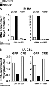
NFATc2 opposes the association of CSL to the Hey2 promoter in osteoblasts. Binding of NFATc2 and CSL to the Hey2 promoter was tested by ChIP in RosaNotch calvarial osteoblasts transduced with Ad-CMV-caNFATc2 tagged with HA (NFATc2) or control Ad-CMV-GFP (Control) and co-transduced with Ad-CMV-CRE (CRE) to induce Notch or control Ad-CMV-GFP (GFP). Cells were cultured to confluence, DNA cross-linked to associated proteins, and fragmented by sonication. DNA-protein complexes were obtained by immunoprecipitation (I.P.) with an HA (A) or a CSL (B) antibody or control IgG, and DNA was purified following reversal of the cross-linking reaction. Immunoprecipitated DNA and fragmented input DNA were amplified by qPCR with primers flanking Csl consensus sequences that regulate the activity of the Hey2 promoter. Data are expressed as enrichment of DNA by immunoprecipitation with the HA or CSL antibody relative to control IgG, corrected for input DNA levels. Values are means of three qPCR reactions.
FIGURE 6.
FIGURE 6.
NICD and NFATc2 regulate osteoblast function. RosaNotch calvarial osteoblasts were transduced with Ad-CMV-caNFATc2 (NFATc2, black bars) or control Ad-CMV-GFP (Control, white bars) and co-transduced with Ad-CMV-CRE (CRE) to induce Notch or control Ad-CMV-GFP (GFP), and cells were cultured for 7–14 days in conditions favoring osteoblast differentiation. In A, following 7 days of culture, total RNA was extracted and amplified by qRT-PCR. Data are expressed as ratio of Dlx5, Runx2, Alpl, Bsp, osteocalcin, Col1a1, and osteopontin copy number, corrected for Rpl38 expression, relative to corrected expression in control cells co-transduced with Ad-CMV-GFP. Values are means ± S.E., n = 4, and representative results from two independent experiments are shown. In B, cells were cultured for 7 days and extracted with Triton X-100 for determination of alkaline phosphatase activity, expressed as nanomoles of p-nitrophenol/min/μg of total protein. Values are means from four independent experiments ± S.E. In C, cells cultured for 14 days were fixed and stained with Alizarin Red, digital images were acquired, and the mineralized area was quantified with the measure function of ImageJ software. A representative culture from two independent experiments is shown. Values are means ± S.E., n = 6. *, significantly different between NFATc2 and control. +, significantly different between CRE and GFP, p < 0.05. In A and B, selected data from control cells co-transduced with CRE or GFP were published previously (14).

Similar articles

Cited by

References

    1. Fortini M. E. (2009) Notch signaling: the core pathway and its posttranslational regulation. Dev. Cell 16, 633–647 - PubMed
    1. D'Souza B., Miyamoto A., Weinmaster G. (2008) The many facets of Notch ligands. Oncogene 27, 5148–5167 - PMC - PubMed
    1. Ehebauer M., Hayward P., Martinez-Arias A. (2006) Notch signaling pathway. Sci. STKE 2006, cm7. - PubMed
    1. Kovall R. A. (2008) More complicated than it looks: assembly of Notch pathway transcription complexes. Oncogene 27, 5099–5109 - PubMed
    1. Iso T., Kedes L., Hamamori Y. (2003) HES and HERP families: multiple effectors of the Notch signaling pathway. J. Cell. Physiol. 194, 237–255 - PubMed

Publication types

LinkOut - more resources