. 2013 Jan 11;288(2):886-93.
doi: 10.1074/jbc.M112.417089. Epub 2012 Nov 19.

Functional expression of SCL/TAL1 interrupting locus (Stil) protects retinal dopaminergic cells from neurotoxin-induced degeneration

Affiliations

Functional expression of SCL/TAL1 interrupting locus (Stil) protects retinal dopaminergic cells from neurotoxin-induced degeneration

Jingling Li et al. J Biol Chem. .

Abstract

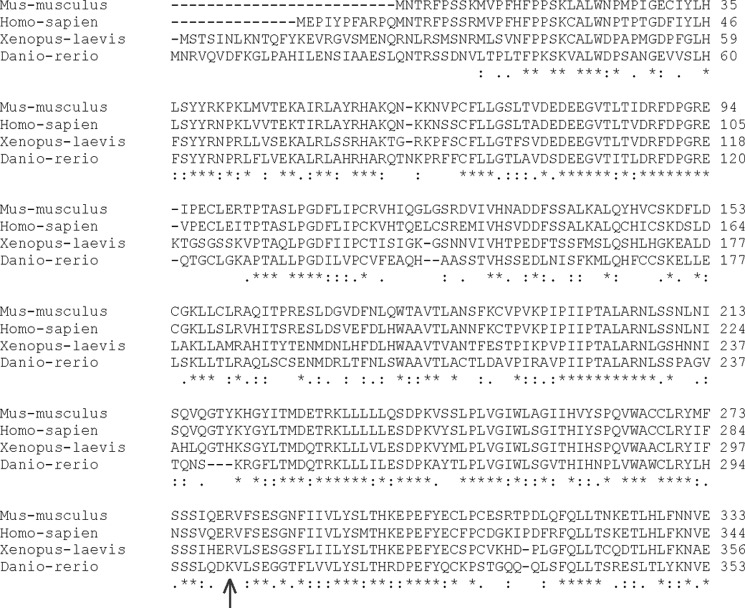
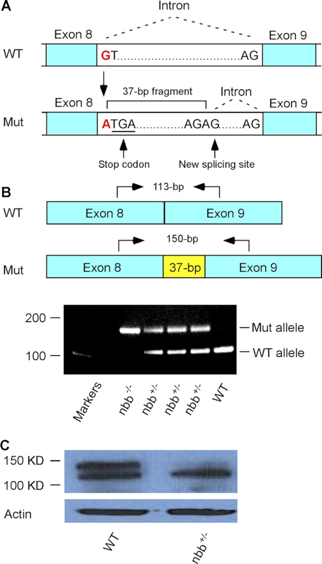
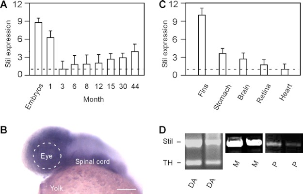
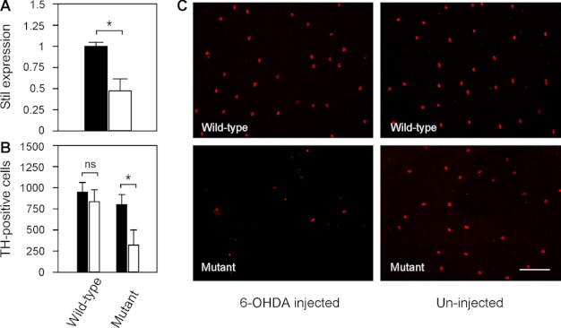
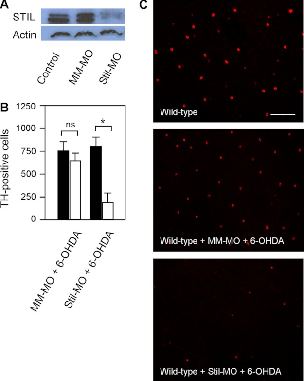
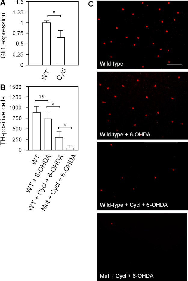
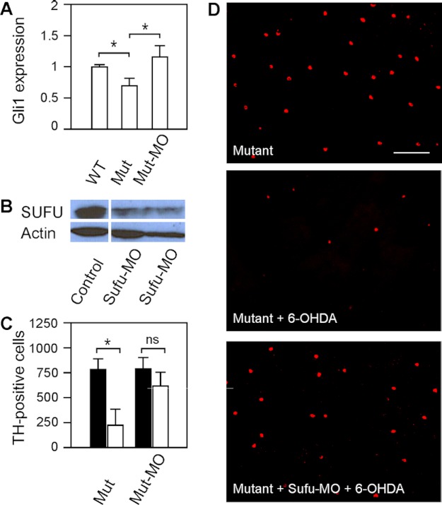
We previously isolated a dominant mutation, night blindness b (nbb), which causes a late onset of retinal dopaminergic cell degeneration in zebrafish. In this study, we cloned the zebrafish nbb locus. Sequencing results revealed that nbb is a homolog of the vertebrate SCL/TAL1 interrupting locus (Stil). The Stil gene has been shown to play important roles in the regulation of vertebrate embryonic neural development and human cancer cell proliferation. In this study, we demonstrate that functional expression of Stil is also required for neural survival. In zebrafish, decreased expression of Stil resulted in increased toxic susceptibility of retinal dopaminergic cells to 6-hydroxydopamine. Increases in Stil-mediated Shh signaling transduction (i.e. by knocking down the Shh repressor Sufu) prevented dopaminergic cell death induced by neurotoxic insult. The data suggest that the oncogene Stil also plays important roles in neural protection.


Figures

FIGURE 1.
FIGURE 1.
Comparison of STIL protein sequences in different vertebrate species. The online CLUSTALW tool was used to align partial sequences of STIL proteins (N terminus). Amino acids are numbered on the right side of the sequence. In nbb mutants, the predicted STIL protein is truncated after amino acid Lys-301 (arrow). Asterisk, fully conserved; colon, strongly conserved; period, weakly conserved.
FIGURE 2.
FIGURE 2.
Analysis of zebrafish nbb mutation. A, a diagram that shows the mutation at the beginning of intron 8–9. The substitution of nucleotides from G to A altered the donor splicing site, resulting in a 37-bp insertion with a premature stop codon. B, RT-PCR analysis of Stil expression in wild-type, heterozygous, and homozygous nbb mutants. In the diagram, arrows indicate RT-PCR primer sites used to amplify the region at exon 8–9 junction from cDNA. In heterozygous mutants (nbb+/−), both wild-type (113-bp) and mutant (150-bp) alleles were amplified. In homozygous mutants (nbb−/−), only the 150-bp fragment was amplified. In wild-type embryos, the 113-bp fragment was amplified. C, Western blot of retinal lysates from wild-type and heterozygous nbb mutant fish. The mutant fish showed decreased STIL protein expression. Mut, mutant.
FIGURE 3.
FIGURE 3.
The expression of Stil in developing embryos and adult zebrafish. A, qRT-PCR analysis of Stil expression at different ages. The lowest expression in 3-month-old fish was normalized to 1. The highest expression was detected in developing embryos (at 1 day post-fertilization). Data represent the means ± S.E. (n > 30 in embryos; n = 9 in each juvenile and adult stage). B, in situ hybridization against Stil mRNA at 1 day post-fertilization. Stil was highly expressed in the brain and spinal cord. Scale bar, 100 μm. C), qRT-PCR analysis of Stil expression in different tissues and organs isolated from adult zebrafish (6 months old). The lowest expression in the heart was normalized to 1. Data represent the means ± S.E. (n = 9). D, single-cell RT-PCR analysis of Stil expression in isolated DA-IPCs (DA), Müller glia cells (M), and photoreceptor cells (P). TH primers were used to confirm the DA cell type.
FIGURE 4.
FIGURE 4.
Toxicity of DA-IPCs to 6-OHDA in wild-type and mutant retinas. A, relative Stil mRNA expression in wild-type and nbb mutant retinas. The expression of Stil in wild-type retinas was normalized to 1 (black bar). Note the decrease of Stil expression in nbb mutant retinas (white bar). Data represent the means ± S.E. (n = 4 in each group). *, p < 0.01. B, number of anti-TH positive retinal DA-IPCs after injection of subtoxic 6-OHDA (white bars) or PBS (black bars). In wild-type fish, injection of subtoxic 6-OHDA produced no effects on DA-IPCs. In nbb retinas, the same treatment resulted in degeneration of DA-IPCs. Data represent the means ± S.E. (n = 3 in each group). ns, not significant; *, p < 0.01. C, fluorescent images of flat-mount wild-type and nbb mutant retinas labeled with anti-TH antibodies. Note the decreased number of DA-IPCs in mutant retinas after subtoxic 6-OHDA treatment. The images were taken at the nasal retina adjacent to the optic nerve. Scale bar, 100 μm.
FIGURE 5.
FIGURE 5.
Effects of STIL knockdown on DA-IPCs in wild-type retinas. A, Western blot of retinal lysates probed with anti-STIL antibodies. STIL was detected in wild-type and mismatched MO-treated (MM-MO) retinas, but not in Stil-MO-treated retinas (SM-MO). B, number of anti-TH positive DA-IPCs in MM-MO- and Stil-MO-treated wild-type retinas that received subtoxic 6-OHDA (white bars) or PBS (black bars) injections. Note the decrease in the number of DA-IPCs in Stil-MO-injected retinas after 6-OHDA treatment. Data represent the means ± S.E. (n = 5 in each group). ns, not significant; *, p < 0.01. C, fluorescent images of 6-OHDA-treated flat-mount retinas labeled with anti-TH antibodies. Note the decreased number of DA-IPCs in Stil-MO-treated retinas after 6-OHDA treatment. The images were taken at the nasal retina adjacent to the optic nerve. Scale bar, 100 μm.
FIGURE 6.
FIGURE 6.
Effects of 6-OHDA on DA-IPCs in wild-type retinas treated with Shh signaling antagonist cyclopamine. A, RT-PCR analysis of Gli1 expression in control and cyclopamine-treated retinas. The injection of cyclopamine resulted in decreased Shh-targeted Gli1 gene expression. Data represent the means ± S.E. (n = 3 in each group). *, p < 0.01. B, number of anti-TH positive DA-IPCs in control, 6-OHDA-treated, and cyclopamine (Cycl)- and 6-OHDA-co-treated wild-type and heterozygous mutant retinas. Note the decrease in the number of DA-IPCs in co-treated retinas. Data represent the means ± S.E. (n = 9 in each group). ns, not significant; *, p < 0.01. C, fluorescent images of flat-mount control and treated retinas labeled with anti-TH antibodies. Note the decreased number of DA-IPCs in cyclopamine and 6-OHDA co-treated retinas. The images were taken at the nasal retina adjacent to the optic nerve. Scale bar, 100 μm. Mut, mutant.
FIGURE 7.
FIGURE 7.
Effects of 6-OHDA on DA-IPCs in mutant retinas after Shh up-regulation by Sufu knockdown. A, relative Gli1 mRNA expression in wild-type, nbb mutant, and Sufu-MO-treated nbb mutant retinas. The expression of Gli1 in wild-type retinas was normalized to 1. The expression of Gli1 decreased in mutant retinas but increased when the expression of Sufu was inhibited. Data represent the means ± S.E. (n = 3 in each group). *, p < 0.01. B, Western blot of retinal lysates probed with anti-SUFU antibodies. Note the decrease of Sufu expression in Sufu-MO treated retinas. C, number of anti-TH positive DA-IPCs in untreated and Sufu-MO-treated nbb retinas in response to subtoxic 6-OHDA (white bars) or PBS (black bars) injections. In nbb mutants, subtoxic 6-OHDA treatment resulted in significant losses of DA-IPCs. Treatment with Sufu-MO protected DA-IPCs from degeneration after exposure to 6-OHDA. Data represent the means ± S.E. (n = 8 in each group). *, p < 0.01; ns, not significant. (D) Fluorescent images of flat-mount nbb retinas labeled with anti-TH antibodies. Note the decreased number of DA-IPCs in 6-OHDA-treated mutant retinas and the rescue after Sufu-MO treatments. The images were taken at the nasal retina adjacent to the optic nerve. Scale bar, 100 μm. Mut, mutant.

Similar articles

Cited by

References

    1. Fuchs Y., Steller H. (2011) Programmed cell death in animal development and disease. Cell 147, 742–758 - PMC - PubMed
    1. Rosello A., Warnes G., Meier U. (2012) Cell death pathways and autophagy in the central nervous system and its involvement in neurodegeneration, immunity and central nervous system infection: to die or not to die–that is the question. Clin. Exp. Immunol. 168, 52–57 - PMC - PubMed
    1. Parsadanian A. S., Cheng Y., Keller-Peck C. R., Holtzman D. M., Snider W. D. (1998) Bcl-XL is antiapoptotic regulator for postnatal CNS neurons. J. Neurosci. 18, 1009–1019 - PMC - PubMed
    1. Arbour N., Vanderluit J. L., Le Grand J. N., Jahani-Asl A., Ruzhynsky V. A., Cheung E. C., Kelly M. A., MacKenzie A. E., Park D. S., Opferman J. T., Slack R. S. (2008) Mcl-1 is a key regulator of apoptosis during CNS development and after DNA damage. J. Neurosci. 28, 6068–6078 - PMC - PubMed
    1. Scott D. A., Tabarean I., Tang Y., Cartier A., Masliah E., Roy S. (2010) A pathologic cascade leading to synaptic dysfunction in α-synuclein-induced neurodegeneration. J. Neurosci. 30, 8083–8095 - PMC - PubMed

Publication types

MeSH terms

LinkOut - more resources