. 2012 Nov 26;199(5):755-70.
doi: 10.1083/jcb.201202155. Epub 2012 Nov 19.

Mitotic chromosomes are compacted laterally by KIF4 and condensin and axially by topoisomerase IIα

Affiliations

Mitotic chromosomes are compacted laterally by KIF4 and condensin and axially by topoisomerase IIα

Kumiko Samejima et al. J Cell Biol. .

Abstract

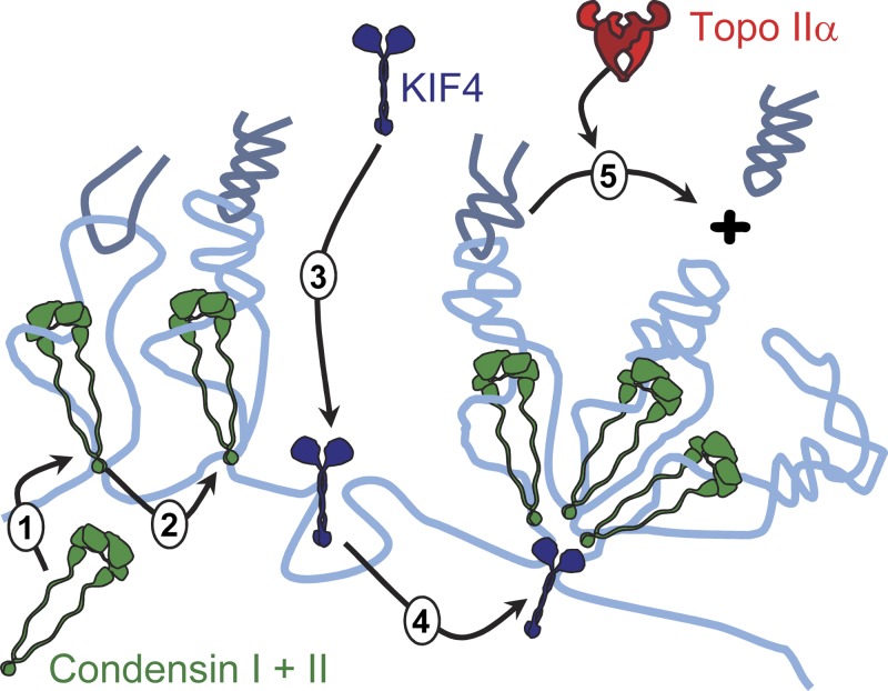
Mitotic chromosome formation involves a relatively minor condensation of the chromatin volume coupled with a dramatic reorganization into the characteristic "X" shape. Here we report results of a detailed morphological analysis, which revealed that chromokinesin KIF4 cooperated in a parallel pathway with condensin complexes to promote the lateral compaction of chromatid arms. In this analysis, KIF4 and condensin were mutually dependent for their dynamic localization on the chromatid axes. Depletion of either caused sister chromatids to expand and compromised the "intrinsic structure" of the chromosomes (defined in an in vitro assay), with loss of condensin showing stronger effects. Simultaneous depletion of KIF4 and condensin caused complete loss of chromosome morphology. In these experiments, topoisomerase IIα contributed to shaping mitotic chromosomes by promoting the shortening of the chromatid axes and apparently acting in opposition to the actions of KIF4 and condensins. These three proteins are major determinants in shaping the characteristic mitotic chromosome morphology.


Figures

Figure 1.
Figure 1.
KIF4 partially colocalizes with SMC2 and topo IIα and affects condensin localization on the chromosome axis. (A) SMC2OFF cells expressing SMC2-TrAP were stained with guinea pig anti-topo IIα, mouse anti-SBP, and rabbit anti-KIF4 plus DAPI for the DNA. Bar, 5 µm. (B and C) Asynchronously growing SMC2OFF:SMC2-GFP-STS cells were transfected with either control or KIF4 siRNA oligos. After 20–24 h, these cells were fixed in PFA/PBS and stained by anti–CENP-T antibody and DAPI. Bars, 2 µm. (D) Quantification of B and C. Intensity of SMC2-GFP signal was measured at 5 kinetochores (Kt), 5 chromosome arms (Arm), and 5 areas in the cytosol in each of 20 cells. (E–G) Asynchronously growing SMC2OFF:SMC2-GFP-STS cells (E), CAP-HOFF:CAP-H-GFP-TrAP cells (F), or CAP-D3OFF:CAP-D3-GFP-TrAP cells (G) were treated as in B and C and stained with DAPI. Bars, 2 µm.
Figure 2.
Figure 2.
Interdependence of chromosome axis localization for KIF4 and SMC2. (A and B) KIF4ON/OFF, (C) SMC2OFF, and (D) Topo IIαOFF cells were stained with anti-KIF4, anti-SMC2, or anti-topo IIα antibodies plus DAPI. (E) Summary of localization dependencies. Bar, 5 µm.
Figure 3.
Figure 3.
KIF4 is highly mobile between chromosomes and cytosol. (A) Schematic presentation of the FRAP experiment. (B) Stills from the FRAP experiment of GFP-KIF4wt expressed in KIF4OFF cells blocked in metaphase using MG132. Bar, 5 µm. (C and D) FRAP analysis. Error bars show SD.
Figure 4.
Figure 4.
Proteomic analysis of KIF4OFF and SMC2OFF isolated chromosomes. (A) Dependency of proteins on KIF4 for their association with mitotic chromosomes. Proteins reduced in mitotic chromosomes isolated from KIF4OFF cells (relative to those on chromosomes isolated from wild-type cells) appear lower on the y axis. Proteins enriched on chromosomes relative to cytoplasm appear to the right on the x axis. This is Classifier II of Ohta et al. (2010a), and uses values from those experiments. Proteins most strongly depleted in this experiment include the chromatin proteins HMGN2 (a), HMGN1 (b), DEK (c), H3.3 (d), and H1.03 (e) and cytoplasmic proteins cytidine deaminase (f) and CKS1B (g). (B) Similar analysis of SMC2 dependency based on the published proteomic analysis of isolated chromosomes from wild-type and SMC2OFF cells (Ohta et al., 2010a). (C and D) Diagrams showing the dependency of selected chromosomal (C) and centromeric (D) proteins for their association with isolated mitotic chromosomes in the presence and absence of KIF4. Bars point downward when the corresponding protein is decreased in KIF4OFF chromosomes.
Figure 5.
Figure 5.
Architecture of chromosomes is compromised after KIF4 depletion. (A) KIF4ON/OFF prometaphase chromosomes. Asynchronously growing cells were subjected to hypotonic treatment (75 mM KCl for 5 min), fixed in 4% PFA in the same buffer, and stained with DAPI. Bar, 1 µm. (B) KIF4ON/OFF cells with a CENP-H-GFP knock-in (top) and or integrated LacO array and expressing LacI-GFP (bottom) were fixed and stained as in A. Bars, 1 µm. (C) Interkinetochore distance. CENP-H-GFP knock-in cells from B (top) were treated either with MG132 (tension +) or with colcemid (tension −) for 3 h and fixed in 4% PFA/PBS. n = 50 from 10 cells. Wider bars represent the mean distance. The difference is statistically significant (D = 0.42, P = 0.0003; KS test) only in the presence of tension. D = 0.1, P = 0.96 (Tension −). (D) Sister chromatid spacing. KIF4ON/OFF LacO–bearing cells expressing LacI-GFP (B, bottom) were treated as in C. n > 20. Differences were not statistically significant: D = 0.31, P = 0.11 (Tension +, KS test); D = 0.25, P = 0.45 (Tension −). Data are shown from a single representative experiment out of two repeats. (E) Intrinsic metaphase structure assay of KIF4ON/OFF chromosomes as described in the text. 100 cells were scored per sample. Bar, 5 µm. Data are shown from a single representative experiment out of two repeats. (F) Summary of chromosomal phenotypes exhibited after depletion of KIF4, SMC2, or Topo IIα. *, interkinetochore distance of SMC2ON/OFF cells (Vagnarelli et al., 2006).
Figure 6.
Figure 6.
Topo IIα is required for axial shortening during mitosis. (A) Cells treated with Dox for 43 h to induce expression of a Topo IIα–specific shRNA (1 and 3) were swollen in 75 mM KCl, fixed in methanol acetic acid, and stained by DAPI. This was done plus or minus addition of colcemid for a further 5 h (2 and 4). Bar, 5 µm. (B) For the double condensin/KIF4 knockdown, SMC2ON/OFF cells (lane 2) were transfected with KIF4 siRNA oligonucleotides (lane 3), with addition of doxycycline (30 h) resulting in the double depletion (lane 4). α-Tubulin was used as a loading control. (C) For the double condensin/topo IIα knockdown, topo IIα inducible shRNA cells were treated with KIF4 siRNA oligos (lane 2), with addition of doxycycline (48 h) resulting in the double depletion (lane 4). α-Tubulin was used as a loading control. (D) Chromosome width of cells in C was measured at randomly selected noncentromeric positions. The mean is shown by a heavy bar. D- and P-values were obtained by: The difference is statistically significant between control and KIF4-depleted (D = 0.64, P < 0.0001; KS test) or Topo IIα–depleted (D = 0.52, P < 0.0001 cells), but not double-depleted (D = 0.14, P = 0.71) cells. Data are shown from a single representative experiment out of two repeats.
Figure 7.
Figure 7.
KIF4 and SMC2 act in parallel to shape mitotic chromosomes and are opposed by Topo IIα. (A) SMC2ON/OFF cells treated as shown were washed in either PBS or 75 mM KCl and fixed in methanol/acetic acid. (B) SMC2ON/OFF cells were transfected with either control or KIF4 or Topo IIα siRNA as in Fig. 6, B–D. Simultaneous transfection of the SMC2ON/OFF cells with KIF4 and Topo IIα siRNA oligos gave the triple knockout. Cells were washed in PBS or 75 mM KCl (hypotonic treatment), fixed in methanol acetic acid, and stained with DAPI. Bars, 5 µm.
Figure 8.
Figure 8.
KIF4 motor domain is required for its chromosomal functions. (A) Schematic representation of KIF4 and the fragments expressed in KIF4OFF cells. Right, chromosome axis localization is shown as + or − for each GFP fusion protein. (B) KIF4OFF cells expressing GFP-KIF4wt or GFP-KIF4520-1226 were treated with 75 mM KCl, fixed in PFA/75 mM KCl, and stained with DAPI. Bars, 5 µm. (C) Interkinetochore distance in the presence of tension. KIF4ON/OFF cells or KIF4OFF cells expressing GFP-KIF4wt or GFP-KIF4520-1226 were treated as in Fig. 5 C and stained with anti CENP-T antibody. More than 50 kinetochore pairs were measured per cell line. D- and P-values were obtained by KS test. Compared with KIF4ON, D = 0.58, P < 0.001 (KIF4OFF); D = 0.30, P = 0.01 (GFP-KIF4wt); and D = 0.49, P < 0.001 (GFP-KIF4520-1226). Compared with KIF4OFF, D = 0.26, P = 0.058 (GFP-KIF4520-1226). Data are shown from a single representative experiment out of two repeats. (D) Intrinsic metaphase structure assay. KIF4ON/OFF cells or KIF4OFF cells expressing GFP-KIF4wt or GFP-KIF4520-1226 were treated as for Fig. 5 E. 100 cells were scored for each cell line. Data are shown from a single representative experiment out of two repeats.
Figure 9.
Figure 9.
Morphological and structural consequences of depleting condensin, KIF4, and topo IIα on mitotic chromosomes. Shaping of mitotic chromosomes requires coordinated action of KIF4, condensins, and topo IIα. KIF4 and SMC2 are required for lateral compaction, whereas topo IIα is required for axial shortening. (a–d) Chromosomes prepared under the most “native” conditions consistent with visualization of individual chromosomes showed relatively minor perturbations after depletion of condensin or KIF4, but were significantly defective in the intrinsic metaphase structure assay. The morphological defects became more apparent when chromosomes were treated in hypotonic buffer (e–l).
Figure 10.
Figure 10.
Interplay between KIF4, condensin, and topo IIα in shaping mitotic chromosomes. A model for interactions between KIF4, condensin, and topo IIα during mitotic chromosome formation.

Comment in

References

    1. Adolph K.W., Cheng S.M., Laemmli U.K. 1977a. Role of nonhistone proteins in metaphase chromosome structure. Cell. 12:805–816 10.1016/0092-8674(77)90279-3 - DOI - PubMed
    1. Adolph K.W., Cheng S.M., Paulson J.R., Laemmli U.K. 1977b. Isolation of a protein scaffold from mitotic HeLa cell chromosomes. Proc. Natl. Acad. Sci. USA. 11:4937–4941 10.1073/pnas.74.11.4937 - DOI - PMC - PubMed
    1. Aizawa H., Sekine Y., Takemura R., Zhang Z., Nangaku M., Hirokawa N. 1992. Kinesin family in murine central nervous system. J. Cell Biol. 119:1287–1296 10.1083/jcb.119.5.1287 - DOI - PMC - PubMed
    1. Baxter J., Aragón L. 2012. A model for chromosome condensation based on the interplay between condensin and topoisomerase II. Trends Genet. 28:110–117 10.1016/j.tig.2011.11.004 - DOI - PubMed
    1. Baxter J., Sen N., Martínez V.L., De Carandini M.E., Schvartzman J.B., Diffley J.F., Aragón L. 2011. Positive supercoiling of mitotic DNA drives decatenation by topoisomerase II in eukaryotes. Science. 331:1328–1332 10.1126/science.1201538 - DOI - PubMed

Publication types

MeSH terms