. 2012;8(11):e1003012.
doi: 10.1371/journal.ppat.1003012. Epub 2012 Nov 15.

The capping domain in RalF regulates effector functions

Affiliations

The capping domain in RalF regulates effector functions

Eric Alix et al. PLoS Pathog. 2012.

Abstract

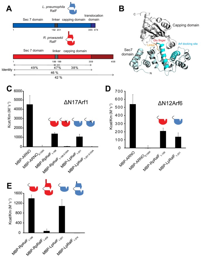
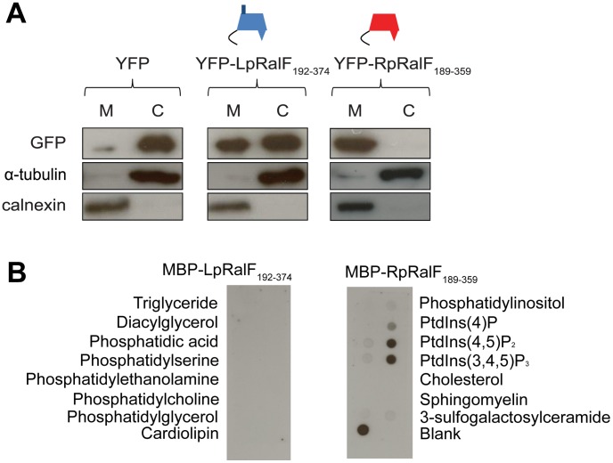
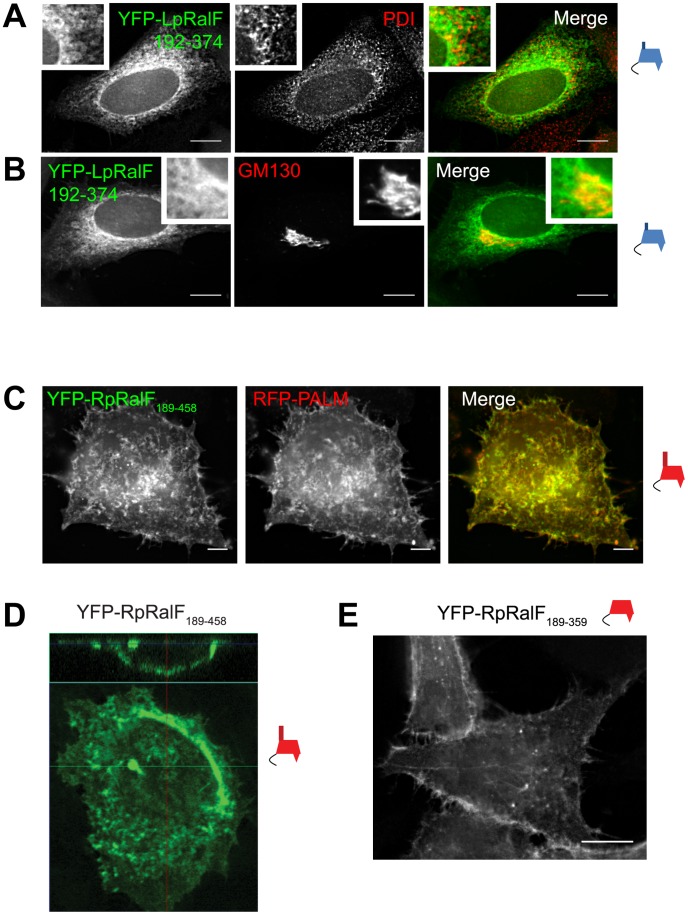
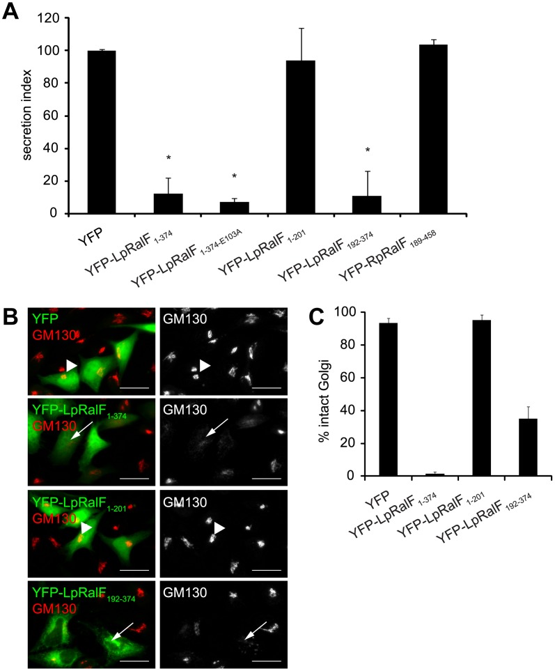
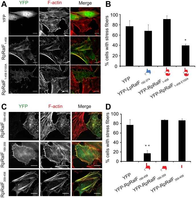
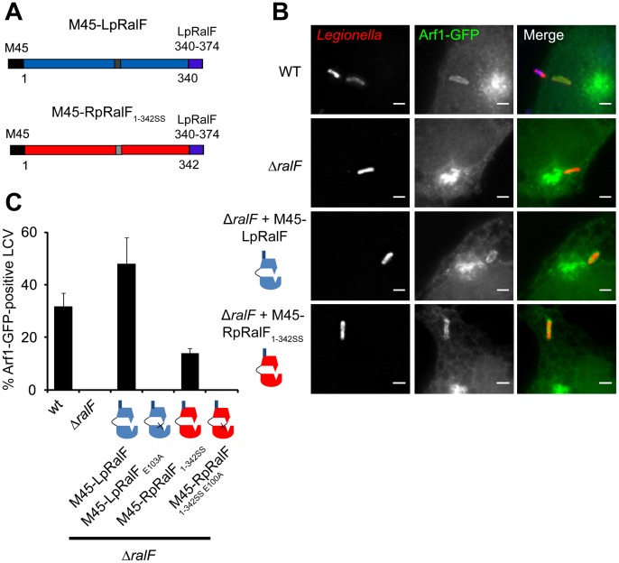
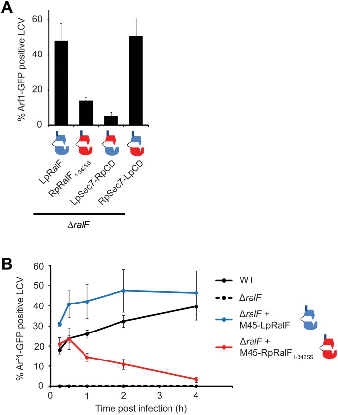
The Legionella pneumophila effector protein RalF functions as a guanine nucleotide exchange factor (GEF) that activates the host small GTPase protein ADP-ribosylation factor (Arf), and recruits this host protein to the vacuoles in which this pathogen resides. GEF activity is conferred by the Sec7 domain located in the N-terminal region of RalF. Structural studies indicate that the C-terminal region of RalF makes contacts with residues in the Sec7 domain important for Arf interactions. Theoretically, the C-terminal region of RalF could prevent nucleotide exchange activity by blocking the ability of Arf to interact with the Sec7 domain. For this reason, the C-terminal region of RalF has been termed a capping domain. Here, the role of the RalF capping domain was investigated by comparing biochemical and effector activities mediated by this domain in both the Legionella RalF protein (LpRalF) and in a RalF ortholog isolated from the unrelated intracellular pathogen Rickettsia prowazekii (RpRalF). These data indicate that both RalF proteins contain a functional Sec7 domain and that the capping domain regulates RalF GEF activity. The capping domain has intrinsic determinants that mediate localization of the RalF protein inside of host cells and confer distinct effector activities. Localization mediated by the capping domain of LpRalF enables the GEF to modulate membrane transport in the secretory pathway, whereas, the capping domain of RpRalF enables this bacterial GEF to modulate actin dynamics occurring near the plasma membrane. Thus, these data reveal that divergence in the function of the C-terminal capping domain alters the in vivo functions of the RalF proteins.


Conflict of interest statement

The authors have declared that no competing interests exist.

Figures

Figure 1
Figure 1. Legionella and Rickettsia RalF proteins are autoinhibited by their capping domain in vitro.
A) Alignment of the Legionella and Rickettsia RalF full length proteins. B) Structural organization of LpRalF protein. Ribbon rendering showing LpRalF Sec7 domain (blue), linker (orange) and capping domain (white) (Protein Data Bank accession code 1XSZ, image generated with PyMOL (http://pymol.sourceforge.net)) . The Sec7 catalytic glutamic acid side chain is represented as a stick with oxygen atoms shown in red. C) LpRalF and RpRalF Sec7 domains activate His-ΔN17Arf1 in vitro. Efficiency of His-ΔN17Arf1 nucleotide exchange catalyzed by the indicated MBP-tagged proteins. Kcat/Km values were obtained as described in Materials and Methods. Average and standard deviation are calculated from three independent experiments. D) Efficiency of His-ΔN12Arf6 nucleotide exchange catalyzed by MBP-tagged LpRalF and RpRalF Sec7 domains. Kcat/Km values were obtained as described in Materials and Methods. Average and standard deviation are calculated from three independent experiments. E) The RalF capping domain regulates GEF activity. Comparison of Kcat/Km values for His-ΔN17Arf1 nucleotide exchanged catalyzed by the Sec7 domain and the full length RalF proteins. Average and standard deviation are calculated from three independent experiments.
Figure 2
Figure 2. LpRalF and RpRalF capping domains associate with membranes by different mechanisms.
A) LpRalF and RpRalF capping domains are associated with the cell membrane fraction. HEK293 cells were transfected with YFP-LpRalF192–374, YFP-RpRalF189–359 or YFP alone. 24 hours after transfection, cells were lysed and centrifuged at 100,000 g for 1 h. The pellet (M) fraction was resuspended in a volume identical to the supernatant (C) fraction. Samples were blotted with GFP, calnexin (membrane marker) or α-tubulin (cytosol marker) antibodies. B) Protein-lipid overlay assay. The binding of MBP-LpRalF and RpRalF capping domains to indicated lipids immobilized on nitrocellulose membranes was analyzed using an anti-MBP antibody.
Figure 3
Figure 3. LpRalF and RpRalF capping domains associate with different subcellular compartments.
A) and B) Fluorescence microscopy of HeLa cells transfected with YFP-LpRalF192–374. Cells were fixed 24 hours after transfection, then stained with anti-PDI (A) or anti-GM130 (B) antibodies as indicated. Bar = 10 µm. C) Fluorescence microscopy of HeLa cells transfected with YFP-RpRalF189–458 and RFP-PALM. Bar = 5 µm. D) Confocal microscopy shows that RpRalF189–458 localizes at the plasma membrane. E) RpRalF capping domain is sufficient to target plasma membrane. Fluorescence microscopy of HeLa cells transfected with YFP-RpRalF189–359. Bar = 10 µm.
Figure 4
Figure 4. Legionella RalF capping domain disrupts secretion and the Golgi apparatus.
A) LpRalF capping domain disrupts secretion. Vectors encoding the indicated YFP-tagged L. pneumophila or R. prowazekii RalF constructs were co-transfected into CHO cells along with a plasmid encoding SEAP. Alkaline phosphatase secretion from CHO cells was plotted as the ratio of SEAP in the culture medium to cell-associated SEAP (Secretion Index). Vector alone (YFP) served as a negative control. These data are from at least 3 independent experiments done in triplicate. The results are normalized so the cells expressing the empty plasmid have a secretion value of 100% (* P<0.001). B) Fluorescence microscopy of Hela cells ectopically expressing YFP alone, YFP-LpRalF1–374, YFP-LpRalF1–201 or YFP-LpRalF192–374. Cells were fixed 24 hours after transfection, then stained with anti-GM130 antibody (red). Arrows indicate disrupted Golgi, arrowheads indicate intact Golgi. Bar = 25 µm. C) Golgi disruption was quantified in cells expressing YFP alone, YFP-LpRalF1–374, YFP-LpRalF1–201 or YFP-LpRalF192–374. These data were obtained from three independent experiments. Standard deviations are represented.
Figure 5
Figure 5. RpRalF modulates actin dynamics.
A) Hela cells transfected with plasmids encoding YFP, YFP-RpRalF1–458 or YFP-RpRalF1–458E100A were fixed 24 hours after transfection, then stained with Texas Red Phalloidin. Bar = 10 µm. B) Actin stress fibers disruption quantification. Cells were transfected with indicated YFP-tagged RpRalF or LpRalF constructs. 24 h after transfection, cells were fixed and actin was stained with phalloidin. The proportion of cells containing stress fibers was quantified for each construct. The standard deviation is derived from 3 independent experiments (* P<0.01, compared to YFP alone). C) and D) The RpRalF capping domain and a proline-rich region direct effector functions. C) Localization of ectopically expressed RpRalF189–458, RpRalF189–359 and RpRalF359–458. Hela cells were transfected with YFP-tagged RpRalF189–458, RpRalF189–359 or RpRalF359–458. 24 h after transfection, cells were fixed and stained with Texas Red phalloidin. Bar = 10 µm. D) RpRalF189–359 and RpRalF359–458 domains are both required for stress fibers disruption. Cells were transfected with indicated YFP-tagged RpRalF constructs. 24 h after transfection, cells were fixed and actin was stained with phalloidin. The proportion of cells containing stress fibers was quantified for each construct. The standard deviation is derived from 3 independent experiments (* * P<0.001).
Figure 6
Figure 6. RpRalF inefficiently restores Arf1 recruitment to vacuoles containing Legionella ΔralF.
A) Representation of the constructs used in the complementation experiment. B) HEK293 cells stably expressing Arf1-GFP were infected with different strains of L. pneumophila (wt, ΔralF mutant and ΔralF mutant complemented with M45-LpRalF, M45-LpRalFE103A, M45-RpRalF1–342SS or M45-RpRalF1–342SSE100A,). Cells were fixed 1 h post-infection, extracellular bacteria were stained in blue, and total bacteria in red. Bar = 1 µm. C) Quantification of Arf1-GFP recruitment to the LCV by the indicated L. pneumophila strains. Represented is the average of 3 experiments where 50 vacuoles were counted. Standard deviations are indicated.
Figure 7
Figure 7. RalF capping domain is the critical determinant for efficient Arf1 recruitment.
A) HEK293 cells stably expressing Arf1-GFP were infected with L. pneumophila ΔralF complemented with the indicated constructs. Arf1-GFP recruitment to the LCV was quantified 1 h post-infection. Represented is the average of 3 experiments where 50 vacuoles were counted. Standard deviations are indicated. B) Kinetics of Arf1-GFP recruitment to the LCV by Legionella wt, ΔralF, or ΔralF expressing M45-LpRalF or M45-RpRalF1–342SS. Represented is the average of 3 experiments where 50 vacuoles were counted. Standard deviations are indicated.

References

    1. Donaldson JG, Jackson CL (2011) ARF family G proteins and their regulators: roles in membrane transport, development and disease. Nat Rev Mol Cell Biol 12: 362–375. - PMC - PubMed
    1. Nie Z, Hirsch DS, Randazzo PA (2003) Arf and its many interactors. Curr Opin Cell Biol 15: 396–404. - PubMed
    1. D'Souza-Schorey C, Chavrier P (2006) ARF proteins: roles in membrane traffic and beyond. Nat Rev Mol Cell Biol 7: 347–358. - PubMed
    1. Kahn RA, Cherfils J, Elias M, Lovering RC, Munro S, et al. (2006) Nomenclature for the human Arf family of GTP-binding proteins: ARF, ARL, and SAR proteins. J Cell Biol 172: 645–650. - PMC - PubMed
    1. Casanova JE (2007) Regulation of Arf activation: the Sec7 family of guanine nucleotide exchange factors. Traffic 8: 1476–1485. - PubMed

Publication types

Substances