. 2012;8(11):e1003022.
doi: 10.1371/journal.pgen.1003022. Epub 2012 Nov 15.

Disruption of mouse Cenpj, a regulator of centriole biogenesis, phenocopies Seckel syndrome

Affiliations

Disruption of mouse Cenpj, a regulator of centriole biogenesis, phenocopies Seckel syndrome

Rebecca E McIntyre et al. PLoS Genet. 2012.

Abstract

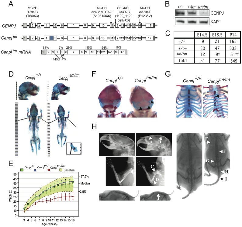
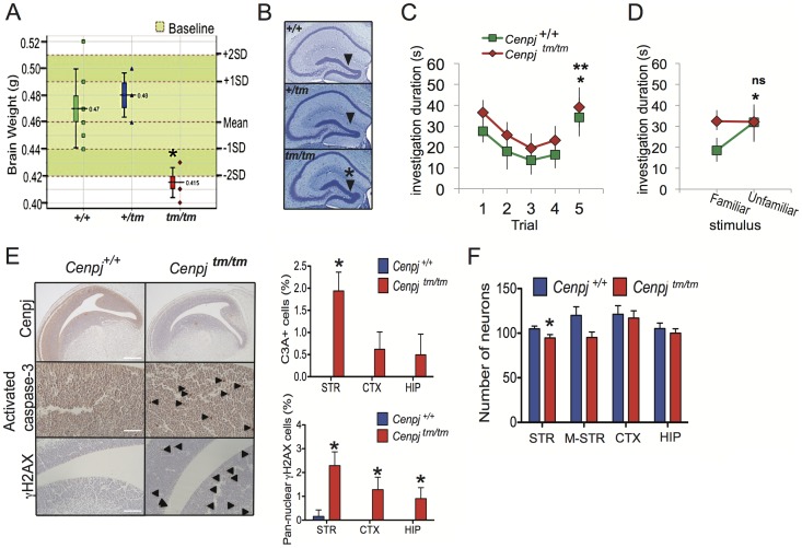
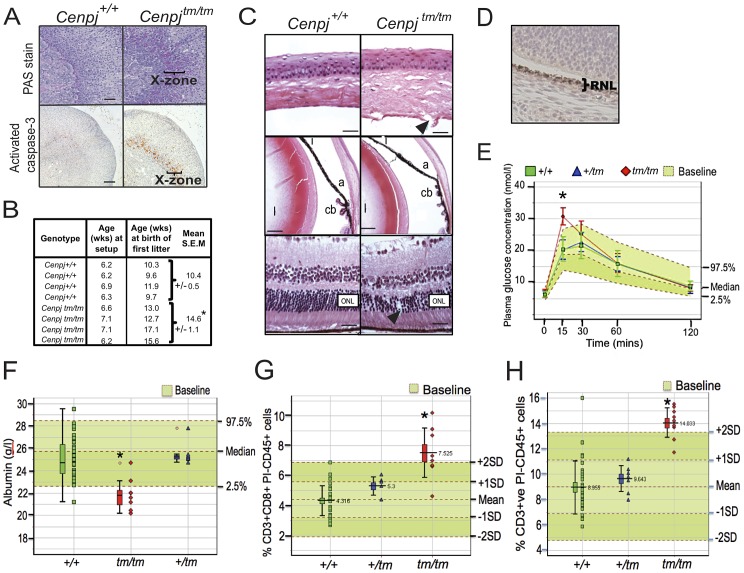
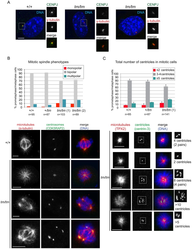
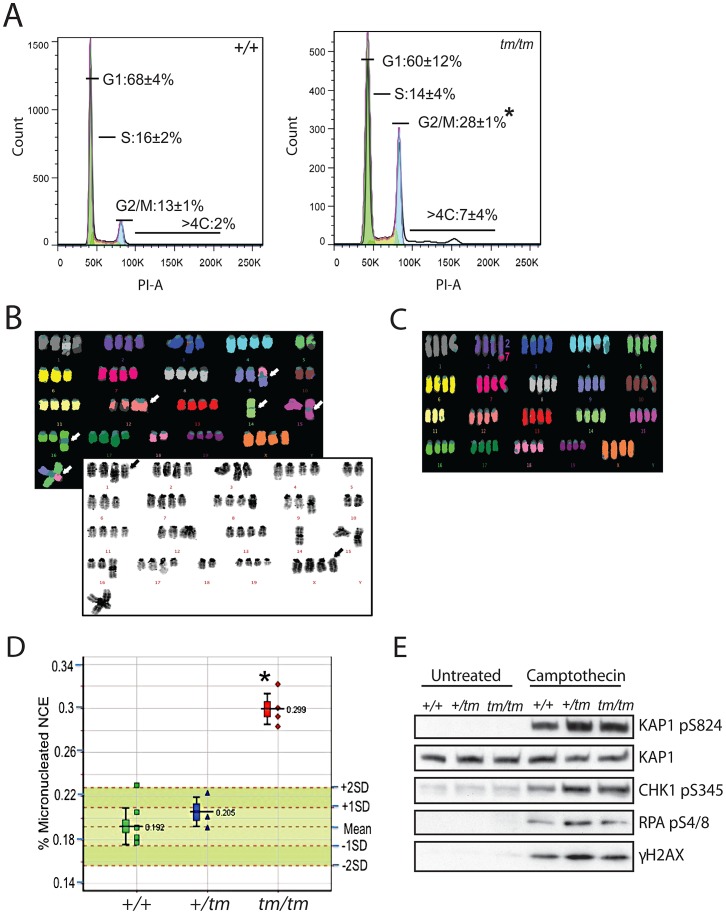
Disruption of the centromere protein J gene, CENPJ (CPAP, MCPH6, SCKL4), which is a highly conserved and ubiquitiously expressed centrosomal protein, has been associated with primary microcephaly and the microcephalic primordial dwarfism disorder Seckel syndrome. The mechanism by which disruption of CENPJ causes the proportionate, primordial growth failure that is characteristic of Seckel syndrome is unknown. By generating a hypomorphic allele of Cenpj, we have developed a mouse (Cenpj(tm/tm)) that recapitulates many of the clinical features of Seckel syndrome, including intrauterine dwarfism, microcephaly with memory impairment, ossification defects, and ocular and skeletal abnormalities, thus providing clear confirmation that specific mutations of CENPJ can cause Seckel syndrome. Immunohistochemistry revealed increased levels of DNA damage and apoptosis throughout Cenpj(tm/tm) embryos and adult mice showed an elevated frequency of micronucleus induction, suggesting that Cenpj-deficiency results in genomic instability. Notably, however, genomic instability was not the result of defective ATR-dependent DNA damage signaling, as is the case for the majority of genes associated with Seckel syndrome. Instead, Cenpj(tm/tm) embryonic fibroblasts exhibited irregular centriole and centrosome numbers and mono- and multipolar spindles, and many were near-tetraploid with numerical and structural chromosomal abnormalities when compared to passage-matched wild-type cells. Increased cell death due to mitotic failure during embryonic development is likely to contribute to the proportionate dwarfism that is associated with CENPJ-Seckel syndrome.


Conflict of interest statement

The authors have declared that no competing interests exist.

Figures

Figure 1
Figure 1. Generation of a mouse model of CENPJ-Seckel syndrome.
A. The CENPJ gene spans 40 kb and comprises 17 exons. The 3′ and 5′ untranslated regions are depicted in grey. Mutations in CENPJ have been associated with either primary microcephaly (MCPH) or Seckel syndrome (SECKEL). The mutation in intron 11 that has been associated with Seckel syndrome results in the generation of three transcripts: one lacking exon 12, one lacking 11 and 12 and one lacking exons 11,12 and 13. Disruption of mouse Cenpj by insertion of a cassette (depicted by the blue square) between exons 4 and 5 results in low levels of splicing over the cassette and cryptic splicing between exons 3 and 6 or 4 and 6; the latter two transcripts are predicted to result in truncated proteins. The allele was designated Cenpjtm1a(EUCOMM)Wtsi and abbreviated to Cenpjtm. Percentages show mean expression of Cenpj across exon boundaries as determined by quantitative RT-PCR relative to Gapdh for Cenpjtm/tm relative to Cenpj+/+ for RNA extracted from n = 3 murine embryonic fibroblast (MEF) lines. B. Immunoblot to show reduction in Cenpj levels in protein extracted from Cenpjtm/tm (tm/tm), Cenpj+/tm (+/tm), and Cenpj+/+(+/+) MEFs. KAP1 was used as a loading control. C. Table shows frequency of Cenpjtm/tm mice born from heterozygote intercrosses. Cenpjtm/tm showed partial embryonic lethality as shown by their reduced frequency at E18.5 and P14 (25% expected, *P = 0.02, **P = 0.0001, χ2 test). D. Representative images of E18.5 skeletal preparations of Cenpj+/+ and Cenpjtm/tm embryos. Staining with alcian blue (cartilage) and alizarin red (bone). Cenpjtm/tm embryo with a sloping forehead and polysyndactylism of digit one of the left hindpaw (inset). E. Bodyweights of male Cenpjtm/tm (n = 8), Cenpj+/tm (n = 7), Cenpj+/+(n = 40) and baseline wild-type controls (n = 912) from 3–16 weeks of age. Data show that Cenpjtm/tm are significantly smaller than Cenpj+/+ mice at all ages (P = 2.2×10⁻¹⁶, Mann-Whitney-Wilcoxon test). F. Skeletal preparations of E18.5 Cenpjtm/tm embryos showed irregular ossification of the cranium and G. sternum. H. X-Rays show that adult Cenpjtm/tm mice may present with a flatter, sloping forehead (A), mild elevation of the parietal bone (B), a short humerus with a prominent deltoid tuberosity (C), prominent medial epicondyle (D), an irregular ribcage (E), short lumbar and sacral vertebrae (F), an abnormal pelvis (G), extra sacrocaudal transitional vertebrae (H), short, abnormal/fused caudal vertebrae 2/3 – caudal vertebrae 7/8 (I) and reduced intervertebral joint space (J).
Figure 2
Figure 2. Neuropathological abnormalities.
A. Cenpjtm/tm mouse brain weights were two standard deviations below that of control mice (n = 144 baseline control mice). *P = 0.0002, t-test, Cenpjtm/tm, n = 6 and Cenpj +/+ n = 10. The lower whisker extends to the lowest datum still within 1.5 Inter-quartile range (IQR) of the lower quartile. The upper whisker extends to the highest datum still within 1.5 IQR of the upper quartile B. The dentate gyrus was significantly shorter in Cenpjtm/tm mice (n = 3) when compared to Cenpj +/+ control mice (n = 30), *P = 0.01, t-test. Scale bar 1 mm C. Social recognition test. When tested for habituation-dishabituation, both Cenpjtm/tm (n = 7) and Cenpj +/+ (n = 7) mice recognized a novel stimulus mouse as shown by a decline in investigation time over the first four trials that was recovered on trial five, when they were exposed to a novel mouse (trial four vs. trial five, * P = 0.0033 and ** P = 0.0014, two-way ANOVA followed by post-hoc t-test). D. A discrimination test was performed 24 h later the habituation-dishabituation test. When given a choice between the familiar (same stimulus animal used for trials one to four) and a new unfamiliar mouse 24 h later, Cenpjtm/tm mice could not discriminate as shown by the similar investigation time for both stimulus animals (Cenpj +/+ P = 0.0326, Cenpjtm/tm P = 0.957, t-test). E. Representative images of immunohistochemical stainings of E14.5 embryo sections. Cenpj was highly expressed in areas of active neurogenesis within the telencephalon. Scale bar 400 µm. There was a generalized increase in cleaved (activated) caspase-3-positive (scale bar 100 µm) and Ser139-phosphorylated H2AX (γH2AX; scale bar 200 µm) cells throughout embryos, images of striatum are shown.. The number of cells positive (as a percentage of total in two different 75 µm2 areas) for cleaved (activated) caspase-3 (C3A+) and pan-nuclear Ser139-phosphorylated H2AX (γH2AX) was increased in areas of active neurogenesis within the striatum and cortex. *P<0.05; Mann-Whitney with continuity correction, Cenpjtm/tm n = 3 and Cenpj +/+ n = 3. Data shows mean and SEM. F. Neuron densities were counted in three different areas (75 µm2) of active neurogenesis for each of the striatum (STR), cortex (CTX) and pro-hippocampus (HIP) and three areas of 150 µm2 in the mid-striatum (M-STR) of E14.5 embryos, *P = 0.0008, t-test, Cenpjtm/tm n = 3 and Cenpj +/+ n = 3. Data shows mean and SEM.
Figure 3
Figure 3. Delayed onset to puberty and ocular, endocrine, haematological, and plasma abnormalities.
A. Periodic acid-Schiff (PAS) staining and cleaved (activated) caspase-3 immunostaining of adrenal sections from 16 week-old virgin female Cenpjtm/tm mice (n = 3) confirmed corticomedullary pigmentation and ongoing apoptosis in the X-zone, respectively (representative images, scale bars 100 µm). B. Breeding records of Cenpjtm/tm females set up with Cenpjtm/tm males at 6–7 weeks of age showed that Cenpjtm/tm females produce their first litter around four weeks later than Cenpj +/+ females. *P = 0.012, t-test. C. Top panel shows normal cornea from a Cenpj +/+ mouse. Cenpjtm/tm mice had disruption of the Descemet's membrane and corneal endothelium (arrow). Middle panel shows normal anterior segment from a Cenpj +/+ mouse. The angle was displaced anteriorly in eyes from Cenpjtm/tm mice and ciliary process morphology was abnormal. (a, angle; i, iris; cb, ciliary body; l, lens). Bottom panel shows normal retina from a Cenpj +/+ mouse eye. The retina photoreceptor cells of Cenpjtm/tm mice were reduced in number and showed columnar disorganized (arrow). (ONL, outer nuclear layer). D. Immunohistochemical staining for Cenpj in Cenpj +/+ embryo eye (E14.5; RNL retinal neuroblast layer). E. Intra-peritoneal glucose tolerance test to show that female Cenpjtm/tm mice have a 15 minute delay in response to glucose challenge (n = 4 Cenpjtm/tm vs. n = 32 Cenpj +/+, *P = 2×10⁻⁵, t-test). Graph also shows n = 9 Cenpjtm/+ and n = 670 baseline wildtype controls. F. Plasma albumin levels were decreased in Cenpjtm/tm males (n = 8 Cenpjtm/tm vs. n = 35 Cenpj +/+, *P = 4.9×10⁻⁵, t-test). Graph also shows n = 7 Cenpjtm/+ and n = 768 baseline wildtype controls. G. Flow cytometric analysis of peripheral blood leukocytes in Cenpjtm/tm mice revealed an increase in the number of CD8+CD3+ and H. total CD3+ cells. Data shows total counts per 30 000 propidium-iodide (PI) negative, CD45-positive cells from male mice. For n = 9 Cenpjtm/tm vs. n = 30 Cenpj +/+: CD3+CD8+ *P = 0.0002 and CD3 *P = 2.9×10⁻⁵, Mann-Whitney-Wilcoxon test. Graphs also show n = 7 Cenpj +/tm and n = 356 baseline wildtype controls. For all ‘Box and Whisker’ plots, the lower whisker extends to the lowest datum still within 1.5 Inter-quartile range (IQR) of the lower quartile. The upper whisker extends to the highest datum still within 1.5 IQR of the upper quartile.
Figure 4
Figure 4. Centrosome and mitotic spindle abnormalities are elevated in Cenpj-deficient cells.
A. Images show examples of Cenpj staining in centrosomes of Cenpj +/+ and Cenpjtm/tm mouse embryonic fibroblasts (MEFs). Cells were stained with antibodies against Cenpj (green in merge) and the centrosomal protein γ-tubulin (red in merge). Framed areas are shown at higher magnification. B. Graph shows mitotic spindle phenotypes in MEFs derived from Cenpj +/+, Cenpj +/tm and two independent Cenpjtm/tm embryos (littermates, +/+ MEFs passage 4, +/tm and tm/tm MEFs passage 3): tm/tm (1) and tm/tm (2). Number of mitotic cells scored are shown for each genotype. Examples for monopolar and multipolar spindle are shown. Note cell on bottom panels forming a bipolar spindle by clustering supernumerary centrosomes. Cells were stained with antibodies against α-tubulin (green in merge) and the centrosomal protein, Cdk5RAP2 (red in merge). C. Graph shows centriole numbers in mitotic MEFs of indicated genotypes (littermates, +/+ MEFs passage 4, +/tm and tm/tm MEFs passage 3). Cells were arrested in mitosis with monastrol that caused monopolar spindle formation and facilitated visualization of centrioles. Note that mitotic cells should normally contain a total of 4 centrioles, but even in wild-type cells we occasionally detect 3 centrioles probably due to insufficient spatial resolution, so 3 or 4 centrioles were considered a single class. Data were collected from two independent experiments; bars show mean ±SD, number of mitotic cells scored are shown for each genotype. Images below depict examples for cells with different centriole numbers (top cell with 4 centrioles is normal, all other cells have too few or too many centrioles). Cells were stained with antibodies against the microtubule-binding protein Tpx2 (green in merge) and the centriolar protein, centrin-3 (red in merge). Framed areas are shown at higher magnification. Scale bars = 5 µm.
Figure 5
Figure 5. Genomic instability is associated with abnormal ploidy of Cenpjtm/tm cells rather than an impaired DNA damage response.
A. Cell cycle analysis of Cenpjtm/tm mouse embryonic fibroblasts (MEFs) by flow cytometry showed an increase in the percentage of cells in G2 (4C) and cells containing >4C DNA content when compared to Cenpj +/+ cells. Percentages represent means of n = 3 independent MEF lines per genotype (each pair of +/+ and tm/tm cells were passage-matched (passage<5) and derived from littermates), *P<0.05, t-test. PI, propidium iodide. B. Example multiplex fluorescent in situ hybridization (M-FISH; top) and DAPI banded (bottom) karyotype of a Cenpjtm/tm MEF metaphase (passage 4). The karyotype is near tetraploid, with centric fusions (white arrows) and chromosomes that have apparently lost their centromeres (black arrows). C. Example M-FISH of a Cenpjtm/tm MEF metaphase (passage 4) showing near tetraploid karyotype with a translocation (t(2;7)). D. Adult Cenpjtm/tm (n = 4) mice showed increased genomic instability when compared to Cenpj +/+ mice (n = 6) as determined by the increased prevalence of micronucleated normochromatic erythrocytes using a flow cytometric assay of micronucleus formation. *P = 0.000004, t-test. The lower whisker extends to the lowest datum still within 1.5 Inter-quartile range (IQR) of the lower quartile. The upper whisker extends to the highest datum still within 1.5 IQR of the upper quartile. E. Immunoblots show normal activation of DNA damage response markers in Cenpj-deficient MEFs (passage 2) before and after treatment with the DNA damaging agent camptothecin (1 µM for 1 h). KAP1 was used as a loading control.

Similar articles

Cited by

References

    1. Majewski F, Goecke T (1982) Studies of microcephalic primordial dwarfism I: approach to a delineation of the Seckel syndrome. American journal of medical genetics 12: 7–21. - PubMed
    1. Faivre L, Le Merrer M, Lyonnet S, Plauchu H, Dagoneau N, et al. (2002) Clinical and genetic heterogeneity of Seckel syndrome. American journal of medical genetics 112: 379–383. - PubMed
    1. Harsha Vardhan BG, Muthu MS, Saraswathi K, Koteeswaran D (2007) Bird-headed dwarf of Seckel. Journal of the Indian Society of Pedodontics and Preventive Dentistry 25 Suppl: S8–9. - PubMed
    1. Al-Dosari MS, Shaheen R, Colak D, Alkuraya FS (2010) Novel CENPJ mutation causes Seckel syndrome. J Med Genet 47: 411–414. - PubMed
    1. Kalay E, Yigit G, Aslan Y, Brown KE, Pohl E, et al. (2011) CEP152 is a genome maintenance protein disrupted in Seckel syndrome. Nature genetics 43: 23–26. - PMC - PubMed

Publication types

MeSH terms

Substances

Supplementary concepts