

Unilateral lumbosacral dislocation: case report and a comprehensive review
- PMID: 23166575
- PMCID: PMC3497577
- DOI: 10.2174/1874325001206010473
Unilateral lumbosacral dislocation: case report and a comprehensive review
Abstract
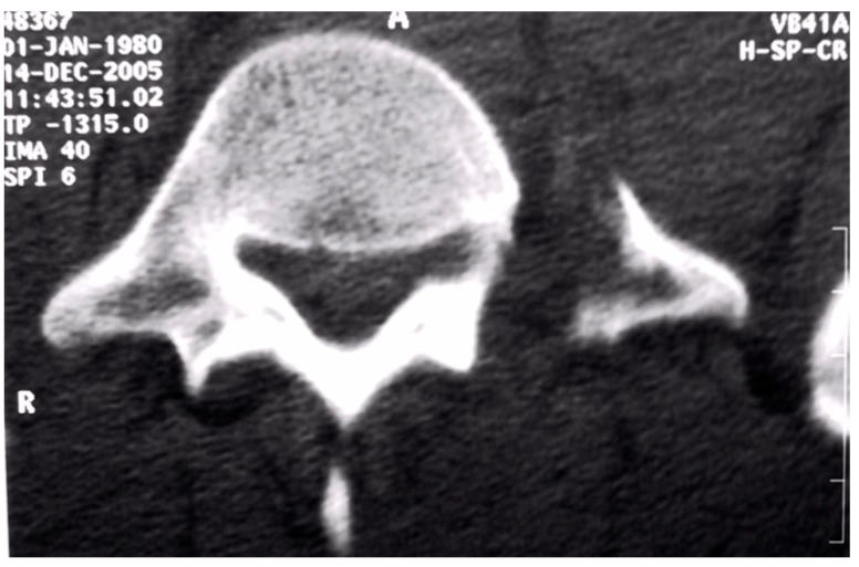
Lumbosacral fracture-dislocation is a rare occurrence. There are more than 73 cases reported in the English literature. We report on the imaging findings and surgical treatment in a patient suffered of unilateral traumatic L5-S1 dislocation associated with severe disruption of the posterior ligamentous complex. The patient underwent open reduction and stabilization of L4-S1 vertebrae with posterior instrumentation system. Open reduction and internal fixation was mandatory as post-traumatic ligamentous insufficiency would lead to abnormal motion. Operative treatment managed to produce a solid arthrodesis and restore stability of the lumbosacral junction. Follow-up revealed excellent results. This study reports a rare injury of the lumbosacral junction, and the literature concerning this unusual condition is extensively reviewed.
Keywords: Unilateral lumbosacral dislocation; case report; review.; surgery.
Figures


References
-
- Tsirikos A, Saifuddin A, Noordeen MH, Tucker SK. Traumatic lumbosacral dislocation: report of two cases. Spine. 2004;29:E164–8. - PubMed
-
- Zoltan JD, Gilula LA, Murphy WA. Unilateral facet dislocation between the fifth lumbar and first sacral vertebrae. J Bone Joint Surg. 1979;61A:767–9. - PubMed
-
- Kramer KM, Levine AM. Unilateral facet dislocation of the lumbosacral junction. J Bone Joint Surg. 1989;71A:1258–61. - PubMed
-
- Shen FH, Crowl A, Shuler TE, Feldenzer JA, Leivy SW. Delayed recognition of lumbosacral fracture dislocation in the multitrauma patient, the triad of transverse process fractures unilateral renal contusion and lumbosacral fracture dislocation. J Orthop Trauma . 2004;56:700–5. - PubMed
-
- Vialle R, Wolff S, Pauthier F, et al. Traumatic lumbosacral dislocation: four cases and review of literature. Clin Orthop. 2004; 419:91–7. - PubMed
Publication types
LinkOut - more resources
Full Text Sources