Comparative Study
. 2012;7(11):e48601.
doi: 10.1371/journal.pone.0048601. Epub 2012 Nov 15.

Pregnancy is associated with decreased cardiac proteasome activity and oxidative stress in mice

Affiliations
Comparative Study

Pregnancy is associated with decreased cardiac proteasome activity and oxidative stress in mice

Andrea Iorga et al. PLoS One. 2012.

Abstract

During pregnancy, the heart develops physiological hypertrophy. Proteasomal degradation has been shown to be altered in various models of pathological cardiac hypertrophy. Since the molecular signature of pregnancy-induced heart hypertrophy differs significantly from that of pathological heart hypertrophy, we investigated whether the cardiac proteasomal proteolytic pathway is affected by pregnancy in mice. We measured the proteasome activity, expression of proteasome subunits, ubiquitination levels and reactive oxygen production in the hearts of four groups of female mice: i) non pregnant (NP) at diestrus stage, ii) late pregnant (LP), iii) one day post-partum (PP1) and iv) 7 days post-partum (PP7). The activities of the 26 S proteasome subunits β1 (caspase-like), and β2 (trypsin-like) were significantly decreased in LP (β1∶83.26 ± 1.96%; β2∶74.74 ± 1.7%, normalized to NP) whereas β5 (chymotrypsin-like) activity was not altered by pregnancy but significantly decreased 1 day post-partum. Interestingly, all three proteolytic activities of the proteasome were restored to normal levels 7 days post-partum. The decrease in proteasome activity in LP was not due to the surge of estrogen as estrogen treatment of ovariectomized mice did not alter the 26 S proteasome activity. The transcript and protein levels of RPN2 and RPT4 (subunits of 19 S), β2 and α7 (subunits of 20 S) as well as PA28α and β5i (protein only) were not significantly different among the four groups. High resolution confocal microscopy revealed that nuclear localization of both core (20S) and RPT4 in LP is increased ∼2-fold and is fully reversed in PP7. Pregnancy was also associated with decreased production of reactive oxygen species and ubiquitinated protein levels, while the de-ubiquitination activity was not altered by pregnancy or parturition. These results indicate that late pregnancy is associated with decreased ubiquitin-proteasome proteolytic activity and oxidative stress.


Conflict of interest statement

Competing Interests: The authors have declared that no competing interests exist.

Figures

Figure 1
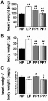
Figure 1. The increased in heart weight of LP mice is reversed post-partum.
Heart weights (HW, A), body weights (BW, B) and HW/BW (C) in non-pregnant (NP, black bars, n = 8), late pregnant (LP, white bars, n = 11), one day post-partum (PP1, grey bars, n = 6) and seven days post-partum (PP7, shaded bars, n = 5) mice. Values are mean ± SEM, *p<0.05 and **p<0.001 vs. NP, #p<0.05 and ##p<0.001 vs. LP, p<0.05 and ∧∧p<0.001 vs. PP1.
Figure 2
Figure 2. Proteasome activity of 26 S, but not 20 S is reduced in late pregnancy.
Activity of different proteasomal beta subunits of the 26 S (A) and 20 S (B) was measured after initiating the reaction with: Z-LLE-AMC (β1), Boc-LSTR-AMC (β2) and Suc-LLVY-AMC (β5) for non pregnant (NP, black bars), late pregnant (LP, white bars), one day post-partum (PP1, grey bars) and seven days post-partum (PP7, shaded bars). The fluorescence values in arbitrary units are represented as mean ± SEM *p<0.05 vs. NP (n = 4 mice per group) and are normalized to NP levels. The raw proteasome activity values for the NP group are as follows (in nmol/min/mg protein): for the 26 S ATP-dependent activities, β1 was 0.11±0.01, β2 was 0.04±0.01 and β5 was 0.16±0.01, while for the 20 S activity, β1 was 0.21±0.03, β2 was 0.15±0.02 and β5 was 0.12±0.01.
Figure 3
Figure 3. Proteasome activity of the 26S is unaffected by estrogen treatment.
Activity of the different proteasomal beta subunits of the 26 S were measured after initiating the reaction with: Z-LLE-AMC (β1), Boc-LSTR-AMC (β2) and Suc-LLVY-AMC (β5) in ovariectomized female mice treated with placebo (Placebo, black bars) or with 17 β-estradiol for 10 days (E2, white bars). The fluorescence values in arbitrary units are normalized to Placebo levels and represented as mean ± SEM (n = 4 mice per group).
Figure 4
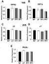
Figure 4. Transcript levels of proteasome 19S and 20S subunits, as well as the regulatory subunit PA28α, are not modified in late pregnancy.
Relative transcript expression of the cardiac proteasome measured by Real-Time qPCR in non pregnant (NP, black bars), in late pregnancy (LP, white bars), 1 day post-partum (PP1, grey bars) and 7 days post-partum (PP7, shaded bars) for RPN2 and RPT4, which are subunits of 19 S (A–B), β2 and α7, which are subunits of 20 S (C–D) and the proteasome regulatory subunit PA28α (E). GAPDH was used as the internal reference gene (data not shown). Values are mean ± SEM as normalized to NP (n = 3–5 per group).
Figure 5
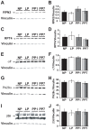
Figure 5. Protein levels of RPT4, RPN2, α7, PA28α and β5i are unaffected by pregnancy.
Immunoblotting of whole heart lysates (100 µg) from non pregnant (NP, black bars), late pregnant (LP, white bars), 1 day post-partum (PP1, grey bars) and 7 days post-partum (PP7, shaded bars) with anti-RPN2 (A–B), -RPT4 (C–D), -α7 (E–F), -PA28α (G–H) and -β5i (I–J) antibodies. In (I), the upper 30 kDa band is the β5i containing the pro-peptide. The bar graphs represent the quantification of fluorescent signal intensity normalized to Vinculin. For β5i both bands were taken into consideration in the quantification of protein levels. Vinculin was used as the loading control (n = 4 per group). Values are mean ± SEM in arbitrary units.
Figure 6
Figure 6. Increased nuclear labeling of core Subunits and RPT4 in late pregnancy was reversed one week postpartum.
A. Representative single confocal sections of cardiomyocytes dissociated from non pregnant (NP), late pregnant (LP), one day post-partum (PP1) and seven days post-partum (PP7) are co-immunostained with anti-core (green) and -RPT4 (red) antibodies. The nuclear overlay of core and RPT4 are also shown at higher resolution. These results are representative of the labeling pattern observed in myocytes from 3 different animals in each group. B. Quantification of nuclear fluorescence labeling in the four groups mentioned above for core (green bars) and RPT4 (red bars) from at least 20–25 cells per group (n = 3 mice/group). Only the nucleus in the confocal plane of focus was taken into account. **denotes p<0.001 vs. NP, #p<0.05 vs. LP and p<0.05 vs. PP1.
Figure 7
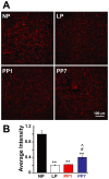
Figure 7. Superoxide production is decreased with pregnancy and remains low seven days after parturition.
A. Representative dihydroethidium (DHE) staining of transverse heart sections in non pregnant (NP), late pregnant (LP), 1 day post-partum (PP1) and 7 days post-partum (PP7). Red staining indicates the presence of reactive oxygen species (ROS). B. Quantification of the DHE staining for detection of ROS in non pregnant (NP, black bar), late pregnant (LP, white bar), 1 day post-partum (PP1, grey bar) and 7 days post-partum (PP7, shaded bar). Values are mean ± SEM as normalized to NP (n = 3 per group), and **denotes p<0.001 vs. NP, #p<0.05 vs. LP and p<0.05 vs. PP1.
Figure 8
Figure 8. Pregnancy is associated with decreased polyubiquitinated protein levels, but not de-ubiqutination levels.
A. Representative Western Blot of polyubiquitinated proteins (using the FK2 antibody) in whole heart lysates (100 µg) from non pregnant (NP), late pregnant (LP), 1 day post-partum (PP1) and 7 days post-partum (PP7). B. PonceauS was used as the loading control (n = 4 per group). C. Quantification of the polyubiquitinated proteins by Western Blot in non-pregnant (NP, black bar), late pregnant (LP, white bar), 1 day post-partum (PP1, grey bar) and 7 days post-partum (PP7, shaded bar). D. Polyubiquitination levels in NP, LP, PP1 and PP7 as determined by ELISA (using the FK1 antibody). E. De-ubiquitination activity levels in NP, LP, PP1 and PP7. Values are mean ± SEM and are normalized to NP, n = 4 per group and * denotes p<0.05 vs. NP.

References

    1. Mone SM, Sanders SP, Colan SD (1996) Control mechanisms for physiological hypertrophy of pregnancy. Circulation 94: 667–72. - PubMed
    1. Dorn GW, Robbins J, Sugden PH (2003) Phenotyping hypertrophy: eschew obfuscation. Circ Res 92: 1171–5. - PubMed
    1. Daniels SR, Meyer RA, Liang YC, Bove KE (1988) Echocardiographically determined left ventricular mass index in normal children, adolescents and young adults. J Am Coll Cardiol 12: 703–8. - PubMed
    1. Pluim BM, Zwinderman AH, van der LA, van der Wall EE (2000) The athlete’s heart. A meta-analysis of cardiac structure and function. Circulation 101: 336–44. - PubMed
    1. Schannwell CM, Zimmermann T, Schneppenheim M, Plehn G, Marx R, et al. (2002) Left ventricular hypertrophy and diastolic dysfunction in healthy pregnant women. Cardiology 97: 73–8. - PubMed

Publication types

MeSH terms