. 2012;7(11):e49255.
doi: 10.1371/journal.pone.0049255. Epub 2012 Nov 14.

ERK1/2 and p38 MAPKs are complementarily involved in estradiol 17ß-D-glucuronide-induced cholestasis: crosstalk with cPKC and PI3K

Affiliations

ERK1/2 and p38 MAPKs are complementarily involved in estradiol 17ß-D-glucuronide-induced cholestasis: crosstalk with cPKC and PI3K

Andrea C Boaglio et al. PLoS One. 2012.

Abstract

Objective: The endogenous, cholestatic metabolite estradiol 17ß-D-glucuronide (E(2)17G) induces endocytic internalization of the canalicular transporters relevant to bile formation, Bsep and Mrp2. We evaluated here whether MAPKs are involved in this effect.

Design: ERK1/2, JNK1/2, and p38 MAPK activation was assessed by the increase in their phosphorylation status. Hepatocanalicular function was evaluated in isolated rat hepatocyte couplets (IRHCs) by quantifying the apical secretion of fluorescent Bsep and Mrp2 substrates, and in isolated, perfused rat livers (IPRLs), using taurocholate and 2,4-dinitrophenyl-S-glutathione, respectively. Protein kinase participation in E(2)17G-induced secretory failure was assessed by co-administering selective inhibitors. Internalization of Bsep/Mrp2 was assessed by confocal microscopy and image analysis.

Results: E(2)17G activated all kinds of MAPKs. The PI3K inhibitor wortmannin prevented ERK1/2 activation, whereas the cPKC inhibitor Gö6976 prevented p38 activation, suggesting that ERK1/2 and p38 are downstream of PI3K and cPKC, respectively. The p38 inhibitor SB203580 and the ERK1/2 inhibitor PD98059, but not the JNK1/2 inhibitor SP600125, partially prevented E(2)17G-induced changes in transporter activity and localization in IRHCs. p38 and ERK1/2 co-inhibition resulted in additive protection, suggesting complementary involvement of these MAPKs. In IPRLs, E(2)17G induced endocytosis of canalicular transporters and a rapid and sustained decrease in bile flow and biliary excretion of Bsep/Mrp2 substrates. p38 inhibition prevented this initial decay, and the internalization of Bsep/Mrp2. Contrarily, ERK1/2 inhibition accelerated the recovery of biliary secretion and the canalicular reinsertion of Bsep/Mrp2.

Conclusions: cPKC/p38 MAPK and PI3K/ERK1/2 signalling pathways participate complementarily in E(2)17G-induced cholestasis, through internalization and sustained intracellular retention of canalicular transporters, respectively.


Conflict of interest statement

Competing Interests: The authors have declared that no competing interests exist.

Figures

Figure 1
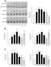
Figure 1. E217G activates the MAPK signalling pathway.
(A) left panel: representative Western blottings of phospho (p)-p38, p-ERK1/2, p-JNK1/2 and total forms of all these MAPK types were obtained from whole cellular lysates of primary-cultured rat hepatocytes incubated with E217G (200 µM) for 10 to 60 min, or with E217G (200 µM) for 20 min in cells pretreated with the PI3K inhibitor wortmanin (WM, 100 nM) or with the cPKC inhibitor Gö6976 (Gö, 1 µM) for 15 min. A (right panel), and B and C panels show phosphorylation status of all MAPK types evaluated (calculated as the p-MAPK to total MAPK ratio for each experimental condition). An arbitrary value of 100 was assigned to the band of highest densitometric intensity in every Western blot before the ratio was calculated. The results are shown as mean ± SEM (n = 5). *P<0.05 vs. control (cells treated only with DMSO), and #P<0.05 vs. E217G (20 min).
Figure 2
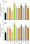
Figure 2. Effect of the inhibition of p38, ERK1/2 and JNK1/2, or the coinhibition of cPKC-ERK1/2, PI3K-p38, or p38-ERK1/2, on E217G-induced impairment of the canalicular accumulation of the Bsep and Mrp2 fluorescent substrates in IRHCs.
IRHCs were incubated with E217G (200 µM, 20 min) (or DMSO in controls), with or without pretreatment for 15 min with the JNK1/2 inhibitor SP600125 (1 µM), the ERK1/2 inhibitor PD98059 (PD; 5 µM), and/or the p38 inhibitor SB203580 (SB; 1 µM), together or not with the cPKC inhibitor Gö6976 (Gö; 1 µM) or PI3K inhibitor wortmanin (WM; 100 nM). Canalicular accumulation CGamF (Bsep substrate, panel A) and GS-MF (Mrp2 substrate, panel B) was determined as the percentage of couplets displaying visible fluorescence in their canalicular vacuoles from a total of at least 200 couplets per preparation. The results are expressed as percentages of the control group and are shown as mean ± SEM (n = 3–4). *P<0.05 vs. E217G, and #P<0.05 vs. E217G-WM, E217G-Gö, E217G-PD or E217G-SB.
Figure 3
Figure 3. Effect of inhibition of p38 or ERK1/2, and coinhibition of cPKC-ERK1/2, PI3K-p38, or p38-ERK1/2 on E217G-induced retrieval of Bsep and Mrp2 in IRHCs.
The upper panels show representative confocal immunofluorescence images of the localization of Bsep and Mrp2 in DMSO-treated (control) or E217G (200 µM)-treated IRHCs, with or without the p38 inhibitor SB203580 (SB; 1 µM) or the ERK1/2 inhibitor PD98059 (PD; 5 µM), in combination or not with the cPKC inhibitor Gö6976 (Gö; 1 µM) or PI3K inhibitor wortmanin (WM; 100 nM). The lower panels show the densitometric analysis of the fluorescence intensity along a line (8 µm) perpendicular to the center of the canalicular vacuole (from +4 to −4 µm). The statistical analysis of the profiles of fluorescence showed a significant change in the E217G-treated group (P<0.05; number of analyzed canalicular vacuoles >10), but this reverted to normal in the E217G-SB, E217G-PD, E217G-PD-SB, E217G-Gö-PD and E217G-WM-SB groups for Bsep and Mrp2.
Figure 4
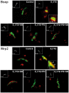
Figure 4. Effect of E217G on colocalization of Rab11a with Mrp2 or Bsep in IRHCs.
Immunofluorescence confocal images showing staining of Mrp2 or Bsep (green) and Rab11a (red) in IRHCs treated with DMSO (control), the ERK1/2 inhibitor PD98059 (PD; 5 µM), the p38 inhibitor SB203580 (SB; 1 µM), or both inhibitors together. Colocalization of Rab11a with Mrp2 or Bsep in the E217G-treated group is indicated by orange-yellow fluorescence in merged images. Insets depict F-actin staining, which was used to demarcate the limits of the canalicular vacuoles.
Figure 5
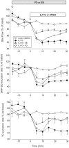
Figure 5. Effect of inhibition of p38 or ERK1/2 on E217G-induced decrease of bile flow and biliary secretion of the Mrp2 and Bsep substrates DNP-SG and taurocholate, respectively, in the perfused rat liver (IPRL) model.
IPRLs were treated with a portal bolus of E217G (2 µmol/liver), or with the E217G vehicle DMSO (control), in the presence and absence of the ERK1/2 inhibitor PD98059 (PD; 5 µM) or the p38 inhibitor SB203580 (SB; 250 nM). The effect of the treatments on (A) bile flow, (B) DNP-SG excretion, (C) and taurocholate excretion are shown. Results are expressed as the mean ± SEM (n = 4).
Figure 6
Figure 6. Effect of inhibition of p38 or ERK1/2 on E217G-induced retrieval of Bsep and Mrp2 in perfused rat livers at the end of the perfusion period.
The upper panels show representative confocal images showing co-staining of Mrp2 or Bsep (green) with F-actin (red; used to visualize the bile canaliculus limits), illustrative of the endocytic internalization of Mrp2 and Bsep induced by E217G (2 µmol/liver), and its protection by the ERK1/2 inhibitor PD98059 (PD; 5 µM) or the p38 inhibitor SB203580 (SB; 250 nM). The lower panels show a densitometric analysis of the intensity of fluorescence associated with Bsep and Mrp2 along a 6 µm line perpendicular to the canaliculus (from −3 µm to +3 µm from the canalicular center), corresponding to the confocal images in the upper panels.
Figure 7
Figure 7. Schematic representation of the signalling events involved in E217G-induced cholestasis by endocytic internalization and further retention of canalicular transporters relevant to bile formation (Bsep, Mrp2).
p38, acting downstream of cPKC, triggers endocytic internalization of the apical carriers presumably towards apical early endosomes (AEE), the first intracellular endosomal compartment receiving internalized proteins from the apical membrane, in a microtubule-independent manner (solid arrow). These transporters traffic to, and accumulate into, apical recycling endosomes (ARE), from where they can be retargeted to the apical membrane during the recovery of the cholestatic process, in a microtubule-dependent manner (dashed arrows). Activation of the PI3K/Akt/ERK1/2 signalling pathway halts this latter process, thus explaining the increased colocalization of Bsep/Mrp2 with Rab11a, an ARE marker. This prolongs the cholestatic effect of E217G by impeding the fast, spontaneous retargeting of intracellular transporters that would lead to a rapid recovery from the cholestatic injury.

Similar articles

Cited by

References

    1. Crocenzi FA, Zucchetti AE, Boaglio AC, Barosso IR, Sanchez Pozzi EJ, et al. (2012) Localization status of hepatocellular transporters in cholestasis. Front Biosci 17: 1201–1218. - PubMed
    1. Vore M, Liu Y, Huang L (1997) Cholestatic properties and hepatic transport of steroid glucuronides. Drug Metabol Rev 29: 183–203. - PubMed
    1. Stieger B, Fattinger K, Madon J, Kullak-Ublick GA, Meier PJ (2000) Drug- and estrogen-induced cholestasis through inhibition of the hepatocellular bile salt export pump (Bsep) of rat liver. Gastroenterology 118: 422–430. - PubMed
    1. Kan KS, Monte M, Parslow RA, Coleman R (1989) Oestradiol 17β-glucuronide increases tight junctional permeability in rat liver. Biochem J 261: 297–300. - PMC - PubMed
    1. Crocenzi FA, Mottino AD, Cao J, Veggi LM, Pozzi EJ, et al. (2003) Estradiol-17β-D-glucuronide induces endocytic internalization of Bsep in rats. Am J Physiol Gastrointest Liver Physiol 285: G449–459. - PubMed

Publication types

MeSH terms

Substances