. 2012;7(11):e49524.
doi: 10.1371/journal.pone.0049524. Epub 2012 Nov 14.

Identification of novel translational urinary biomarkers for acetaminophen-induced acute liver injury using proteomic profiling in mice

Affiliations

Identification of novel translational urinary biomarkers for acetaminophen-induced acute liver injury using proteomic profiling in mice

Rachel P L van Swelm et al. PLoS One. 2012.

Abstract

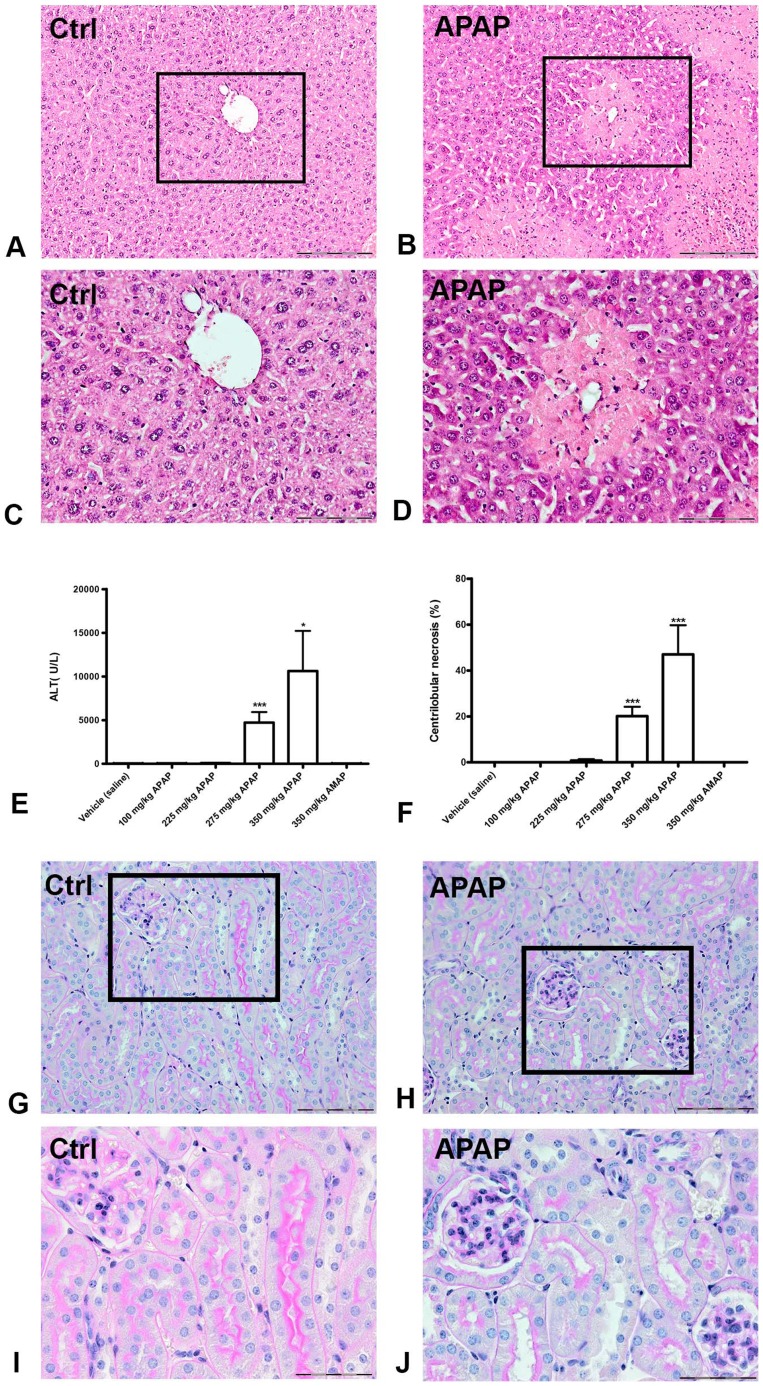
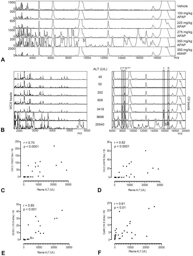
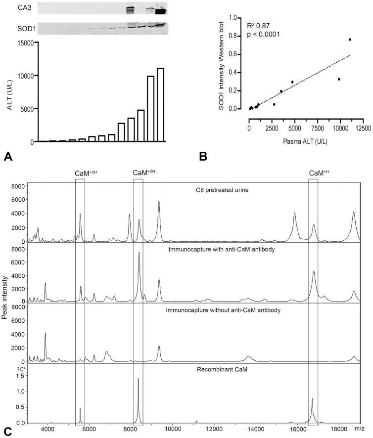
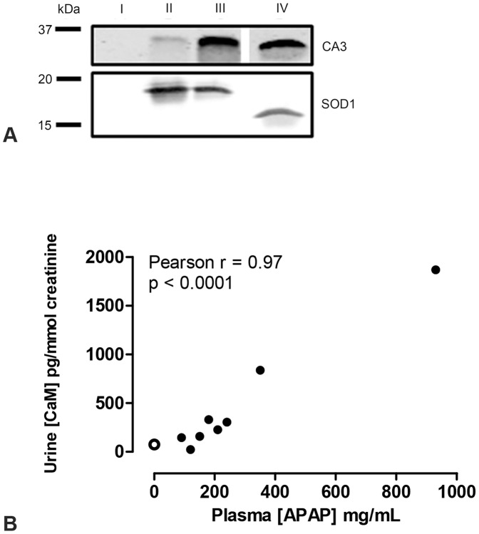
Drug-induced liver injury (DILI) is the leading cause of acute liver failure. Currently, no adequate predictive biomarkers for DILI are available. This study describes a translational approach using proteomic profiling for the identification of urinary proteins related to acute liver injury induced by acetaminophen (APAP). Mice were given a single intraperitoneal dose of APAP (0-350 mg/kg bw) followed by 24 h urine collection. Doses of ≥275 mg/kg bw APAP resulted in hepatic centrilobular necrosis and significantly elevated plasma alanine aminotransferase (ALT) values (p<0.0001). Proteomic profiling resulted in the identification of 12 differentially excreted proteins in urine of mice with acute liver injury (p<0.001), including superoxide dismutase 1 (SOD1), carbonic anhydrase 3 (CA3) and calmodulin (CaM), as novel biomarkers for APAP-induced liver injury. Urinary levels of SOD1 and CA3 increased with rising plasma ALT levels, but urinary CaM was already present in mice treated with high dose of APAP without elevated plasma ALT levels. Importantly, we showed in human urine after APAP intoxication the presence of SOD1 and CA3, whereas both proteins were absent in control urine samples. Urinary concentrations of CaM were significantly increased and correlated well with plasma APAP concentrations (r = 0.97; p<0.0001) in human APAP intoxicants, who did not present with elevated plasma ALT levels. In conclusion, using this urinary proteomics approach we demonstrate CA3, SOD1 and, most importantly, CaM as potential human biomarkers for APAP-induced liver injury.


Conflict of interest statement

Competing Interests: The authors have declared that no competing interests exist.

Figures

Figure 1
Figure 1. APAP-induced liver injury and kidney histology in mice.
Hematoxylin and eosin staining of representative liver slides from a vehicle-treated mouse (A and C) and an APAP-treated mouse (B and D). Panels A and B show a 10× magnification, and a 20× magnification of the framed area is given in panels C and D, respectively. Centrilobular necrosis can be observed in liver slides after APAP treatment. Plasma ALT levels (E) and the percentage of centrilobular necrosis (F) increased significantly in mice receiving 275 and 350 mg/kg APAP. Periodic acid-Schiff staining of representative kidney slides from a vehicle-treated mouse (G and I) and an APAP-treated mouse (H and J) show no difference in histology. Panels G and H demonstrate a 20× magnification and a 40× magnification is given for the framed areas in panels I and J. The scalebar represents 200 µm in the slides with 10× magnification, 100 µm with 20× magnification and 50 µm with 40× magnification. ** P<0.01, *** P<0.001 compared to vehicle treated mice. ALT: alanine aminotransferase; AMAP: 3-acetamidophenol; APAP: acetaminophen.
Figure 2
Figure 2. Urinary protein profiles of APAP-induced liver injury in mice.
Representative urine protein profiles of m/z values versus peak intensity illustrate an APAP dose-related increase in urinary protein excretion (A). ALT-dependent increases in protein peaks were observed in urine samples pretreated with WCX beads or C8 beads (B). The protein masses of 15.9 kDa and 16.8 kDa are indicated by (I) and (II), respectively. Double charged forms are indicated by (+2H). The correlation between the relative peak intensity of two representative urinary CA3 fragments (C & D), SOD1 (E), and CaM (F) and plasma ALT was determined using the Spearman's rank correlation coefficient (r) in mice with APAP dose ≥275 mg/kg body weight. ALT: alanine aminotransferase; APAP: acetaminophen; CA3: carbonic anhydrase 3; CaM: calmodulin; SOD1: superoxide dismutase 1; WCX: weak cation exchange.
Figure 3
Figure 3. Identification of CA3, SOD1 and CaM in mouse urine.
Western blots show the relation between urinary SOD1 and CA3, and plasma ALT levels in individual mice (n = 13; panel A), of which urinary SOD1 intensity on Western blot was analyzed by linear regression analysis (B). Immunoprecipitation demonstrated the specific protein profile of CaM, i.e. the mass peak for CaM at 16.8 kDa (CaM+H) and its double and triple charged form (CaM+2H and CaM+3H), in mouse urine after APAP treatment (C). ALT: alanine aminotransferase; APAP: acetaminophen; CA3: carbonic anhydrase 3; CaM: calmodulin; SOD1: superoxide distmutase 1.
Figure 4
Figure 4. Detection of SOD1, CA3 and CaM in human urine samples.
Presence of CA3 and SOD1 was assessed by Western blot in urine samples of masterpool control (I), severe APAP intoxication sample 1 (II) and 2 (III) and a positive control (IV) (panel A). Using an ELISA assay, the urinary concentration of CaM was correlated with plasma APAP concentrations using the Pearson correlation (r) test in patients with an APAP-intoxication, but without elevated plasma ALT values (B). The open data point represents the masterpool control urine sample. ALT: alanine aminotransferase; APAP acetaminophen; CA3: carbonic anhydrase 3; CaM: calmodulin; SOD1: superoxide dismutase 1.

References

    1. Stine JG, Lewis JH (2011) Drug-induced liver injury: a summary of recent advances. ExpertOpinDrug Metab Toxicol 7: 875–890. - PubMed
    1. Ozer J, Ratner M, Shaw M, Bailey W, Schomaker S (2008) The current state of serum biomakers of hepatotoxicity. Toxicology 245: 194–205. - PubMed
    1. Watkins PB, Seligman PJ, Pears JS, Avigan MI, Senior JR (2008) Using controlled clinical trials to learn more about acute drug-induced liver injury. Hepatology 48: 1680–1689. - PubMed
    1. Banks AT, Zimmerman HJ, Ishak KG, Harter JG (1995) Diclofenac-associated hepatotoxicity: analysis of 180 cases reported to the Food and Drug Administration as adverse reactions. Hepatology 22: 820–827. - PubMed
    1. Visser K, van der Heijde DM (2009) Risk and management of liver toxicity during methotrexate treatment in rheumatoid and psoriatic arthritis: a systematic review of the literature. Clin Exp Rheumatol 27: 1017–1025. - PubMed

Publication types

MeSH terms