. 2012;7(11):e49701.
doi: 10.1371/journal.pone.0049701. Epub 2012 Nov 14.

Paeoniflorin protects against ischemia-induced brain damages in rats via inhibiting MAPKs/NF-κB-mediated inflammatory responses

Affiliations

Paeoniflorin protects against ischemia-induced brain damages in rats via inhibiting MAPKs/NF-κB-mediated inflammatory responses

Ruo-Bing Guo et al. PLoS One. 2012.

Abstract

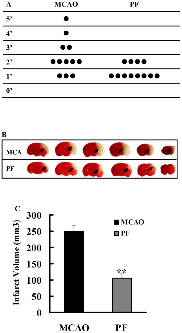
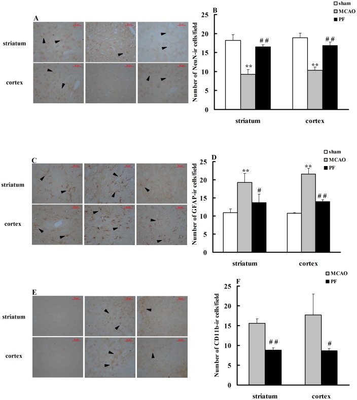
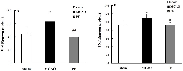
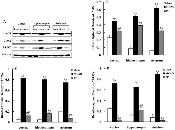
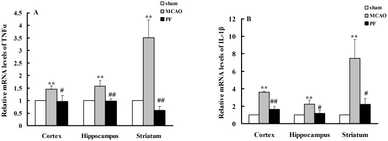
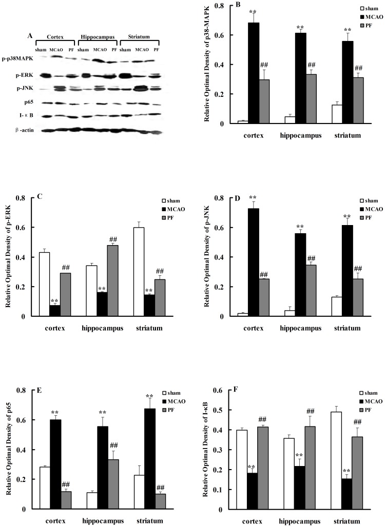
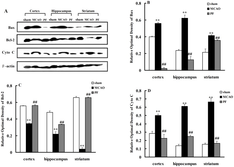
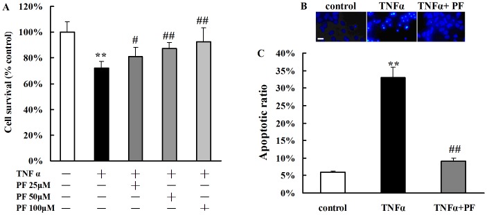
Paeoniflorin (PF), the principal component of Paeoniae Radix prescribed in traditional Chinese medicine, has been reported to exhibit many pharmacological effects including protection against ischemic injury. However, the mechanisms underlying the protective effects of PF on cerebral ischemia are still under investigation. The present study showed that PF treatment for 14 days could significantly inhibit transient middle cerebral artery occlusion (MCAO)-induced over-activation of astrocytes and microglia, and prevented up-regulations of pro-inflamamtory mediators (TNFα, IL-1β, iNOS, COX(2) and 5-LOX) in plasma and brain. Further study demonstrated that chronic treatment with PF suppressed the activations of JNK and p38 MAPK, but enhanced ERK activation. And PF could reverse ischemia-induced activation of NF-κB signaling pathway. Moreover, our in vitro study revealed that PF treatment protected against TNFα-induced cell apoptosis and neuronal loss. Taken together, the present study demonstrates that PF produces a delayed protection in the ischemia-injured rats via inhibiting MAPKs/NF-κB mediated peripheral and cerebral inflammatory response. Our study reveals that PF might be a potential neuroprotective agent for stroke.


Conflict of interest statement

Competing Interests: The authors have declared that no competing interests exist.

Figures

Figure 1
Figure 1. Effects of PF on the size of cerebral infarct and the neurological deficits.
(A) The neurological score of MCAO groups and PF groups (n = 12). (B) Representative photographs showing the cerebral infarct of rat brain slices measured by 2, 3, 5-triphenyltetrazolium chloride (TTC) staining(n = 8). Black arrow indicated “infarcted area”. (C) The infarct volume of MCAO groups and PF groups. MCAO, rats treated with saline for 14 days after transient MCAO; PF, PF (5 mg. kg−1) was administered for 14 days after transient MCAO. All data were expressed as mean ± SD. **P<0.01 vs MCAO.
Figure 2
Figure 2. PF treatment inhibits activations of astrocytes and microglia, and prevents the loss of neuron.
(A) Microphotographs of NeuN-ir cells in the cortex and striatum of rats with ×40 objective. (B) Stereological counts of NeuN-ir cells in the rat brain. Sham, rats received surgery without vessel occlusion. (C) Microphotographs of GFAP-ir cells in the cortex and striatum of rats with ×40 objective. (D) Stereological counts of GFAP-ir cells in the rat brain. (E) Microphotographs of CD11b-ir cells in the cortex and striatum of rats with ×40 objective. (F) Stereological counts of CD11b-ir cells in the rat brain. Sham, rats received surgery without vessel occlusion; MCAO, rats treated with saline for 14 days after transient MCAO; PF, PF (5 mg. kg−1) was administered for 14 days after transient MCAO. n = 4–6. All data were expressed as mean ± SD. Black arrows indicated the positive staining. * P<0.05,** P<0.01 vs sham; #P<0.05, ##P<0.01 vs MCAO. Scale bar, 50 µm.
Figure 3
Figure 3. PF treatment decreases the levels of IL-1β(A) and TNFα(B) in plasma.
Sham, rats received surgery without vessel occlusion; MCAO, rats treated with saline for 14 days after transient MCAO; PF, PF (5 mg. kg−1) was administered after MCAO for 14 days. n = 9. All data were expressed as mean ± SD. * P<0.05,** P<0.01 vs sham; #P<0.05, ##P<0.01 vs MCAO.
Figure 4
Figure 4. PF treatment inhibits the protein expressions of iNOS, COX-2 and 5-LOX in the brain.
(A) represents the protein expression of iNOS, COX-2 and 5-LOX in the cortex, hippocampus and striatum of rats. (B–D) indicates the relative optical density of these proteins expression. Sham, rats received surgery without vessel occlusion; MCAO, rats treated with saline for 14 days after transient MCAO; PF, PF (5 mg. kg−1) was administered for 14 days after MCAO. n = 4. All data were expressed as mean ± SD. ** P<0.01 vs sham; ## P<0.01 vs MCAO.
Figure 5
Figure 5. PF treatment decreases the mRNA expressions of TNFα (A) and IL-1β (B) in the brain.
Sham, rats received surgery without vessel occlusion; MCAO, rats treated with saline for 14 days after transient MCAO; PF, PF (5 mg. kg−1) was administered for 14 days after MCAO; n = 4. All data were expressed as mean ± SD. ** P<0.01 vs sham, # P<0.05; ## P<0.01 vs MCAO.
Figure 6
Figure 6. Effects of PF on MAPK and NF-κB signaling effectors expression.
p-P38 MAPK, p-ERK, p-JNK, p65 and IκB protein expressions in the cortex, hippocampus and striatum of rats were indicated in (A). The relative optical densities were indicated in (B–F). Sham, rats received surgery without vessel occlusion; MCAO, rats treated with saline for 14 days after transient MCAO; PF, PF (5 mg. kg−1) was administered for 14 days after MCAO. n = 4. All data were expressed as mean ± SD. ** P<0.01 vs sham; ## P<0.01 vs MCAO.
Figure 7
Figure 7. PF treatment decreases expressions of cytochrome c and Bax, but increases the expressions of Bcl-2.
Cytochrome c, Bax and Bcl-2 protein expressions in the cortex, hippocampus and striatum of rats were indicated in (A). The relative optical densities were indicated in (B–D). Sham, rats received surgery without vessel occlusion; MCAO, rats treated with saline for 14 days after transient MCAO; PF, PF (5 mg. kg−1) was administered for 14 days after MCAO. n = 4. All data were expressed as mean ± SD. ** P<0.01 vs sham; ## P<0.01 vs MCAO.
Figure 8
Figure 8. PF treatment protects TNFα-induced cytotoxicity in hippocampal neurons as assessed by MTT(A) and staining with Hoechst33342 (B and C).
A and C. TNFα stimulation increased cell apoptosis and cell death in hippocampal neurons. PF inhibited the apoptotic ratio of neurons and promoted cell survival. B. Fluorescence photomicrographs of neurons with Hoechst 33242 staining. Bar = 50 µm. **P<0.01 vs. control group, # P<0.05, ## P<0.01 vs. TNFα group. Data are means ± S.E.M. n = 4.

References

    1. Auriel E, Bornstein NM (2010) Neuroprotection in acute ischemic stroke–current status. J Cell Mol Med 14 9: 2200–2202. - PMC - PubMed
    1. del Zoppo GJ (2010) The neurovascular unit, matrix proteases, and innate inflammation. Ann N Y Acad Sci 1207: 46–49. - PMC - PubMed
    1. Girouard H, Iadecola C (2006) Neurovascular coupling in the normal brain and in hypertension, stroke, and Alzheimer disease. J Appl Physiol 100 1: 328–335. - PubMed
    1. Mander P, Borutaite V, Moncada S, Brown GC (2005) Nitric oxide from inflammatory-activated glia synergizes with hypoxia to induce neuronal death. J Neurosci Res 79 1–2: 208–215. - PubMed
    1. Vexler ZS, Tang XN, Yenari MA (2006) Inflammation in adult and neonatal stroke. Clin Neurosci Res 6 5: 293–313. - PMC - PubMed

Publication types

MeSH terms