. 2012;7(11):e49851.
doi: 10.1371/journal.pone.0049851. Epub 2012 Nov 14.

IFN regulatory factor 8 is a key constitutive determinant of the morphological and molecular properties of microglia in the CNS

Affiliations

IFN regulatory factor 8 is a key constitutive determinant of the morphological and molecular properties of microglia in the CNS

Carsten Minten et al. PLoS One. 2012.

Erratum in

  • PLoS One. 2013;8(5). doi:10.1371/annotation/492fdf80-c999-4947-b569-96af8cb4e9d9

Abstract

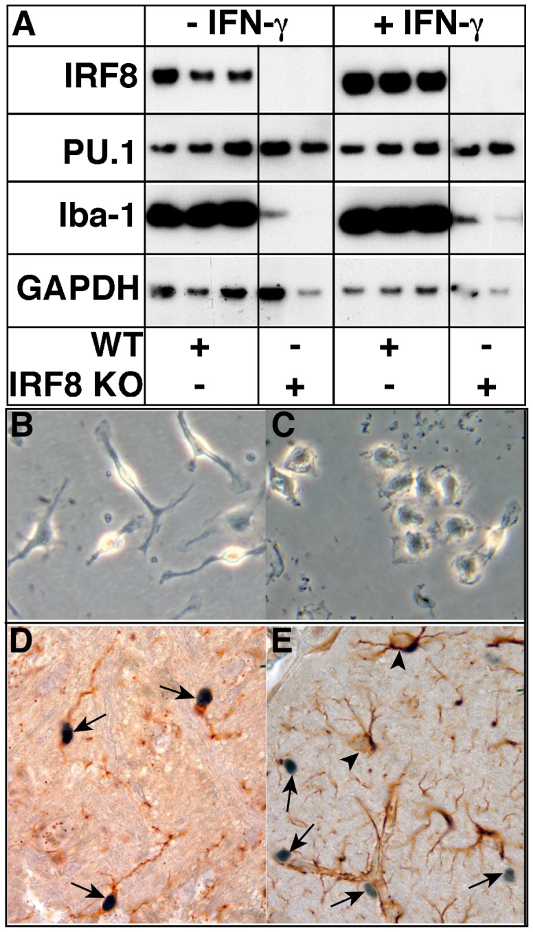
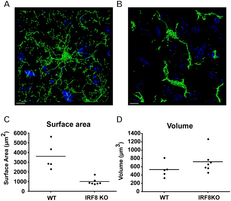
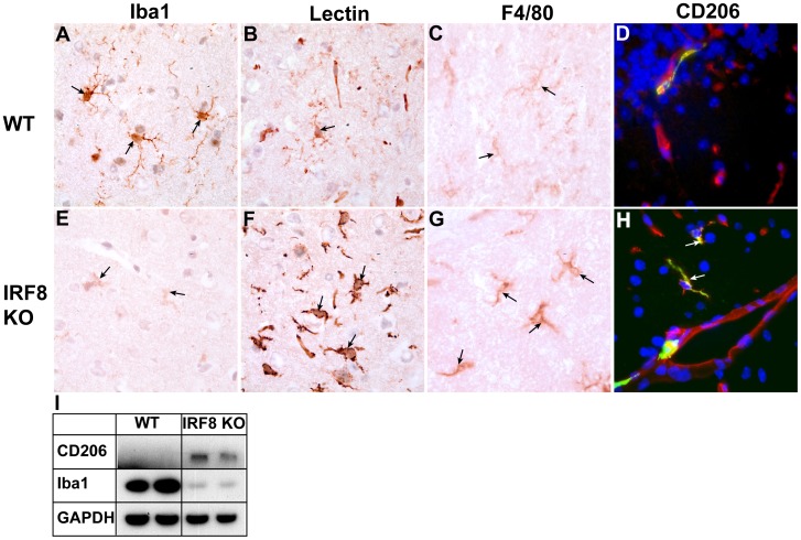
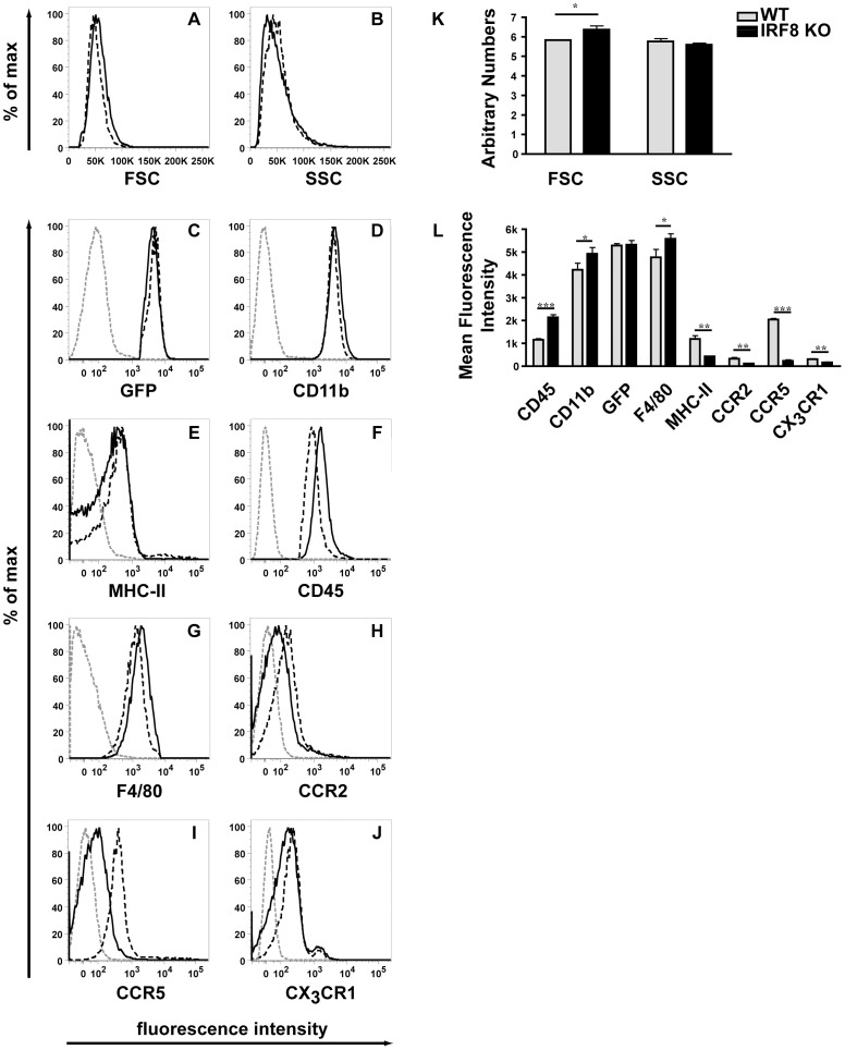
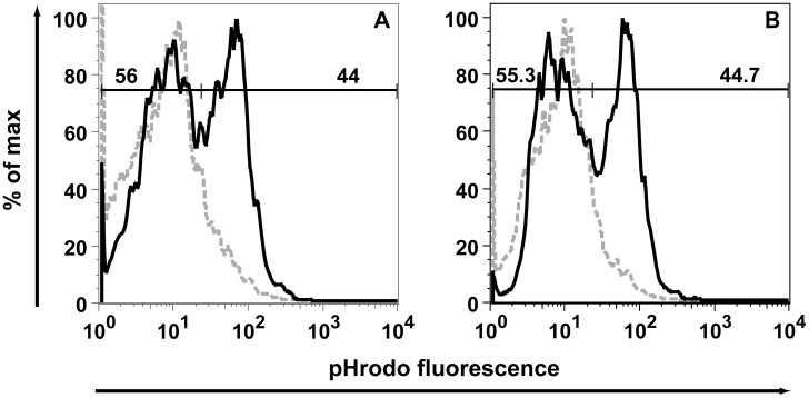
IFN regulatory factor (IRF) 8 is a transcription factor that has a key role in the cellular response to IFN-γ and is pivotal in myeloid cell differentiation. Whether IRF8 plays a role in the development and function of microglia, the tissue-resident myeloid cells of the brain, is unknown. Here, we show IRF8 is a constitutively produced nuclear factor in microglia, which suggested that IRF8 might also be a key homeostatic transcriptional determinant of the microglial cell phenotype. In support of this, in mice with a targeted disruption of the IRF8 gene, microglia were increased in number and showed gross alterations in morphology and surface area. In situ analysis of some key myeloid markers revealed that IRF8-deficient microglia had significantly reduced levels of Iba1, but increased levels of CD206 (mannose receptor) and F4/80 as well as increased tomato lectin binding. Analysis of microglia ex vivo revealed IRF8-deficient microglia had significantly increased levels of CD45, CD11b and F4/80, but significantly decreased levels of the chemokine receptors CCR2, CCR5 and CX3CR1. The known involvement of some of these molecular markers in membrane dynamics and phagocytosis led us to examine the phagocytic capacity of cultured IRF8-deficient microglia, however, this was found to be similar to wild type microglia. We conclude IRF8 is a constitutively produced nuclear factor in resident microglia of the CNS being a crucial transcriptional determinant of the phenotype of these cells in the healthy brain.


Conflict of interest statement

Competing Interests: The authors have declared that no competing interests exist.

Figures

Figure 1
Figure 1. Comparative features of cultured microglia from WT versus IRF8-deficient mice and localization of IRF8 in the brain.
Microglial cell cultures were prepared from the brain of neonatal mice as described in the Materials and Methods. Western blot analysis was performed on lysates of WT and IRF8-deficient primary microglia following treatment with (100 U/ml) or without IFN-γ for 4 h. GAPDH was used as a loading control (A). Morphological appearance of WT (B) or IRF8-deficient (C) microglia in primary culture (original magnification panels B&C, 400X). Immunohistochemical detection of IRF8 (D & E, arrows) combined with histochemical staing for tomato lectin binding (D) or GFAP immunohistochemistry (E, arrowheads). performed on brain sections from healthy adult mice. IRF8 staining is mostly confined to the nucleus of the cells (original magnification panel D, 1000X).
Figure 2
Figure 2. IRF8-deficient microglia have a marked reduction in ramification and surface area.
Representative confocal images of eGFP positive WT (A) and IRF8-deficient (B) microglia in the cortex, counterstained with DAPI (blue) were obtained from 100 µm vibratome sections on a Zeiss LSM 510 Meta confocal microscope. The 3D analysis (surface rendering) was performed using Bitplane Imaris software. Size bar = 10 µm. Quantification of single-cell surface area (C) and volume (D) for microglia in the cortex of WT (n = 5) or IRF8-deficient (n = 7) mice. Values are shown with the bar representing the mean. IRF8-deficient microglia showed a significant (p<0.0025; Mann-Whitney U test) reduction in cell surface area and although cellular volume of these cells was increased slightly this was not significant.
Figure 3
Figure 3. The levels of some key myeloid markers are altered in the brain of IRF8-deficient mice.
Immunostaining was performed on brain sections from healthy, adult WT (A–D) or IRF8-deficient (E–H) mice as described in the Materials and Methods. Panels A, B, C, E, F, G show cortex while panels D, H show cerebellum (original magnification all panels 1000X). For immunofluorescence (G, H) DAPI was used to stain nuclei. Whole brain lysates were prepared from healthy, adult mice and 20 µg of protein analysed by western blotting (I).
Figure 4
Figure 4. GFP+ cells in the CNS are exclusively CD11b+ microglia.
Flow cytometry was performed on cells isolated from the brain of healthy adult WT (A, B) and IRF8-deficient mice as described in the Materials and Methods. For analysis, cells were scatter-gated to exclude dead cells and GFP+ cells were selected (A). Greater than 99% of GFP+ cells expressed CD11b in WT and IRF8-deficient mice (B), consistent with a microglial phenotype. Isotype matched antibodies were used to determine background staining (data not shown).
Figure 5
Figure 5. Surface levels of various molecules are altered on IRF8-deficient microglia.
Flow cytometry (A-J) was performed on microglial cells isolated from the brain of adult WT (black dashed line) and IRF8-deficient (black line) mice as described in the Materials and Methods. For analysis, cells were scatter-gated to exclude dead cells and were selected for GFP, CD11b expression. An isotype matched antibody was used as a negative control (grey dashed line). Quantification of forward and sideward scatter; (K) or mean fluorescent intensity of histograms (L). The histograms represent means +/− SD from three separate experiments. For significance: ***P<0.001, **P<0.01, *P<0.05; by two-tailed t-test.
Figure 6
Figure 6. IRF8-deficient microglia show no defect in E.coli particle phagocytosis.
Flow cytometry analysing pHrodo fluorescent E.coli particle uptake after 3 h by WT (A) and IRF8-deficient (B) primary microglia (grey dashed line = 4°C control assay, black continuous line = 37°C). Cells were gated for eGFP and CD11b (CD11b-PerCP-Cy5.5) and pHrodo fluorescent E.coli particle uptake was determined by measuring signal increase in the red fluorescence channel (fl2).

Similar articles

Cited by

References

    1. Prinz M, Mildner A (2010) Microglia in the CNS: Immigrants from another world. Glia 59: 177–187. - PubMed
    1. Ransohoff RM, Cardona AE (2010) The myeloid cells of the central nervous system parenchyma. Nature 468: 253–262. - PubMed
    1. Ellis SL, Gysbers V, Manders PM, Li W, Hofer MJ, et al. (2010) The cell-specific induction of CXC chemokine ligand 9 mediated by IFN-gamma in microglia of the central nervous system is determined by the myeloid transcription factor PU.1. J Immunol 185: 1864–1877. - PMC - PubMed
    1. Wang H, Morse HC 3rd (2009) IRF8 regulates myeloid and B lymphoid lineage diversification. Immunol Res 43: 109–117. - PMC - PubMed
    1. Taniguchi T, Ogasawara K, Takaoka A, Tanaka N (2001) IRF family of transcription factors as regulators of host defense. Ann Rev Immunol 19: 623–655. - PubMed

Publication types