. 2012;7(11):e50262.
doi: 10.1371/journal.pone.0050262. Epub 2012 Nov 16.

Sensitivity to lysosome-dependent cell death is directly regulated by lysosomal cholesterol content

Affiliations

Sensitivity to lysosome-dependent cell death is directly regulated by lysosomal cholesterol content

Hanna Appelqvist et al. PLoS One. 2012.

Abstract

Alterations in lipid homeostasis are implicated in several neurodegenerative diseases, although the mechanisms responsible are poorly understood. We evaluated the impact of cholesterol accumulation, induced by U18666A, quinacrine or mutations in the cholesterol transporting Niemann-Pick disease type C1 (NPC1) protein, on lysosomal stability and sensitivity to lysosome-mediated cell death. We found that neurons with lysosomal cholesterol accumulation were protected from oxidative stress-induced apoptosis. In addition, human fibroblasts with cholesterol-loaded lysosomes showed higher lysosomal membrane stability than controls. Previous studies have shown that cholesterol accumulation is accompanied by the storage of lipids such as sphingomyelin, glycosphingolipids and sphingosine and an up regulation of lysosomal associated membrane protein-2 (LAMP-2), which may also influence lysosomal stability. However, in this study the use of myriocin and LAMP deficient fibroblasts excluded these factors as responsible for the rescuing effect and instead suggested that primarily lysosomal cholesterol content determineD the cellular sensitivity to toxic insults. Further strengthening this concept, depletion of cholesterol using methyl-β-cyclodextrin or 25-hydroxycholesterol decreased the stability of lysosomes and cells became more prone to undergo apoptosis. In conclusion, cholesterol content regulated lysosomal membrane permeabilization and thereby influenced cell death sensitivity. Our data suggests that lysosomal cholesterol modulation might be used as a therapeutic strategy for conditions associated with accelerated or repressed apoptosis.


Conflict of interest statement

Competing Interests: The authors have declared that no competing interests exist.

Figures

Figure 1
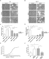
Figure 1. Cholesterol modulation in human fibroblasts is associated with alterations of the lysosomal compartment.
Human wt fibroblasts were treated with U18666A or quinacrine to induce cholesterol accumulation, and NPC1-mutant fibroblasts were treated with methyl-β-cyclodextrin (MβCD) or 25-hydroxy cholesterol (25-HC) to revert cholesterol storage. A) Measurement of unesterified cholesterol (n = 4) and B) representative images of filipin staining (scale bar 10 µm). C and D) Representative histogram from flow cytometric analysis of Lysotracker fluorescence staining. M1 gate denotes the highly fluorescent population. E and F) Quantification of peak channel in the M1 population (seen in C and D; n = 4). Data are presented as the mean ± SD, * p≤0.05.
Figure 2
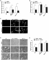
Figure 2. Manipulation of lysosomal cholesterol content modulates the cellular sensitivity to apoptosis.
Cholesterol content of human fibroblasts was modulated using U18666A, quinacrine, methyl-β-cyclodextrin (MβCD) or 25-hydroxy cholesterol (25-HC) before apoptosis was induced using O-methyl-serine dodecylamide hydrochloride (MSDH; 24 h). Phase contrast images of A) wt and B) NPC1-mutant fibroblasts (NPC1mut). Scale bar 20 µm. C) Viability of cultures in A and B, respectively, assessed by the MTT assay (n = 4). Viability is expressed as percentage of untreated cultures. D) Caspase-3 like activity (n = 4–8). E) Representative curve of increase in green fluorescence during photo-oxidation of acridine orange. F) Quantification of lag time (as presented in E; n = 5–6). Data are presented as the mean ± SD, * p≤0.05.
Figure 3
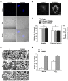
Figure 3. Cholesterol, and not accumulating sphingolipids, is responsible for the apoptosis protection.
Human wt fibroblasts, with or without U18666A treatment, and NPC1-mutant fibroblasts were treated with vehicle (dimethyl sulfoxide; DMSO) or myriocin to inhibit sphingolipid biosynthesis. A) Sphingomyelin (n = 3), B) cholesterol content (n = 4) and C) filipin staining (scale bar 10 μm) of human fibroblasts. D) Phase contrast images of human fibroblasts exposed to O-methyl-serine dodecylamide hydrochloride (MSDH; 24 h). Scale bar 20 μm. E) Viability of cultures in D, assessed by the MTT assay (n = 3). Viability is expressed as percentage of MSDH-untreated cultures. Data are presented as the mean ± SD, * p≤0.05, ns; non-significant.
Figure 4
Figure 4. Cholesterol Accumulation in Cortical Neurons Rescues Cells from Apoptosis Induced by MSDH and Oxidative Stress.
Cortical neurons were treated with U18666A. A) Filipin staining and differential interference contrast microscopy (DIC) images (scale bar 10 µm) and B) a higher magnification of filipin staining (scale bar 10 μM). C) Viability analysis and caspase-3-like activity (n = 3) after 72 h. D) Phase contrast images (scale bar 20 µm) and E) viability analysis (MTT assay; n = 3) of cultures exposed to O-methyl-serine dodecylamide hydrochloride (MSDH) or H2O2, generated by glucose oxidase, with or without pretreatment with U18666A (48 h). Viability is expressed as percentage of untreated control. Data are presented as the mean ± SD, * p≤0.05, ns; non-significant.
Figure 5
Figure 5. Cholesterol Accumulation Protects MEFs from Oxidative Stress-induced Apoptosis, Independent of the Expression of LAMP Proteins.
A) Localization (scale bar 10 μm) and B) expression of lysosome-associated membrane protein-2 (LAMP-2) in wt and NPC1-mutant (NPC1mut) human fibroblasts. Glyceraldehyde-3-phosphate dehydrogenase (GAPDH) was used to verify equal protein loading. One representative blot out of three is shown. C) Phase contrast images (scale bar 5 µm) and D) viability analysis (n = 4) of wt mouse embryonic fibroblasts (MEFs) and MEFs deficient for LAMP-1 (LAMP-1−/−) or LAMP-2 (LAMP-2−/−) 24 h after H2O2 exposure, with or without U18666A pretreatment. Viability was measured by crystal violet staining and expressed as percentage of untreated cultures. Data are presented as the mean ± SD, * p≤0.05) Filipin staining in MEFs, with or without U18666A treatment. Scale bar 10 µm.
Figure 6
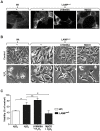
Figure 6. Cholesterol Modulation Influences the Sensitivity of MEFs to Oxidative Stress-induced Apoptosis.
Mouse embryonic fibroblasts (MEFs) deficient for both LAMP-1 and LAMP-2 (LAMPnull) were treated with U18666A or methyl-β-cyclodextrin (MβCD). A) Filipin staining of wt and LAMPnull MEFs (scale bar 10 µm). B) Phase contrast images (scale bar 5 µm) and C) viability (n = 4) of wt and LAMPnull MEFs 24 h after H2O2 exposure. Viability was measured by crystal violet staining and expressed as percentage of untreated cultures. Data are presented as the mean ± SD, * p≤0.05, ns; non-significant.

References

    1. Repnik U, Stoka V, Turk V, Turk B (2012) Lysosomes and lysosomal cathepsins in cell death. Biochim Biophys Acta 1824: 22–33. - PubMed
    1. Guicciardi ME, Deussing J, Miyoshi H, Bronk SF, Svingen PA, et al. (2000) Cathepsin B contributes to TNF-alpha-mediated hepatocyte apoptosis by promoting mitochondrial release of cytochrome c. Journal of Clinical Investigation 106: 1127–1137. - PMC - PubMed
    1. Foghsgaard L, Wissing D, Mauch D, Lademann U, Bastholm L, et al. (2001) Cathepsin B acts as a dominant execution protease in tumor cell apoptosis induced by tumor necrosis factor. Journal of Cell Biology 153: 999–1010. - PMC - PubMed
    1. Roberg K, Kågedal K, Öllinger K (2002) Microinjection of cathepsin D induces caspase-dependent apoptosis in fibroblasts. American Journal of Pathology 161: 89–96. - PMC - PubMed
    1. Johansson AC, Appelqvist H, Nilsson C, Kågedal K, Roberg K, et al. (2010) Regulation of apoptosis-associated lysosomal membrane permeabilization. Apoptosis 15: 527–540. - PMC - PubMed

Publication types

MeSH terms