. 2012:2:874.
doi: 10.1038/srep00874. Epub 2012 Nov 19.

TMV-Gate vectors: gateway compatible tobacco mosaic virus based expression vectors for functional analysis of proteins

Affiliations

TMV-Gate vectors: gateway compatible tobacco mosaic virus based expression vectors for functional analysis of proteins

Sateesh Kagale et al. Sci Rep. 2012.

Abstract

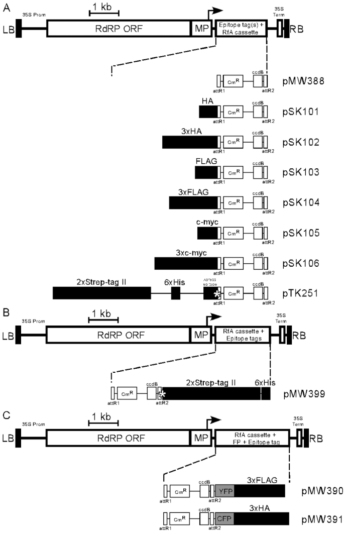
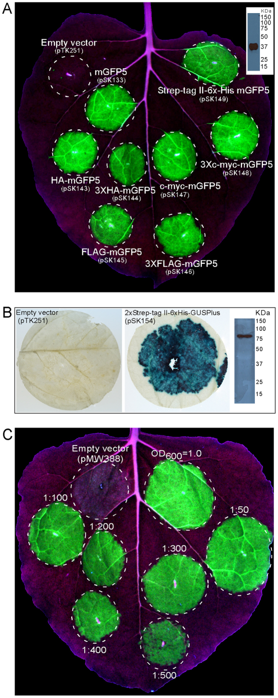
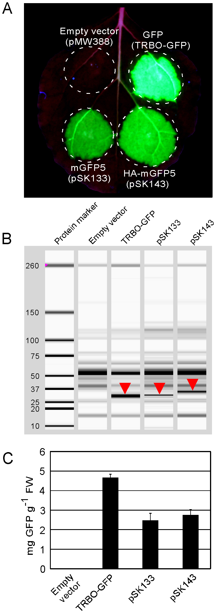
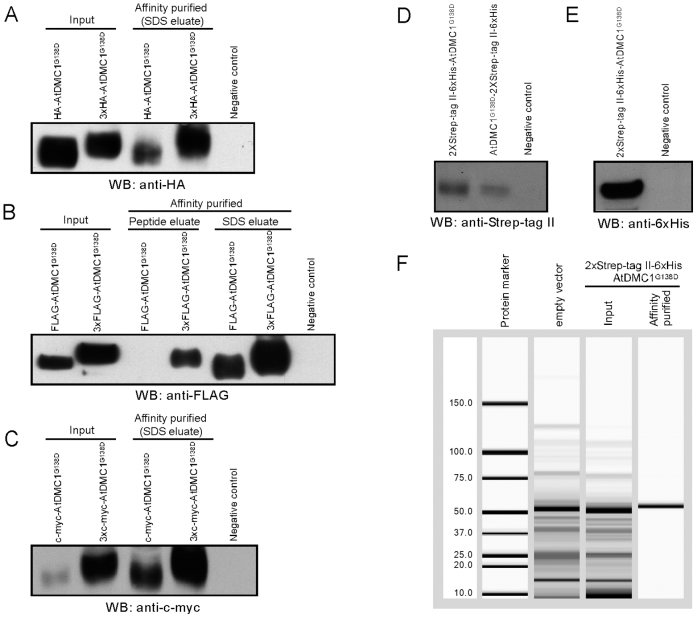
Plant viral expression vectors are advantageous for high-throughput functional characterization studies of genes due to their capability for rapid, high-level transient expression of proteins. We have constructed a series of tobacco mosaic virus (TMV) based vectors that are compatible with Gateway technology to enable rapid assembly of expression constructs and exploitation of ORFeome collections. In addition to the potential of producing recombinant protein at grams per kilogram FW of leaf tissue, these vectors facilitate either N- or C-terminal fusions to a broad series of epitope tag(s) and fluorescent proteins. We demonstrate the utility of these vectors in affinity purification, immunodetection and subcellular localisation studies. We also apply the vectors to characterize protein-protein interactions and demonstrate their utility in screening plant pathogen effectors. Given its broad utility in defining protein properties, this vector series will serve as a useful resource to expedite gene characterization efforts.


Figures

Figure 1
Figure 1. Schematic illustration of the TMV-Gate vectors.
(A) Expression vectors facilitating N-terminal tagging of target protein with various epitope tags, including single- or triple-repeats of HA, FLAG or c-myc, as well as 2xStrep-tag II-6xHis-Xpress epitope. (B) Expression vector facilitating C-terminal tagging of target protein with 2xStrep-tag II-6xHis epitope tags. (C)Expression vectors facilitating C-terminal tagging of target protein with epitope tagged fluorescent proteins, including YFP-3xFLAG or CFP-3xHA. The illustrated vector features include: LB, T-DNA left border; RB, T-DNA right border; 35S Prom, CaMV 35S promoter; 35S term, CaMV 35S terminator; RdRP, RNA-dependent RNA polymerase; MP, movement protein; bent arrow, coat protein promoter; attR1 and attR2, Gateway recombination sites; CmR, chloramphenicol resistance marker; ccdB, E. coli DNA gyrase inhibitor; asterisk in pTK251, enterokinase protease recognition site; asterisk in pMW399, 3C protease recognition site; YFP, yellow fluorescent protein; CFP, cyan fluorescent protein. The epitope tag regions in the map are enlarged 1:20 for readability. DNA sequences for the vectors have been deposited at GenBank with accession numbers as follows: pMW388, JX971627; pSK101, JX971619; pSK102, JX971620; pSK103, JX971621; pSK104, JX971622; pSK105, JX971623; pSK106, JX971624; pTK251, JX971625; pMW399, JX971626; pMW390, JX971628; and pMW391, JX971629.
Figure 2
Figure 2. Transient expression of mGFP5 and GusPlus in N. benthamiana leaves by using TMV-Gate vectors.
(A) N. benthamiana leaf infiltrated with A. tumefaciens carrying empty vector or viral expression vector encoding mGFP5 with or without translational fusion to either HA, 3xHA, FLAG, 3xFLAG, c-myc, 3xc-myc or 2xStrep-tag II-6xHis-Xpress epitope tag(s) was illuminated under UV light at 5 DPI and photographed. Inset shows immunoblot detection of 2xStrep-tag II-6xHis-Xpress epitope-tagged mGFP5 (in silico predicted size of 37.8 kDa) in crude extract using anti-6xHis antibody. (B) N. benthamiana leaf discs from the area infiltrated with A. tumefaciens carrying empty vector (pTK251) or viral expression vector encoding 2xStrep-tag II-6xHis-GusPlus (pSK154) were excised 5 DPI and stained for GUS activity. Inset in right panel shows immunoblot detection of 2xStrep-tag II-6xHis-Xpress epitope-tagged GusPlus (in silico predicted size of 81.5 kDa) in crude leaf extract using anti-6xHis antibody. (C) N. benthamiana leaf infiltrated with various dilutions of a cell suspension of A. tumefaciens at OD600 of 1.0 carrying pSK133 (pMW388 encoding mGFP5) and photographed with UV illumination at 5 DPI.
Figure 3
Figure 3. Quantitative analysis of transient GFP production in N. benthamiana using TRBO or TMV-Gate vectors.
(A) Fluorescence emitted from GFP transiently produced in a N. benthamiana leaf by infiltration with A. tumefaciens carrying TRBO-GFP (Lindbo, 2007b) or TMV-Gate constructs pSK133 encoding mGFP5, pSK143 encoding HA-mGFP5 or pMW388 (empty vector) and photographed with UV illumination at 5 DPI. (B) Assessment of GFP accumulation by automated capillary electrophoresis analysis. N. benthamiana leaf discs from the area outlined in (A) were excised at 5 DPI and corresponding protein extracts were resolved and GFP identified using the Experion capillary electrophoresis system. Position of the GFP band in each sample is indicated by downward pointing triangles in the simulated gel view produced by the Experion software. The shift in the molecular weight of GFP in pSK143 is due to the presence of an HA tag at the N-terminus. (C) Quantification of GFP resolved by automated capillary electrophoresis analysis using the Experion system. Error bars represent standard deviation. FW, fresh weight.
Figure 4
Figure 4. Affinity purification and detection of epitope-tagged AtDMC1G138D transiently expressed in N. benthamiana leaves by using TMV-Gate vectors.
Epitope-tagged recombinant AtDMC1G138D immobilized on anti-HA, anti-FLAG or anti-c-myc antibody-conjugated agarose beads were eluted either by boiling the beads in SDS-PAGE sample buffer (SDS eluate) or by incubating the beads with 3xFLAG peptide (peptide eluate). The Strep-tag II-tagged AtDMC1G138D immobilized on Strep-Tactin matrix was eluted using desthiobiotin. Equal volume aliquots of the input protein extract and eluted AtDMC1G138D proteins were subjected to SDS–PAGE, transferred onto PVDF membrane then subjected to immunoblot analysis. WB, western blot. (A) Immunoblot detection of HA- or 3xHA-tagged AtDMC1G138D protein affinity captured using anti-HA agarose affinity gel. (B) Immunoblot detection of FLAG- or 3xFLAG-tagged AtDMC1G138D protein affinity captured using anti-FLAG agarose affinity gel. (C) Immunoblot detection of c-myc- or 3xmyc-tagged AtDMC1G138D protein affinity captured using anti-c-myc conjugated agarose beads. (D) Immunoblot detection of 2xStrep-tag II-6xHis-Xpress epitope-tagged AtDMC1G138D in crude extract using anti-Strep-tag II antibody. (E) Immunoblot detection of 2xStrep-tag II-6xHis-Xpress epitope-tagged AtDMC1G138D in crude extract using anti-6xHis antibody. (F) Capillary electrophoresis analysis using the Experion system of 2xStrep-tag II-6xHis-Xpress epitope-tagged AtDMC1G138D protein affinity purified by using Strep-Tactin matrix.
Figure 5
Figure 5. Protein-protein interaction of AtDMC1G138D expressed using TMV-Gate vectors.
(A) Yeast two-hybrid analysis demonstrating self-interaction of AtDMC1G138D. (B) Confirmation of AtDMC1G138D self-interaction by pull-down experiment. The FLAG-tagged AtDMC1G138D and 3xHA-tagged AtDMC1G138D immobilized on anti-FLAG and anti-HA agarose beads, respectively, were incubated with total protein extract of N. benthamiana leaf discs transiently expressing c-myc-tagged AtDMC1G138D or c-myc-tagged mGFP5 (negative control) and 3xFLAG-tagged AtDMC1G138D or 3xFLAG-tagged GusPlus (negative control), respectively; corresponding TMV-Gate vectors and recombinant plasmids are listed in Table I. After washing the affinity matrices, proteins were eluted using SDS-PAGE sample buffer, resolved by SDS-PAGE and visualized by immunoblotting with antibodies as indicated. WB, western blot.
Figure 6
Figure 6. Nuclear localizsation of NLS-mCherry expressed using TMV-Gate vectors.
(A) Schematic diagram showing constructs used for analysis of subcellular localisation of NLS-mCherry. mCherry carrying a nuclear localization signal (NLS) was introduced unmodified into pMW388. A NLS-mCherry variant without translational stop codon was linked in-frame with the coding sequence of YFP-3xFLAG or CFP-3xHA in pMW390 and pMW391, respectively. Annotated features as per Figure 1. (B) Epifluorescent microscopy images showing nuclear localisation of NLS-mCherry (upper panel), NLS-mCherry linked to YFP-3xFLAG (middle panel) or NLS-mCherry linked to CFP-3xHA in cells of N. benthamiana leaves infiltrated with A. tumefaciens carrying the constructs described in (A) and imaged 3 DPI. The nucleus was detected by staining cells with DAPI. Images were taken using YFP, CFP, mCherry or DAPI filter sets as indicated. Scale bar, 20 µM. DIC, differential interference contrast.
Figure 7
Figure 7. TMV-Gate vector based expression of XopD effector protein in N. benthamiana leaves.
N. benthamiana leaves were infiltrated with A. tumefaciens strains expressing FLAG-tagged XopD (pSU101) or mGFP5 (negative control; pSK145) and the phenotypes were photographed at 2, 4, 6, 8, 10 and 12 DPI.

Similar articles

Cited by

References

    1. Scholthof H. B., Scholthof K. B. & Jackson A. O. Plant virus gene vectors for transient expression of foreign proteins in plants. Annu Rev Phytopathol 34, 299–323 (1996). - PubMed
    1. Turpen T. H. Tobacco mosaic virus and the virescence of biotechnology. Philos Trans R Soc Lond B Biol Sci 354, 665–673 (1999). - PMC - PubMed
    1. Marillonnet S. et al. In planta engineering of viral RNA replicons: efficient assembly by recombination of DNA modules delivered by Agrobacterium. Proc Natl Acad Sci USA 101, 6852–6857 (2004). - PMC - PubMed
    1. Yusibov V., Shivprasad S., Turpen T. H., Dawson W. & Koprowski H. Plant viral vectors based on tobamoviruses. Curr Top Microbiol Immunol 240, 81–94 (1999). - PubMed
    1. Grimsley N., Hohn B., Hohn T. & Walden R. “Agroinfection,” an alternative route for viral infection of plants by using the Ti plasmid. Proc Natl Acad Sci USA 83, 3282–3286 (1986). - PMC - PubMed

Publication types

MeSH terms