

doi: 10.3980/j.issn.2222-3959.2012.05.16.
Epub 2012 Oct 18.
Retinoblastoma in a young adult mimicking Coats' disease
Affiliations
- PMID: 23166876
- PMCID: PMC3484701
- DOI: 10.3980/j.issn.2222-3959.2012.05.16
Item in Clipboard
Retinoblastoma in a young adult mimicking Coats' disease
Int J Ophthalmol.
2012.
Abstract
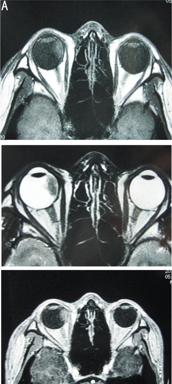
Retinoblastoma is the most common childhood primary intraocular malignancy, with the majority of cases being diagnosed before 5 years of age. Retinoblastoma in adults is extremely rare. Here, we report the case of a 20-year-old man who presented with a 3 year history of blurred vision in the right eye. Imaging did not reveal the typical presentation of retinoblastoma. After considering Coats' disease, a diagnosis of late-presenting retinoblastoma was made through cytological analysis. Diagnosis of retinoblastoma should be considered in the presence of uncertain mass lesions in the fundus of an adult.
Keywords: Coats'disease; adult; cytological analysis; retinoblastoma.
Figures


Similar articles
-
Advanced Coats' disease.Trans Am Ophthalmol Soc. 1991;89:371-476. Trans Am Ophthalmol Soc. 1991. PMID: 1808814 Free PMC article. Review.
-
Coats' retinitis or retinoblastoma in a 3-year-old girl: a case report.Medicina (Kaunas). 2012;48(4):224-7. Medicina (Kaunas). 2012. PMID: 22836296
-
Bilateral Coats' disease with unusual presentation--a case report.Bull Soc Belge Ophtalmol. 2005;(295):35-9. Bull Soc Belge Ophtalmol. 2005. PMID: 15849987
-
Clinical-histopathological correlation in a case of Coats' disease.Diagn Pathol. 2006 Aug 30;1:24. doi: 10.1186/1746-1596-1-24. Diagn Pathol. 2006. PMID: 16942617 Free PMC article.
-
Recent advances in the diagnosis and treatment of Coats' disease.Int Ophthalmol. 2019 Apr;39(4):957-970. doi: 10.1007/s10792-019-01095-8. Epub 2019 Mar 20. Int Ophthalmol. 2019. PMID: 30895419 Review.
Cited by
-
Adult onset retinoblastoma.Indian J Ophthalmol. 2016 Jul;64(7):485-91. doi: 10.4103/0301-4738.190099. Indian J Ophthalmol. 2016. PMID: 27609158 Free PMC article. Review.
-
Retinoblastoma in older patients: A retrospective comparative analysis of 100 consecutive patients based on age.Saudi J Ophthalmol. 2019 Jul-Sep;33(3):243-250. doi: 10.1016/j.sjopt.2019.07.008. Epub 2019 Aug 27. Saudi J Ophthalmol. 2019. PMID: 31686965 Free PMC article.
-
Indications for Pediatric Enucleations of Non-Retinoblastoma Globes in Saudi Arabia with Clinical and Radiological Correlation To Final Histopathological Diagnosis.J Epidemiol Glob Health. 2025 Aug 7;15(1):105. doi: 10.1007/s44197-025-00449-z. J Epidemiol Glob Health. 2025. PMID: 40773126 Free PMC article.
-
Clinicopathological Correlations in Enucleated Globes of Late-Stage Coats Disease with a Review of the Literature.J Epidemiol Glob Health. 2022 Dec;12(4):496-503. doi: 10.1007/s44197-022-00068-y. Epub 2022 Sep 29. J Epidemiol Glob Health. 2022. PMID: 36175755 Free PMC article. Review.
-
MR Imaging Features to Differentiate Retinoblastoma from Coats' Disease and Persistent Fetal Vasculature.Cancers (Basel). 2020 Nov 30;12(12):3592. doi: 10.3390/cancers12123592. Cancers (Basel). 2020. PMID: 33266342 Free PMC article.
References
-
- Karcioglu ZA, Abboud EB, Al-Mesfer SA, Al-Rashed W, Pilapil DH. Retinoblastoma in older children. J AAPOS. 2002;6(1):26–32. - PubMed
LinkOut - more resources
Full Text Sources