. 2013 Apr 1;22(7):1063-75.
doi: 10.1089/scd.2012.0315. Epub 2013 Jan 4.

Cord-blood-derived mesenchymal stromal cells downmodulate CD4+ T-cell activation by inducing IL-10-producing Th1 cells

Affiliations

Cord-blood-derived mesenchymal stromal cells downmodulate CD4+ T-cell activation by inducing IL-10-producing Th1 cells

Silvia Selleri et al. Stem Cells Dev. .

Abstract

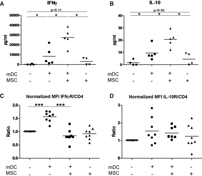
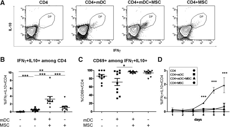
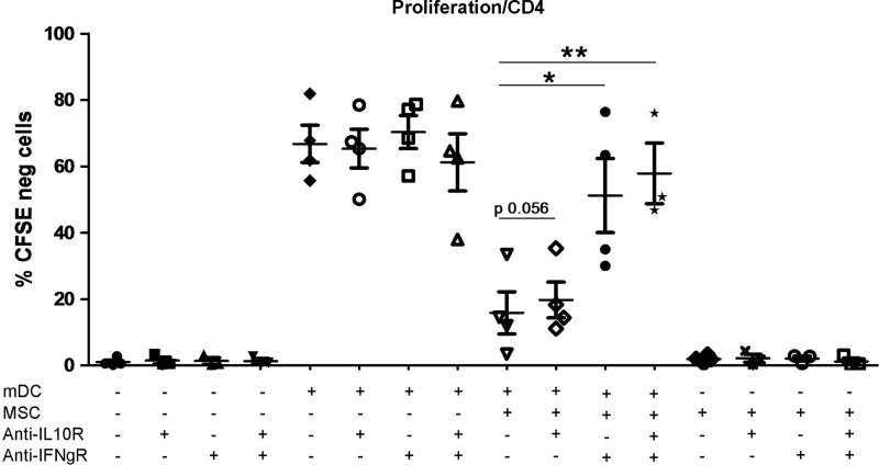
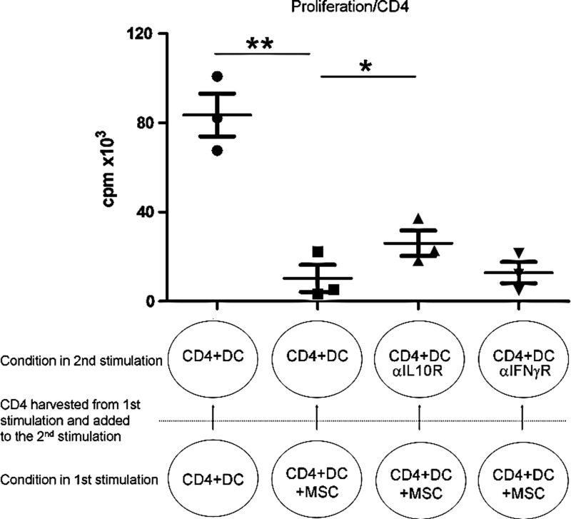
The mechanisms by which mesenchymal stromal cells (MSCs) induce immunomodulation are still poorly understood. In the current work, we show by a combination of polymerase chain reaction (PCR) array, flow cytometry, and multiplex cytokine data analysis that during the inhibition of an alloantigen-driven CD4+ T-cell response, MSCs induce a fraction of CD4+ T-cells to coexpress interferon-γ (IFNγ) and interleukin-10 (IL-10). This CD4+ IFNγ+ IL-10+ cell population shares properties with recently described T-cells originating from switched Th1 cells that start producing IL-10 and acquire a regulatory function. Here we report that IL-10-producing Th1 cells accumulated with time during T-cell stimulation in the presence of MSCs. Moreover, MSCs caused stimulated T-cells to downregulate the IFNγ receptor (IFNγR) without affecting IL-10 receptor expression. Further, the inhibitory effect of MSCs could be reversed by an anti-IFNγR-blocking antibody, indicating that IFNγ is one of the major players in MSC-induced T-cell suppression. Stimulated (and, to a lesser extent, resting) CD4+ T-cells treated with MSCs were able to inhibit the proliferation of autologous CD4+ T-cells, demonstrating their acquired regulatory properties. Altogether, our results suggest that the generation of IL-10-producing Th1 cells is one of the mechanisms by which MSCs can downmodulate an immune response.


Figures

FIG. 1.
FIG. 1.
Mesenchymal stromal cells (MSCs) inhibit CD4+ T-cell proliferation, but do not induce a classical Treg phenotype. Total peripheral blood mononuclear cells (PBMCs) were tested upon stimulation with allogeneic PBMCs in the presence and absence of MSCs. (A) CD4+ T-cell proliferation, analyzed by CFSE dilution, was inhibited by the presence of MSCs. (B) CD4+ T-cells were also analyzed for the presence of Tregs (CD4+ CD25+ CD127 FoxP3+ cells). (C) CD69 expression was analyzed on CD4+ FoxP3 CD25 cells. n=4. *p=0.05, **p=0.01, and ***p=0.0005 (paired t-test). CFSE, 5-(and 6)-carboxyfluorescein diacetate succinimidyl ester.
FIG. 2.
FIG. 2.
PCR array cluster analysis. PCR array data were clustered (A) independently or (B) grouped according to the condition. PCR, polymerase chain reaction.
FIG. 3.
FIG. 3.
Overlapping gene lists. (A) Each condition was compared with resting CD4+ T-cells, and upregulated genes are listed. In (B) and (C), gene lists were compared to the allogeneic mature dendritic cell (mDC)-stimulated CD4+ T-cell condition, and upregulated (B) and downregulated (C) genes are reported. In all analyses, we accepted fold-regulation >1.5 with a p-value<0.05. The obtained lists were analyzed using Venn diagrams. Color images available online at www.liebertpub.com/scd
FIG. 4.
FIG. 4.
MSCs modulate the secretion of IFNγ and IL-10, and the expression of their receptors on allogeneic stimulated CD4+ T-cells. (A) IFNγ and (B) IL-10 concentrations were measured in the supernatants of resting and stimulated CD4+ T-cells in the presence and absence of MSCs. Expression of (C) IFNγR and (D) IL-10R was evaluated by flow cytometry. Values were normalized based on the control condition (resting CD4+ T-cells). n=4 (A, B), n=8 (C, D). *p≤0.05, **p≤0.01, and ***p≤0.005. (A, B): Mann–Whitney test; (B, C): paired t-test. MFI=mean fluorescence intensity; IL-10, interleukin-10; IFNγ, interferon γ.
FIG. 5.
FIG. 5.
MSCs induce an increase in the percentage of CD4+ T-cells coexpressing IL-10 and IFNγ during allogeneic stimulation. Purified CD4+ T-cells were stimulated for 6 days by allogeneic mDCs in the presence and absence of MSCs. In (A), the cytofluorimetric analysis of a representative donor is shown. In (B, C), results on day 6 from 10 donors are shown. (C) Expression of CD69 gated on IFNγ+ IL-10+ CD4+ cells (n=10, analysis on day 6). (D) Time course analysis of the percentage of IFNγ+ IL-10+ CD4+ cells (n=3). All presented data were gated on CD4+ T-cells. *p≤0.05, **p≤0.01, and ***p≤0.005 (paired t-test).
FIG. 6.
FIG. 6.
Treatment with blocking anti-IFNγR antibodies largely restores T-cell proliferation in the presence of MSCs. CD4+ T-cells were stimulated with allogeneic mDCs for 6 days in the presence and absence of MSCs. Where indicated, blocking anti-IL-10R and/or anti-IFNγR antibodies were added everyday to the culture (20 μg/mL each). Data are expressed as the percentage of CD4+ T-cells negative for CFSE. n=4. *p≤0.05, **p≤0.01, paired t-test, 2 tails.
FIG. 7.
FIG. 7.
CD4+ T-cells previously cocultured with allogeneic mDCs and MSCs induce the inhibition of cellular proliferation of autologous CD4+ T-cells stimulated by allogeneic mDCs. After 6 days of culture in the presence of allogeneic mDCs and MSCs, CD4+ T-cells were harvested and added to a second culture where fresh autologous CD4+ T-cells were stimulated by the same allogeneic mDCs. The second culture was carried for 4 days. Where indicated, neutralizing antibodies blocking the activity of the IL-10R or IFNγR were added during the 4 days of the second stimulation. n=3 independent experiments. Statistics: paired t-test, *p≤0.05, **p≤0.01.

References

    1. Bernardo ME. Pagliara D. Locatelli F. Mesenchymal stromal cell therapy: a revolution in Regenerative Medicine? Bone Marrow Transplant. 2012;47:164–171. - PubMed
    1. Baron F. Lechanteur C. Willems E. Bruck F. Baudoux E. Seidel L. Vanbellinghen JF. Hafraoui K. Lejeune M, et al. Cotransplantation of mesenchymal stem cells might prevent death from graft-versus-host disease (GVHD) without abrogating graft-versus-tumor effects after HLA-mismatched allogeneic transplantation following nonmyeloablative conditioning. Biol Blood Marrow Transplant. 2010;16:838–847. - PubMed
    1. Kebriaei P. Isola L. Bahceci E. Holland K. Rowley S. McGuirk J. Devetten M. Jansen J. Herzig R, et al. Adult human mesenchymal stem cells added to corticosteroid therapy for the treatment of acute graft-versus-host disease. Biol Blood Marrow Transplant. 2009;15:804–811. - PubMed
    1. Ringden O. Uzunel M. Rasmusson I. Remberger M. Sundberg B. Lonnies H. Marschall HU. Dlugosz A. Szakos A, et al. Mesenchymal stem cells for treatment of therapy-resistant graft-versus-host disease. Transplantation. 2006;81:1390–1397. - PubMed
    1. Weng JY. Du X. Geng SX. Peng YW. Wang Z. Lu ZS. Wu SJ. Luo CW. Guo R, et al. Mesenchymal stem cell as salvage treatment for refractory chronic GVHD. Bone Marrow Transplant. 2010;45:1732–1740. - PMC - PubMed

Publication types

MeSH terms