

Polycystic kidneys have decreased vascular density: a micro-CT study
- PMID: 23167921
- PMCID: PMC3698948
- DOI: 10.1111/micc.12022
Polycystic kidneys have decreased vascular density: a micro-CT study
Abstract
Objective: Polycystic kidney disease (PKD) is a common cause of end-stage renal failure and many of these patients suffer vascular dysfunction and hypertension. It remains unclear whether PKD is associated with abnormal microvascular structure. Thus, this study examined the renovascular structure in PKD.
Methods: PKD rats (PCK model) and controls were studied at 10 weeks of age, and mean arterial pressure (MAP), renal blood flow, and creatinine clearance were measured. Microvascular architecture and cyst number and volume were assessed using micro-computed tomography, and angiogenic pathways evaluated.
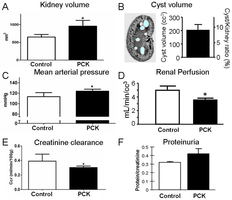
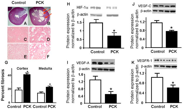
Results: Compared with controls, PKD animals had an increase in MAP (126.4 ± 4.0 vs. 126.2 ± 2.7 mmHg) and decreased clearance of creatinine (0.39 ± 0.09 vs. 0.30 ± 0.05 mL/min), associated with a decrease in microvascular density, both in the cortex (256 ± 22 vs. 136 ± 20 vessels per cm2) and medullar (114 ± 14 vs. 50 ± 9 vessels/cm2) and an increase in the average diameter of glomeruli (104.14 ± 2.94 vs. 125.76 ± 9.06 mm). PKD animals had increased fibrosis (2.2 ± 0.2 fold vs. control) and a decrease in the cortical expression in hypoxia inducible factor 1-α and vascular endothelial growth factor.
Conclusions: PKD animals have impaired renal vascular architecture, which can have significant functional consequences. The PKD microvasculature could represent a therapeutic target to decrease the impact of this disease.
© 2012 John Wiley & Sons Ltd.
Figures

References
-
- Grantham JJ. Clinical practice. Autosomal dominant polycystic kidney disease. N Engl J Med. 2008;359(14):1477–1485. - PubMed
-
- Mochizuki T, Wu G, Hayashi T, Xenophontos SL, Veldhuisen B, Saris JJ, Reynolds DM, Cai Y, Gabow PA, Pierides A, Kimberling WJ, Breuning MH, Deltas CC, Peters DJ, Somlo S. PKD2, a gene for polycystic kidney disease that encodes an integral membrane protein. Science. 1996;272(5266):1339–1342. - PubMed
-
- Hughes J, Ward CJ, Peral B, Aspinwall R, Clark K, San Millan JL, Gamble V, Harris PC. The polycystic kidney disease 1 (PKD1) gene encodes a novel protein with multiple cell recognition domains. Nat Genet. 1995;10(2):151–160. - PubMed
-
- Kelleher CL, McFann KK, Johnson AM, Schrier RW. Characteristics of hypertension in young adults with autosomal dominant polycystic kidney disease compared with the general U.S population. Am J Hypertens. 2004;17(11 Pt 1):1029–1034. - PubMed
Publication types
MeSH terms
Substances
Grants and funding
LinkOut - more resources
Full Text Sources
Other Literature Sources
