

Brain magnetic resonance spectroscopy in episodic hepatic encephalopathy
- PMID: 23168529
- PMCID: PMC3564202
- DOI: 10.1038/jcbfm.2012.173
Brain magnetic resonance spectroscopy in episodic hepatic encephalopathy
Abstract
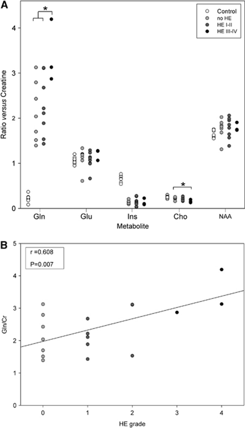
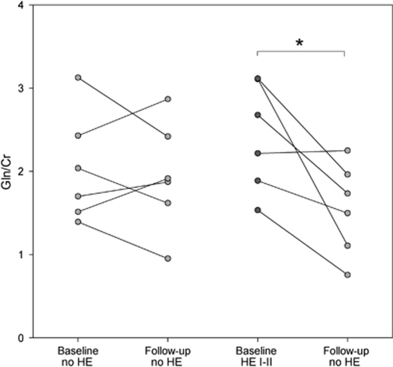
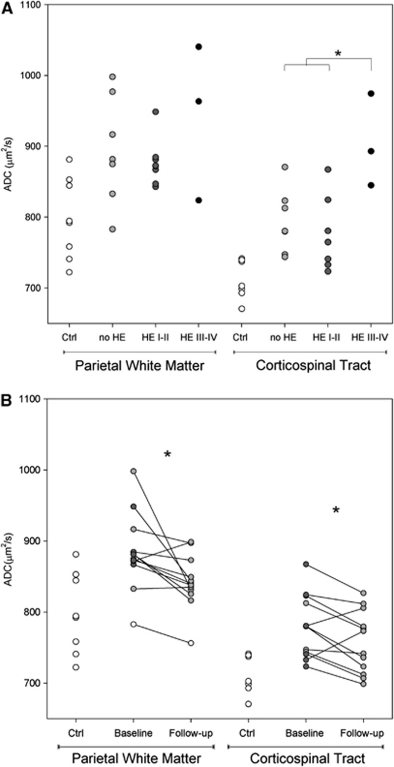
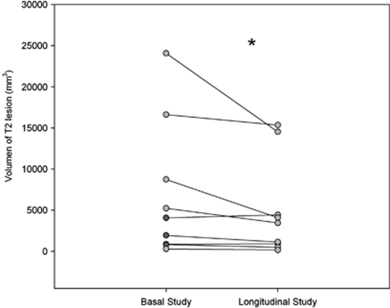
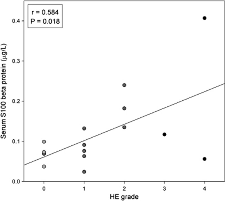
Brain magnetic resonance (MR) study has shown metabolic abnormalities and changes in water distribution of the brain tissue that may relate to the pathogenesis of hepatic encephalopathy (HE). We designed a study to investigate the disturbances in brain water and metabolites during episodic HE using a 3-T MR scanner. Cirrhotic patients with different grades of HE underwent MR during hospitalization (n=18). The MR was repeated at 6 weeks' follow-up (n=14). The results were compared with those of a group of healthy volunteers (n=8). During episodic HE, brain diffusion-weighted imaging showed a high apparent diffusion coefficient (ADC) (12% to 14%) that decreased during follow-up (-1% to -4%). These disturbances were accompanied by high glutamine (581%), low choline (-31%), and low myo-inositol (-86%) peaks on MR spectroscopy. In overt HE, patients showed high glutamine that decreased during follow-up (-22%). In addition, these patients exhibited a rise in plasma S100 beta and enlargement of brain white-matter lesions. In conclusion, several disturbances detected by MR support the presence of impaired brain water homeostasis during episodic HE. Although astrocytes have a major role in this condition, brain edema during episodic HE may be extracellular and does not appear to be directly responsible for the development of neurologic manifestations.
Figures
References
-
- Cordoba J, Minguez B. Hepatic encephalopathy. Semin Liver Dis. 2008;28:70–80. - PubMed
-
- Norenberg MD. Astroglial dysfunction in hepatic encephalopathy. Metab Brain Dis. 1998;13:319–335. - PubMed
-
- Zemtsova I, Gorg B, Keitel V, Bidmon HJ, Schror K, Haussinger D. Microglia activation in hepatic encephalopathy in rats and humans. Hepatology. 2011;54:204–215. - PubMed
-
- Palomero-Gallagher N, Bidmon HJ, Cremer M, Schleicher A, Kircheis G, Reifenberger G, et al. Neurotransmitter receptor imbalances in motor cortex and basal ganglia in hepatic encephalopathy. Cell Physiol Biochem. 2009;24:291–306. - PubMed
Publication types
MeSH terms
Substances
LinkOut - more resources
Full Text Sources
Medical
Molecular Biology Databases
Miscellaneous
