Review
. 2013 Jan 1;5(1):a007898.
doi: 10.1101/cshperspect.a007898.

The β-catenin destruction complex

Affiliations
Review

The β-catenin destruction complex

Jennifer L Stamos et al. Cold Spring Harb Perspect Biol. .

Abstract

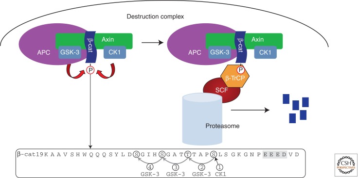
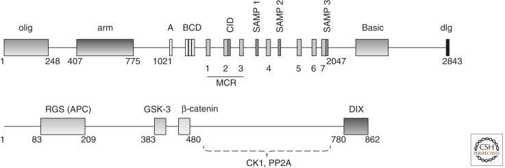
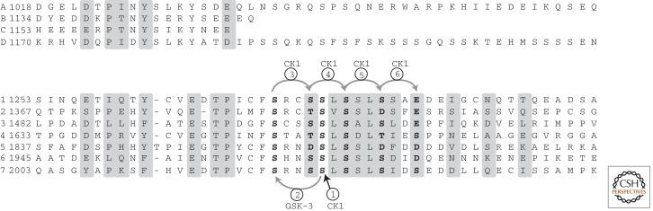
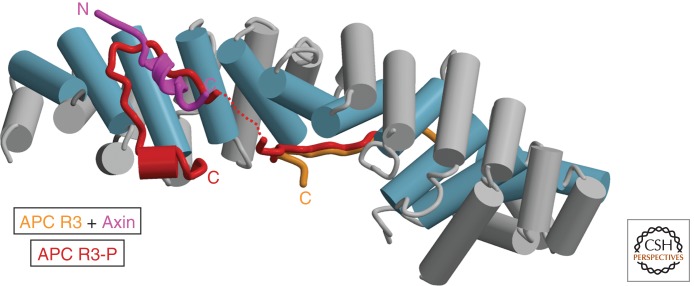
The Wnt/β-catenin pathway is highly regulated to insure the correct temporal and spatial activation of its target genes. In the absence of a Wnt stimulus, the transcriptional coactivator β-catenin is degraded by a multiprotein "destruction complex" that includes the tumor suppressors Axin and adenomatous polyposis coli (APC), the Ser/Thr kinases GSK-3 and CK1, protein phosphatase 2A (PP2A), and the E3-ubiquitin ligase β-TrCP. The complex generates a β-TrCP recognition site by phosphorylation of a conserved Ser/Thr-rich sequence near the β-catenin amino terminus, a process that requires scaffolding of the kinases and β-catenin by Axin. Ubiquitinated β-catenin is degraded by the proteasome. The molecular mechanisms that underlie several aspects of destruction complex function are poorly understood, particularly the role of APC. Here we review the molecular mechanisms of destruction complex function and discuss several potential roles of APC in β-catenin destruction.


Figures

Figure 1.
Figure 1.
Overview of the destruction complex. The phosphorylated sequence in the amino terminus of β-catenin is shown, with CK1 and GSK-3 sites circled. The acidic cluster that primes CK1 phosphorylation is highlighted in gray. SCF, the Skp1/cullin/F-box complex.
Figure 2.
Figure 2.
Primary structures of human Axin and APC. Key protein–protein interaction regions are indicated. Structured domains are RGS, regulator of G-protein signaling homology; DIX, domain common to Dishevelled and Axin; olig, coiled-coil dimerization domain of human APC; and arm, armadillo repeat domain. 15-mer repeats are marked A–D; 20-mer repeats are marked 1–7. The mutation cluster region (MCR) is the site of the majority of APC truncations found in colorectal cancers. Dashed lines indicate that mapping of the CK1 and PP2A sites on Axin are based on deletion studies and have not been verified with purified proteins.
Figure 3.
Figure 3.
Alignment of β-catenin-binding sequences of human APC, showing the 15-mers (top) and 20-mers (bottom). Residues that directly contact β-catenin, as observed in crystal structures of β-catenin bound to 15RA (Spink et al. 2001) and 20R3 (Ha et al. 2004; Xing et al. 2004), are shaded. 20-mer residues phosphorylated by CK1 and GSK-3 are shown in bold, with the phosphorylation order determined by priming rules for each kinase. (From Ha et al. 2004; adapted, with express permission, from the authors.)
Figure 4.
Figure 4.
Competition between phosphorylated APC and Axin for β-catenin. Crystal structures of nonphosphorylated APC R3 (orange) (Ha et al. 2004), Axin (purple) (Xing et al. 2003), and phosphorylated APC R3-P (red) (Ha et al. 2004; Xing et al. 2004) bound to the armadillo repeat domain of β-catenin (blue and gray) were superimposed to show the relationship between the bound ligands. The portion of APC that becomes ordered on phosphorylation by CK1 and GSK-3 (compare red and orange structures) binds to an overlapping surface with Axin, and biochemical studies show that these two ligands compete for binding. This competition is the basis for models suggesting that Axin is displaced from β-catenin when the Axin-bound kinases phosphorylate APC.

References

    1. Aberle H, Bauer A, Stappert J, Kispert A, Kemler R 1997. β-Catenin is a target for the ubiquitin-proteasome pathway. EMBO J 16: 3797–3804 - PMC - PubMed
    1. Albuquerque C, Breukel C, van der Luijt R, Fidalgo P, Lage P, Slors FJ, Leitao CN, Fodde R, Smits R 2002. The “just-right” signaling model: APC somatic mutations are selected based on a specific level of activation of the β-catenin signaling cascade. Hum Mol Genet 11: 1549–1560 - PubMed
    1. Amit S, Hatzubai A, Birman Y, Andersen JS, Ben-Shushan E, Mann M, Ben-Neriah Y, Alkalay I 2002. Axin-mediated CKI phosphorylation of β-catenin at Ser 45: A molecular switch for the Wnt pathway. Genes Dev 16: 1066–1076 - PMC - PubMed
    1. Behrens J, von Kries JP, Kühl M, Bruhn L, Wedlich D, Grosschedl R, Birchmeier W 1996. Functional interaction of β-catenin with the transcription factor LEF-1. Nature 382: 638–642 - PubMed
    1. Behrens J, Jerchow B-A, Würtele M, Grimm J, Asbrand C, Wirtz R, Kühl M, Wedlich D, Birchmeier W 1998. Functional interaction of an Axin homolog, conductin, with β-catenin, APC, and GSK3β. Science 280: 596–599 - PubMed

Publication types

MeSH terms