. 2013 Feb;41(2):466-74.
doi: 10.1124/dmd.112.045674. Epub 2012 Nov 20.

Immunohistochemical and functional characterization of peptide, organic cation, neutral and basic amino acid, and monocarboxylate drug transporters in human ocular tissues

Affiliations

Immunohistochemical and functional characterization of peptide, organic cation, neutral and basic amino acid, and monocarboxylate drug transporters in human ocular tissues

Rajendra S Kadam et al. Drug Metab Dispos. 2013 Feb.

Retraction in

  • Notice of retraction.
    [No authors listed] [No authors listed] Drug Metab Dispos. 2015 Feb;43(2):234. doi: 10.1124/dmd.114.045674err. Drug Metab Dispos. 2015. PMID: 25537846 Free PMC article. No abstract available.

Abstract

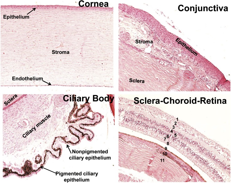
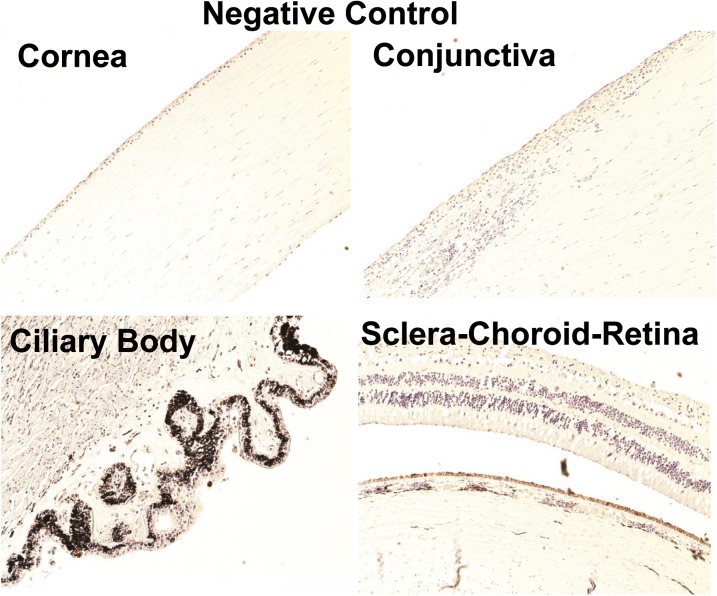
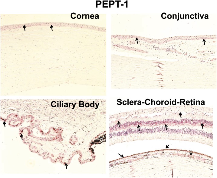
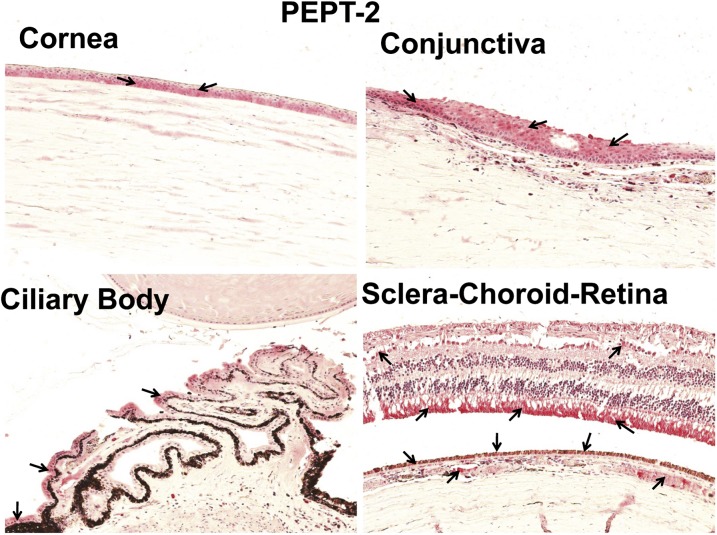
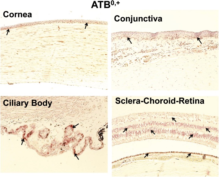
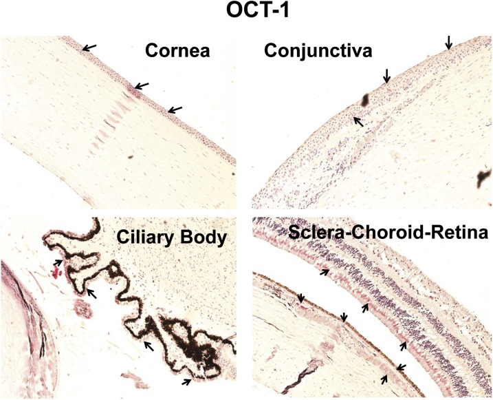
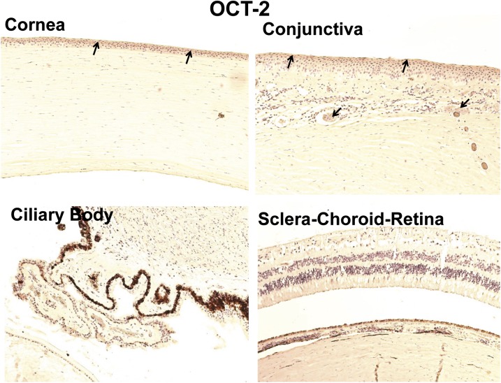
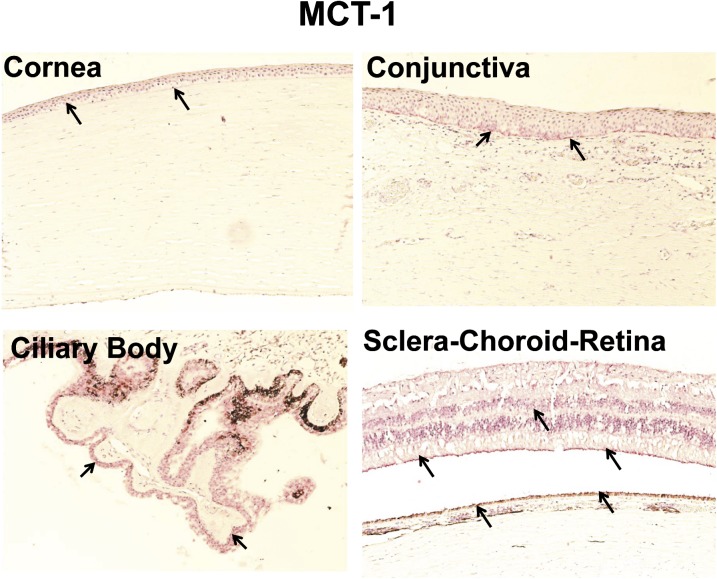
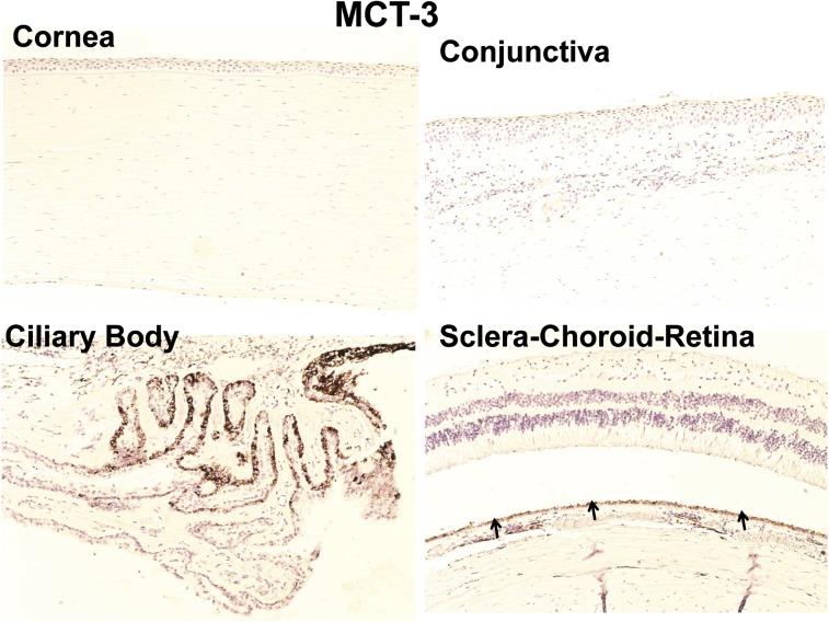
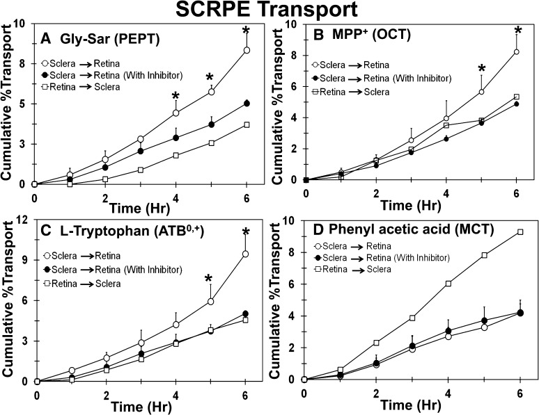
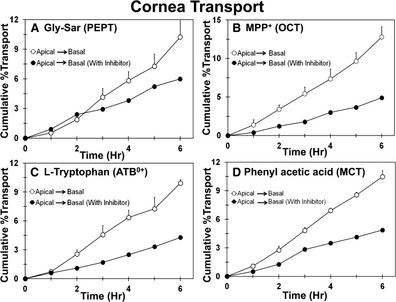
Since there is paucity of information on solute transporters in human ocular tissues, the aim of this study was immunohistochemical and functional characterization of peptide transporters (PEPT), organic cation transporters (OCTs), neutral and basic amino acid transporters (ATB(0,+)), and monocarboxylate transporters (MCTs) in human ocular barriers. Immunohistochemical localization of transporters was achieved using 5-µm-thick paraffin-embedded sections of whole human eyes. In vitro transport studies were carried out across human cornea and sclera-choroid-retinal pigment epithelium (SCRPE) using a cassette of specific substrates in the presence and absence of inhibitors to determine the role of transporters in transtissue solute delivery. Immunohistochemistry showed the expression of PEPT-1, PEPT-2, ATB(0,+), OCT-1, OCT-2, MCT-1, and MCT-3 in human ocular tissues. PEPT-1, PEPT-2, OCT-1, MCT-1, and ATB(0,+) expression was evident in the cornea, conjunctiva, ciliary epithelium, and neural retina. Expression of PEPT-1, PEPT-2, and OCT-1 was evident in choroid tissue as well. OCT-2 expression could be seen in the corneal and conjunctival epithelia, whereas MCT-3 expression was confined to the RPE layer. OCT-2 expression was evident in conjunctival blood vessel walls, whereas PEPT-1, PEPT-2, and OCT-1 were expressed in the choroid. Preliminary transport studies indicated inward transport of Gly-Sar (PEPT substrate), 1-methyl-4-phenylpyridinium (MPP+) (OCT substrate), and l-tryptophan (ATB(0,+) substrate) across cornea as well as SCRPE. For phenylacetic acid (MCT substrate), transporter-mediated inward transport across the cornea and outward transport across SCRPE were evident. Thus, PEPT, OCT, and ATB(0,+) are influx transporters present in human ocular barriers, and they can potentially be used for transporter-guided retinal drug delivery after topical, transscleral, and systemic administrations.


Figures

Fig. 1.
Fig. 1.
Anatomic assessment of human ocular tissues. Human ocular tissue sections were stained with H&E for morphologic assessment. Numbers in sclera-choroid-retina refer to different layers: 1, inner limiting membrane; 2, ganglion cell layer; 3, inner plexiform layer; 4, inner nuclear layer; 5, outer plexiform layer; 6, outer nuclear layer; 7, inner segment of photoreceptor cell; 8 outer segment of photoreceptor cell; 9, retinal pigmented epithelium; 10, choroid; 11, sclera.
Fig. 2.
Fig. 2.
Representative negative control images for immunohistochemical localization of drug transporters in human ocular tissues. For control experiments, the sections were processed in the same manner as test samples, except that the incubation step with primary antibody was omitted. Nuclei were counterstained in blue with hematoxylin.
Fig. 3.
Fig. 3.
Representative figure of immunohistochemical localization of PEPT-1 in human ocular tissues. Arrowhead indicates the localization of PEPT-1 staining. Light staining of PEPT-1 was observed in the epithelial layers of the cornea and conjunctiva and nonpigmented epithelial layer of the ciliary body. In sclera-choroid-retina, strong labeling of PEPT-1 was observed in the outer plexiform layer and light staining was observed in RPE cell layer, smooth muscles of choroid blood vessels, and ganglion cell layer. Nuclei were counterstained in blue with hematoxylin.
Fig. 4.
Fig. 4.
Representative figure of immunohistochemical localization of PEPT-2 in human ocular tissues. Arrowhead indicates the localization of PEPT-2 staining. PEPT-2 clearly localized in the epithelial layers of the cornea and conjunctiva and nonpigmented epithelial layer of the ciliary body. In the sclera-choroid-retina, PEPT-2 was abundantly localized in the outer segment of photoreceptor cells, RPE cell layer, and smooth muscles of choroid blood vessels. Light staining for PEPT-2 was also observed in the ganglion cell layer. Nuclei were counterstained in blue with hematoxylin.
Fig. 5.
Fig. 5.
Representative figure of immunohistochemical localization of ATB0,+ in human ocular tissues. ATB0,+ showed localization near the nucleus. Arrowhead indicates the localization of ATB0,+ staining. ATB0,+ showed light staining in the corneal and conjunctival epithelium and conjunctival stroma. ATB0,+ exhibited abundant expression in the nonpigmented ciliary epithelium and RPE layer. Nuclei were counterstained in blue with hematoxylin.
Fig. 6.
Fig. 6.
Representative figure of immunohistochemical localization of OCT-1 in human ocular tissues. Arrowhead indicates the localization of OCT-1 staining. OCT-1 showed light staining in the corneal epithelium, conjunctival epithelium, and nonpigmented ciliary epithelium. OCT-1 exhibited abundant expression in the inner segment of photoreceptor cells, RPE cell layer, and smooth muscles of choroidal blood vessels. Nuclei were counterstained in blue with hematoxylin.
Fig. 7.
Fig. 7.
Representative figure of immunohistochemical localization of OCT-2 in human ocular tissues. Arrowhead indicates the localization of OCT-2 staining. OCT-2 showed light expression in the corneal and conjunctival epithelia. Nuclei were counterstained in blue with hematoxylin.
Fig. 8.
Fig. 8.
Representative figure of immunohistochemical localization of MCT-1 in human ocular tissues. Arrowhead indicates the localization of MCT-1 staining. MCT-1 showed light expression in corneal and conjunctival basal epithelial cells. MCT-1 showed strong labeling in the nonpigmented ciliary epithelium compared with any other ocular tissues. For choroid-retina, MCT-1 staining was present in the outer segment of photoreceptor cells, inner limiting membrane, and RPE cell layer. Nuclei were counterstained in blue with hematoxylin.
Fig. 9.
Fig. 9.
Representative image for immunohistochemical localization of MCT-3 transporter in human ocular tissues. MCT-3 immunolabeling was observed only in the RPE layer; all other ocular tissues were devoid of labeling with MCT-3 antibody. Nuclei were counterstained in blue with hematoxylin.
Fig. 10.
Fig. 10.
Sclera-to-retina transport of Gly-Sar, MPP+, and l-tryptophan was higher than retina-to-sclera transport and significantly inhibited in the presence of the inhibitor cocktail, whereas there was no effect of inhibitors on the transport of phenylacetic acid. The effect of inhibitors on transport of (A) Gly-Sar, (B) MPP+, (C) l-tryptophan, and (D) phenylacetic acid across the human sclera-choroid-RPE. Data are expressed as mean ± S.D. for n = 4 for sclera to retina direction. For the retina to sclera direction, data are expressed as mean for n = 2. * P ≤ 0.05 when compared with transport in the presence of inhibitor.
Fig. 11.
Fig. 11.
Transport of Gly-Sar, MPP+, l-tryptophan, and phenylacetic acid across the human cornea is inhibited in the presence of an inhibitor cocktail. The effect of inhibitors on transport of (A) Gly-Sar, (B) MPP+, (C) l-tryptophan, and (D) phenylacetic acid across the human cornea. Data are expressed as mean ± S.D. for n = 4 for the apical to basal direction and mean of n = 2 for the apical to basal direction with inhibitors.

Comment in

  • Findings of Research Misconduct.
    [No authors listed] [No authors listed] Fed Regist. 2018 Dec 6;83(234):62875. Fed Regist. 2018. PMID: 30556543 Free PMC article. No abstract available.

Similar articles

Cited by

References

    1. Anand BS, Mitra AK. (2002) Mechanism of corneal permeation of L-valyl ester of acyclovir: targeting the oligopeptide transporter on the rabbit cornea. Pharm Res 19:1194–1202 - PubMed
    1. Basu SK, Haworth IS, Bolger MB, Lee VH. (1998) Proton-driven dipeptide uptake in primary cultured rabbit conjunctival epithelial cells. Invest Ophthalmol Vis Sci 39:2365–2373 - PubMed
    1. Biegel A, Knütter I, Hartrodt B, et al. (2006) The renal type H+/peptide symporter PEPT2: structure-affinity relationships. Amino Acids 31:137–156 - PubMed
    1. Brandsch M, Knütter I, Thunecke F, Hartrodt B, Born I, Börner V, Hirche F, Fischer G, Neubert K. (1999) Decisive structural determinants for the interaction of proline derivatives with the intestinal H+/peptide symporter. Eur J Biochem 266:502–508 - PubMed
    1. Chidlow G, Wood JP, Graham M, Osborne NN. (2005) Expression of monocarboxylate transporters in rat ocular tissues. Am J Physiol Cell Physiol 288:C416–C428 - PubMed

Publication types

MeSH terms

Substances