. 2012 Dec 11;109(50):20467-72.
doi: 10.1073/pnas.1218535109. Epub 2012 Nov 20.

Mutual protection of ribosomal proteins L5 and L11 from degradation is essential for p53 activation upon ribosomal biogenesis stress

Affiliations

Mutual protection of ribosomal proteins L5 and L11 from degradation is essential for p53 activation upon ribosomal biogenesis stress

Sladana Bursać et al. Proc Natl Acad Sci U S A. .

Abstract

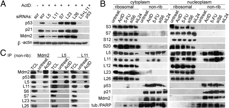
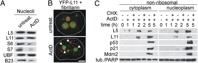
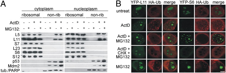
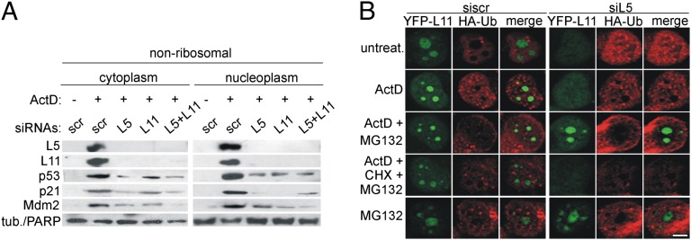
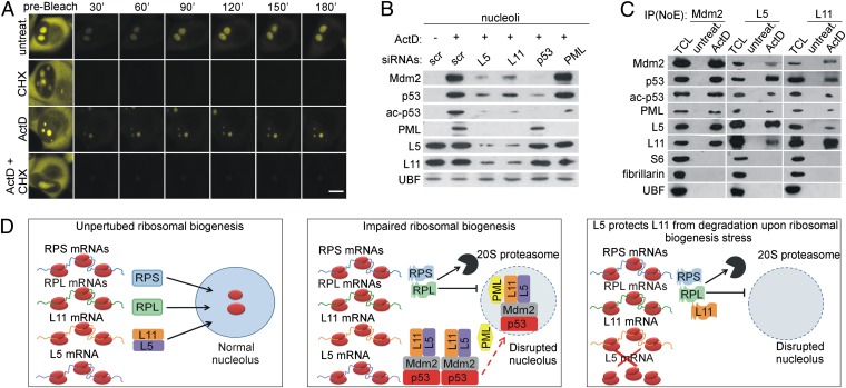
Impairment of ribosomal biogenesis can activate the p53 protein independently of DNA damage. The ability of ribosomal proteins L5, L11, L23, L26, or S7 to bind Mdm2 and inhibit its ubiquitin ligase activity has been suggested as a critical step in p53 activation under these conditions. Here, we report that L5 and L11 are particularly important for this response. Whereas several other newly synthesized ribosomal proteins are degraded by proteasomes upon inhibition of Pol I activity by actinomycin D, L5 and L11 accumulate in the ribosome-free fraction where they bind to Mdm2. This selective accumulation of free L5 and L11 is due to their mutual protection from proteasomal degradation. Furthermore, the endogenous, newly synthesized L5 and L11 continue to be imported into nucleoli even after nucleolar disruption and colocalize with Mdm2, p53, and promyelocytic leukemia protein. This suggests that the disrupted nucleoli may provide a platform for L5- and L11-dependent p53 activation, implying a role for the nucleolus in p53 activation by ribosomal biogenesis stress. These findings may have important implications with respect to understanding the pathogenesis of diseases caused by impaired ribosome biogenesis.


Conflict of interest statement

The authors declare no conflict of interest.

Figures

Fig. 1.
Fig. 1.
Specific requirement for L5 and L11 in p53 up-regulation upon inhibition of ribosome biogenesis in A549 cells. (A) Cells were transfected with the indicated siRNAs or scrambled (scr) negative control siRNA for 48 h and treated with ActD for 16 h. Expression of the indicated proteins was confirmed by immunoblotting. (B) Cells were transfected with the indicated siRNAs for 48 h or treated with ActD or 5-FU for 16 h. Ribosomal and nonribosomal (nonrib) cytoplasmic (Left) or ribosomal and nonribosomal nucleoplasmic fractions (Right) were immunoblotted with the indicated antibodies. The per-cell ratio of the amount of protein loaded onto a gel for cytoplasmic ribosomal, nucleoplasmic ribosomal, cytoplasmic nonribosomal, and nucleoplasmic nonribosomal fractions was 1:10:3:10. Tubulin (tub.) was used as a cytoplasmic marker and PARP as a nucleoplasmic marker. (C) Nonribosomal fractions from untreated and ActD-treated cells were immunoprecipitated with the indicated antibodies. These immunoprecipitates and the total cell lysate (TCL) from ActD-treated A549 cells were immunoblotted with the indicated antibodies.
Fig. 2.
Fig. 2.
De novo protein synthesis of L5 and L11 is required for their accumulation in the ribosome-free form. (A) A549 cells were left untreated or treated with ActD for 5 h. Purified nucleoli were immunoblotted with the indicated antibodies. (B) H1299 cells containing YFP-L11 (green) were treated with ActD for 5 h, stained with antifibrillarin antibodies (red), and analyzed by CLSM. Fibrillarin nucleolar caps are indicated by arrowheads. Nuclei are outlined with dashed lines. (Scale bar, 5 μm.) (C) A549 cells were treated with ActD in the presence or absence of CHX for 1, 2, or 5 h, and nonribosomal fractions were immunoblotted with the indicated antibodies.
Fig. 3.
Fig. 3.
Ribosome-free L5 and L11 are not degraded by proteasomes upon ActD treatment. (A) A549 cells were treated with ActD for 5 h in the presence or absence of MG132. Nonribosomal fractions were immunoblotted with the indicated antibodies. (B) H1299 cells expressing YFP-L11 or YFP-S6 were transfected with HA-Ub for 24 h and then treated as indicated for 6 h, followed by staining with anti-HA antibody (red) and analysis by CLSM. (Scale bar, 5 μm.)
Fig. 4.
Fig. 4.
Accumulation of ribosome-free L5 and L11 upon ActD treatment is due to their mutual protection from proteasomal degradation. (A) A549 cells were transfected with the indicated siRNAs for 48 h and then treated with ActD for 5 h. Nonribosomal fractions were immunoblotted with the indicated antibodies. (B) H1299 cells expressing YFP-L11 (green) were transfected with L5 siRNA for 24 h and then transfected with HA-Ub for 24 h. Cells were then treated as indicated for 6 h, stained with anti-HA antibody (red), and analyzed by CLSM. (Scale bar, 5 μm.)
Fig. 5.
Fig. 5.
Newly synthesized L5 and L11 colocalize with Mdm2, p53, and PML in the nucleolus upon ribosomal stress. (A) H1299 cells expressing YFP-L11 were treated with ActD for 5 h in the presence or absence of CHX and then photobleached using a 488-nm laser. Time-lapse laser scanning microscopy was used to monitor the recovery of the nucleolar fluorescence signal. (Scale bar, 5 μm.) (B) A549 cells were transfected with siRNAs against L5, L11, p53, or PML for 48 h and then left untreated or treated with ActD for 5 h. Purified nucleoli were immunoblotted with the indicated antibodies. (C) Nucleolar extracts (NoE) from untreated or ActD-treated (5 h) A549 cells were immunoprecipitated with antibodies against Mdm2, L5, or L11. These immunoprecipitates and total cell lysate (TCL) from ActD-treated A549 cells were immunoblotted with the indicated antibodies. (D) (Left) Under normal conditions, newly synthesized RPs of 40S (RPS) and 60S (RPL) ribosomal subunits are imported into the nucleolus. (Middle) Upon impairment of ribosomal biogenesis, the majority of RPL and RPS are synthesized, but they are degraded by nuclear 20S proteasomes. In contrast, L5 and L11 are not degraded, and they accumulate in the nonribosomal fraction, where they bind Mdm2. L5 and L11 colocalize with Mdm2, p53, and PML in the nucleolus after impairment of ribosomal biogenesis, where full p53 activation probably takes place. Less efficient import of newly synthesized L5 and L11 into the nucleolus upon inhibition of ribosomal biogenesis may also contribute to their accumulation in the nonribosomal fraction (indicated by dashed red arrow). (Right) In the absence of L5, ribosome-free L11 is degraded by proteasomes upon ribosomal biogenesis stress, suggesting that L5 and L11 protect each other from degradation and explaining their mutual requirement in p53 activation.

References

    1. Vousden KH, Prives C. Blinded by the light: The growing complexity of p53. Cell. 2009;137(3):413–431. - PubMed
    1. Zilfou JT, Lowe SW. Tumor suppressive functions of p53. Cold Spring Harb Perspect Biol. 2009;1(5):a001883. - PMC - PubMed
    1. Robles AI, Harris CC. Clinical outcomes and correlates of TP53 mutations and cancer. Cold Spring Harb Perspect Biol. 2010;2(3):a001016. - PMC - PubMed
    1. Volarevic S, et al. Proliferation, but not growth, blocked by conditional deletion of 40S ribosomal protein S6. Science. 2000;288(5473):2045–2047. - PubMed
    1. Sulic S, et al. Inactivation of S6 ribosomal protein gene in T lymphocytes activates a p53-dependent checkpoint response. Genes Dev. 2005;19(24):3070–3082. - PMC - PubMed

Publication types

MeSH terms