. 2012 Dec 11;109(50):E3434-43.
doi: 10.1073/pnas.1211198109. Epub 2012 Nov 20.

Heptahelical protein PQLC2 is a lysosomal cationic amino acid exporter underlying the action of cysteamine in cystinosis therapy

Affiliations

Heptahelical protein PQLC2 is a lysosomal cationic amino acid exporter underlying the action of cysteamine in cystinosis therapy

Adrien Jézégou et al. Proc Natl Acad Sci U S A. .

Erratum in

  • Proc Natl Acad Sci U S A. 2013 Feb 19;110(8):3197

Abstract

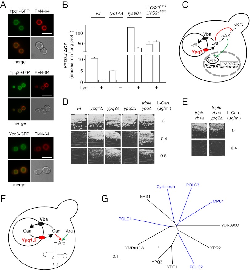
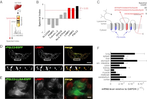
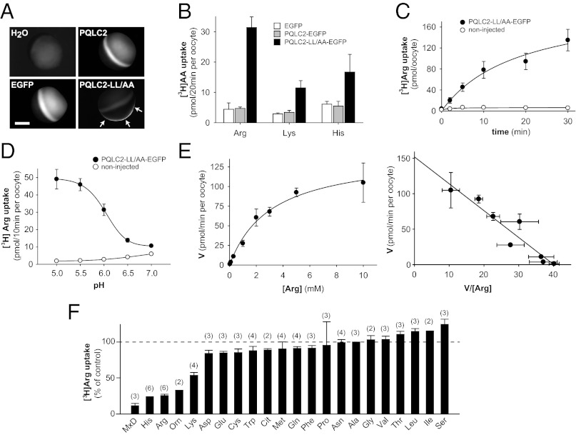
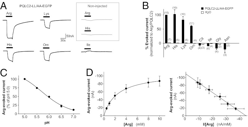
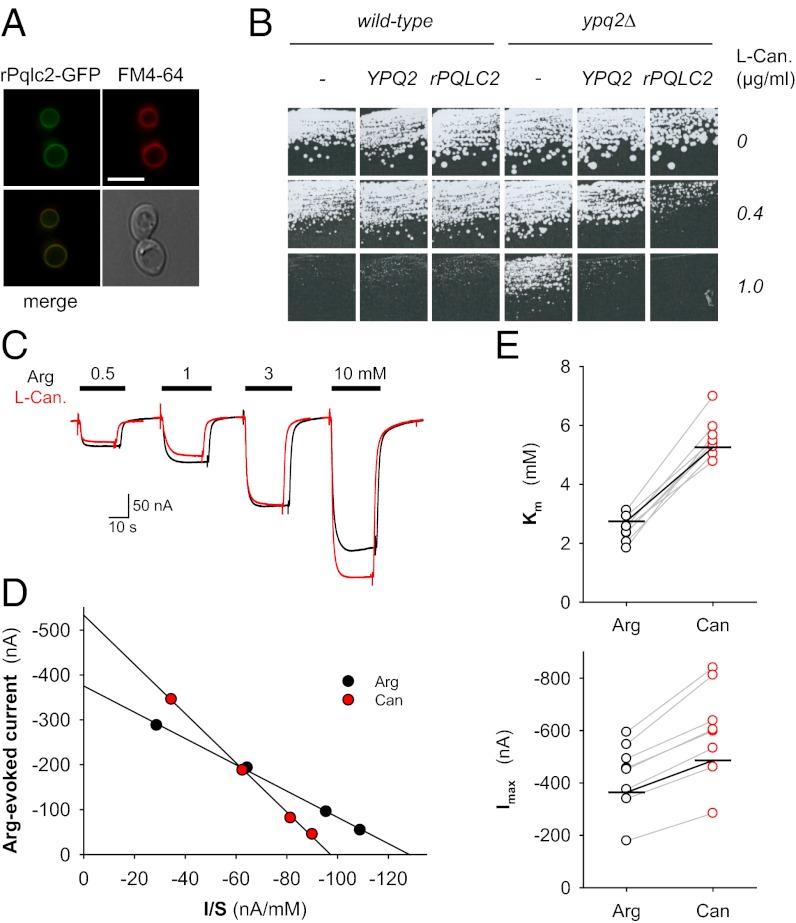
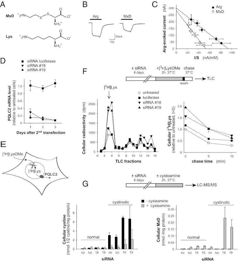
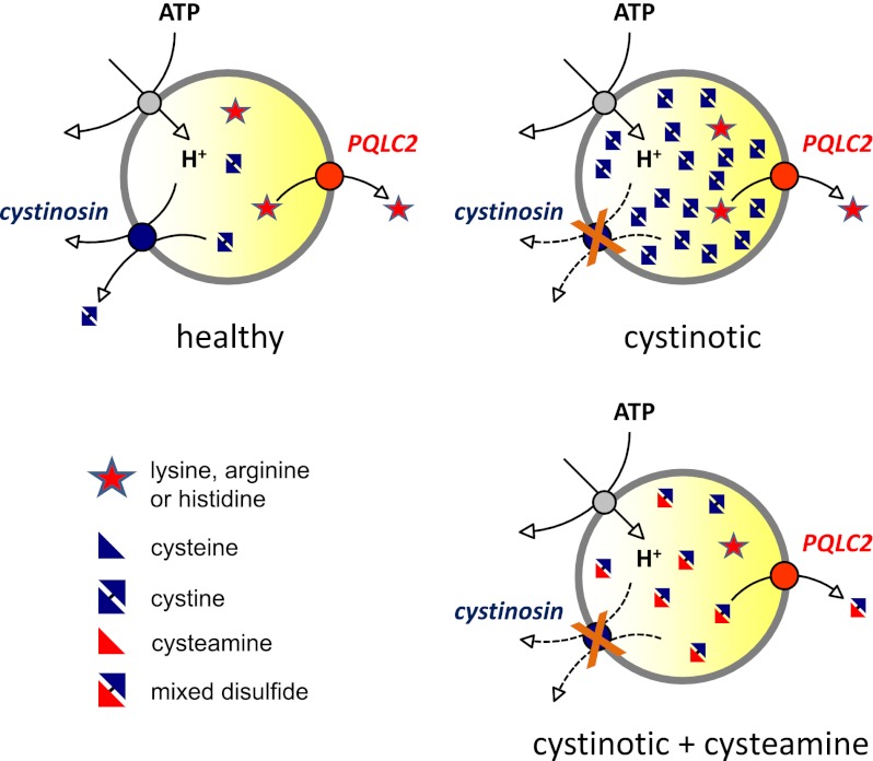
Cystinosin, the lysosomal cystine exporter defective in cystinosis, is the founding member of a family of heptahelical membrane proteins related to bacteriorhodopsin and characterized by a duplicated motif termed the PQ loop. PQ-loop proteins are more frequent in eukaryotes than in prokaryotes; except for cystinosin, their molecular function remains elusive. In this study, we report that three yeast PQ-loop proteins of unknown function, Ypq1, Ypq2, and Ypq3, localize to the vacuolar membrane and are involved in homeostasis of cationic amino acids (CAAs). We also show that PQLC2, a mammalian PQ-loop protein closely related to yeast Ypq proteins, localizes to lysosomes and catalyzes a robust, electrogenic transport that is selective for CAAs and strongly activated at low extracytosolic pH. Heterologous expression of PQLC2 at the yeast vacuole rescues the resistance phenotype of an ypq2 mutant to canavanine, a toxic analog of arginine efficiently transported by PQLC2. Finally, PQLC2 transports a lysine-like mixed disulfide that serves as a chemical intermediate in cysteamine therapy of cystinosis, and PQLC2 gene silencing trapped this intermediate in cystinotic cells. We conclude that PQLC2 and Ypq1-3 proteins are lysosomal/vacuolar exporters of CAAs and suggest that small-molecule transport is a conserved feature of the PQ-loop protein family, in agreement with its distant similarity to SWEET sugar transporters and to the mitochondrial pyruvate carrier. The elucidation of PQLC2 function may help improve cysteamine therapy. It may also clarify the origin of CAA abnormalities in Batten disease.


Conflict of interest statement

The authors declare no conflict of interest.

Figures

Fig. 1.
Fig. 1.
Yeast Ypq1–3 proteins are vacuolar membrane proteins associated with homeostasis of CAAs. (A) Ypq1–3 proteins localize to the vacuolar membrane. Yeast cells of strain 23344c (ura3) were transformed with URA3 plasmids expressing the YPQ1-GFP or YPQ3-GFP fusion gene under its natural promoter or the YPQ3-GFP gene under a galactose-inducible promoter. After growth on minimal medium with proline as a nitrogen source and glucose (Ypq1, Ypq2) or galactose (Ypq3) as a carbon source, cells were analyzed by fluorescence microscopy. The vacuolar membrane was labeled with the lipophilic dye FM4-64. (Scale bars: 5 μm.) (B) YPQ3 gene belongs to the lysine-repressible LYS regulon. Strains of the indicated genotypes were transformed with a centromere-based plasmid expressing the lacZ reporter gene under the control of the YPQ3 gene promoter. Cells were grown on a minimal glucose/ammonium medium with (+) or without (−) lysine (Lys.; 1 mM). β-Galactosidase activities are means of at least two independent experiments. (C) Model of Ypq3 function. Ypq3 may export lysine stored in the vacuole via the Vba1–3 transporters. When present in excess in the cytosol, lysine represses transcription of the YPQ3 gene and the LYS genes involved in lysine biogenesis from α-ketoglutarate (αKG). This repression results from an allosteric inhibition of the Lys20 and Lys21 enzymes by lysine, leading to a decrease in α-aminoadipate semialdehyde (αAS), a pathway intermediate acting as a coinducer of the transcriptional activator Lys14. (D) ypq1Δ and ypq2Δ mutants are resistant to canavanine (Can). Yeast strains of the indicated genotypes were spread on a solid minimal glucose medium with or without canavanine and grown for 3 d. (E) ypq2Δ mutation does not confer resistance to canavanine in a vba1Δ vba2Δ vba3Δ mutant. Conditions were as in D. (F) Model for the role of Ypq1–2 in sensitivity to canavanine, a toxic analog of arginine misincorporated into proteins. Ypq1 and Ypq2 may export canavanine stored in the vacuole via the Vba transporters. (G) Phylogenetic tree of yeast PQ-loop proteins. Selected human PQ-loop proteins are shown in blue for comparison. (Scale bar: 10% sequence divergence.)
Fig. 2.
Fig. 2.
PQLC2 is a ubiquitous lysosomal membrane protein. (A) After purification from rat liver by isopycnic centrifugation on Nycodenz gradients, lysosomes and lysosome-depleted fractions were subjected to hydrophobic protein extraction, SDS/PAGE, and comparative semiquantitative proteomic analysis. (B) Relative protein abundance in the two subcellular fractions was assessed by calculating a lysosome spectral index ranging from −1 (fully excluded) to +1 (fully included), based on normalized spectral counts and the number of positive replicates. The spectral index of PQLC2 is similar to those of lysosomal markers (LAMP1, LAMP2) and well above those of mitochondrial (SLC25A4), peroxisomal (PXMP2), cytoskeleton (tubulin α2), endoplasmic reticulum (SERCA), and plasma membrane (Na,K ATPase) markers. The dotted line represents the 5% significance threshold. (C) Putative membrane topology of PQLC2. PQ-loop motifs are highlighted in blue. The peptides identified by MS are shown in red, along with their spectral counts. (D) WT EGFP-tagged rat PQLC2 (green) was transiently expressed in HeLa cells and compared with LAMP1 immunostaining (red) by deconvolution fluorescence microscopy. EGFP-stained puncta overlap with LAMP1-positive lysosomes and late endosomes in the deconvoluted optical slice. (Lower) Enlarged views of the boxed areas are shown. Arrows indicate colocalization. (Scale bar: 10 μm.) (E) Mutation of a C-terminal dileucine-type sorting motif (underlined in C) prevents PQLC2 delivery to the lysosome. The epifluorescence images highlight the diffuse distribution of the LL290/291AA mutant on the plasma membrane, including microvilli. (Scale bar: 10 μm.) (F) PQLC2 mRNA was quantified in diverse mouse tissues by real-time RT-PCR. Expression levels are compared with the GAPDH transcript using the comparative CT method. Means ± SEMs of six mice are shown. sm., small.
Fig. 3.
Fig. 3.
PQLC2 is a CAA transporter. cRNA-injected Xenopus oocytes were analyzed by epifluorescence microscopy (A) and radiotracer flux measurements (BF). (A) Fluorescence is detected at the plasma membrane for the PQLC2-LL/AA-EGFP construct (arrows), but not for WT PQLC2-EGFP (Upper Right) or free EGFP (Lower Left). The focus was adjusted in the equatorial plane, and images were acquired under identical conditions. (Scale bar: 0.2 mm.) (B and C) Oocytes expressing PQLC2-LL/AA-EGFP, but not WT PQLC2-EGFP or free EGFP, accumulate l-arginine, l-lysine, and l-histidine (0.1 mM) at extracellular pH 5.0. Means ± SEMs from representative pools of five oocytes are shown. (C) Time course of arginine (1 mM) uptake. (D) Arginine (0.1 mM) uptake was measured at distinct pH values. PQLC2-LL/AA is activated in extracellular acidic medium, a condition mimicking the natural environment in the lysosome. (E) Saturation kinetics of l-arginine uptake at pH 5.0. (Right) Graph (Eadie–Hofstee plot) shows that arginine uptake follows Michaelis–Menten kinetics. In this experiment, Km = 3.8 mM and Vmax = 152 pmol/min per oocyte (R2 = 0.901). Means ± SEMs of five to seven oocytes are shown. (F) Substrate selectivity. Inhibitors (10 mM) were added simultaneously to [3H]l-Arg (40 nM) at pH 5.0. Proteinogenic amino acids are indicated by their three-letter code. Cit, citrulline; Orn, l-ornithine. Means ± SEMs of the number of oocytes indicated in parentheses are shown.
Fig. 4.
Fig. 4.
Electrophysiological characterization of PQLC2. PQLC2-LL/AA-EGFP oocytes and water-injected oocytes were recorded under two-electrode voltage clamp at −40 mV and perfused with l-amino acids at pH 5.0, unless otherwise stated. (A) Raw traces from two representative oocytes. In the PQLC2-LL/AA oocyte, CAAs (10 mM), but not isoleucine, evoked an inward current that was absent from the noninjected oocyte. Orn, l-ornithine. (B) Mean steady-state currents ± SEM evoked by various amino acids (10 mM). The number of oocytes analyzed is shown above the bars. Values were normalized for each oocyte to the corresponding l-arginine signal. (C) Extracellular pH dependence of the arginine-evoked current. Means ± SEMs of 9–12 oocytes from three experiments are shown. Where not visible, error bars are smaller than symbols. (D) Saturation kinetics of the arginine response. The steady-state current mediated by PQLC2 follows Michaelis–Menten kinetics. In this experiment, Km = 2.49 mM, Imax = −110 nA, and R2 = 0.994. Means ± SEMs of 7 oocytes from the same batch are shown.
Fig. 5.
Fig. 5.
Defective drug export from the vacuole may account for the yeast canavanine-sensitivity phenotype. (A) PQLC2 localizes to the vacuolar membrane of yeast cells. The 23344c (ura3) strain transformed with a URA3 plasmid expressing the rPQLC2-GFP fusion gene under a galactose-inducible promoter was grown on galactose (3%)/proline (10 mM) medium. Glucose (3%) was added to the cell culture for 2 h before staining with the vacuolar membrane marker FM4-64 and fluorescent microscopy analysis. (Scale bar: 5 μm.) (B) PQLC2 complements the growth phenotype of the ypq2Δ mutant. Strains 23344c (ura3) and EL031 (ypq2Δ ura3) transformed with URA3 plasmids expressing or not expressing the YPQ2-GFP and rPQLC2-GFP fusion genes under a galactose-inducible promoter were spread on a minimal glucose/ammonium medium with or without l-canavanine (Can.) and grown for 6 d at 29 °C. (CE) PQLC2 transports canavanine. Raw current traces evoked by arginine or canavanine and the resulting Eadie–Hofstee plots are shown for a single representative oocyte in C and D, respectively. I/S, current/substrate concentration ratio. (E) Distribution of Km and Imax values determined from paired applications of the two compounds to eight oocytes from two batches is shown. Canavanine shows higher Km (P < 10−6, paired Student t test) and Imax (P < 10−4) values than arginine. Mean values (horizontal marks) are given in the main text.
Fig. 6.
Fig. 6.
PQLC2 exports a key chemical intermediate in cysteamine therapy of cystinosis. (A) Chemical structure of the MxD resembles that of lysine. (B) Current traces evoked by MxD and arginine (10 mM each) on a representative PQLC2-LL/AA-EGFP oocyte at −40 mV and pH 5.0. (C) Saturation kinetics of paired MxD and arginine responses (means ± SEMs of five oocytes from two batches). Km and Imax values are reported in the main text. I/S, current/substrate concentration ratio. (D) Kinetics of PQLC2 mRNA knockdown in human cystinotic fibroblasts after two rounds of siRNA transfection. Two PQLC2-targeted siRNAs are compared with a luciferase-targeted negative control. Means ± SEMs of four measurements are shown. (E and F) PQLC2 gene silencing decreases the clearance of lysine from an intracellular compartment. (E) Scheme depicts how lysosomes are preferentially loaded with amino acids in whole cells using a methyl ester precursor. After loading human fibroblasts with [3H]LysOMe, the fate of the resulting intracellular [3H]Lys pool was monitored by TLC. (F) Plots show representative chromatograms (Left) and representative [3H]Lys clearance kinetics (Right), respectively. PQLC2 gene silencing increases the intracellular [3H]Lys pool. (G) Effect of PQLC2 gene silencing on intracellular cystine and MxD levels. PQLC2 knockdown exacerbates cystine storage (Left) and dramatically increases the level of MxD induced by cysteamine (Right) in human cystinotic fibroblasts, as illustrated in this representative experiment (means ± SEMs of three measurements). luc, luciferase; no, untreated.
Fig. P1.
Fig. P1.
PQLC2 is a lysosomal cationic amino acid exporter that plays a key role in cysteamine therapy of cystinosin. (Upper Left) In healthy subjects, cystinosin and PQLC2 export cystine (the oxidized form of cysteine) and cationic amino acids, respectively, from the lysosomal lumen. Their activity is stimulated by the acidity of the lumen. (Upper Right) In patients with cystinosis, lysosomes accumulate cystine because cystinosin is impaired. (Lower Right) The drug cysteamine can reverse this accumulation by entering lysosomes and reacting with cystine to form a cysteamine-cysteine mixed disulfide, which resembles lysine. The mixed disulfide is then exported by PQLC2, thus depleting cystine from lysosomes and alleviating symptoms.

References

    1. Palmieri F. Diseases caused by defects of mitochondrial carriers: A review. Biochim Biophys Acta. 2008;1777(7-8):564–578. - PubMed
    1. Bröer S, Palacín M. The role of amino acid transporters in inherited and acquired diseases. Biochem J. 2011;436(2):193–211. - PubMed
    1. Sagné C, Gasnier B. Molecular physiology and pathophysiology of lysosomal membrane transporters. J Inherit Metab Dis. 2008;31(2):258–266. - PubMed
    1. Fredriksson R, Nordström KJ, Stephansson O, Hägglund MG, Schiöth HB. The solute carrier (SLC) complement of the human genome: Phylogenetic classification reveals four major families. FEBS Lett. 2008;582(27):3811–3816. - PubMed
    1. Schlessinger A, et al. Comparison of human solute carriers. Protein Sci. 2010;19(3):412–428. - PMC - PubMed

Publication types

MeSH terms