

Low-affinity B cells transport viral particles from the lung to the spleen to initiate antibody responses
- PMID: 23169669
- PMCID: PMC3528531
- DOI: 10.1073/pnas.1206970109
Low-affinity B cells transport viral particles from the lung to the spleen to initiate antibody responses
Abstract
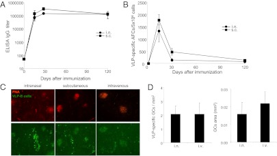
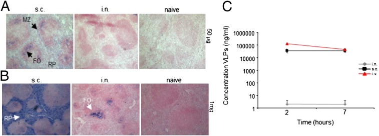
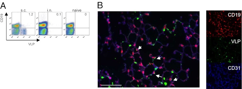
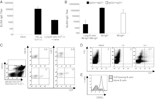
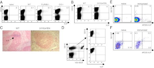
The lung is an important entry site for pathogens; its exposure to antigens results in systemic as well as local IgA and IgG antibodies. Here we show that intranasal administration of virus-like particles (VLPs) results in splenic B-cell responses with strong local germinal-center formation. Surprisingly, VLPs were not transported from the lung to the spleen in a free form but by B cells. The interaction between VLPs and B cells was initiated in the lung and occurred independently of complement receptor 2 and Fcγ receptors, but was dependent upon B-cell receptors. Thus, B cells passing through the lungs bind VLPs via their B-cell receptors and deliver them to local B cells within the splenic B-cell follicle. This process is fundamentally different from delivery of blood or lymph borne particulate antigens, which are transported into B cell follicles by binding to complement receptors on B cells.
Conflict of interest statement
The authors declare no conflict of interest.
Figures
Similar articles
-
Co-delivery of GPI-anchored CCL28 and influenza HA in chimeric virus-like particles induces cross-protective immunity against H3N2 viruses.J Control Release. 2016 Jul 10;233:208-19. doi: 10.1016/j.jconrel.2016.05.021. Epub 2016 May 10. J Control Release. 2016. PMID: 27178810 Free PMC article.
-
Migration of antigen-presenting B cells from peripheral to mucosal lymphoid tissues may induce intestinal antigen-specific IgA following parenteral immunization.J Immunol. 1999 Sep 15;163(6):3064-70. J Immunol. 1999. PMID: 10477570
-
Immune Correlates of Protection Induced by Virus-Like Particles Containing 2009 H1N1 Pandemic Influenza HA, NA or M1 Proteins.Immunol Invest. 2019 May;48(4):355-366. doi: 10.1080/08820139.2018.1544640. Epub 2018 Nov 15. Immunol Invest. 2019. PMID: 30430891
-
Efficient induction of mucosal and systemic immune responses by virus-like particles administered intranasally: implications for vaccine design.Eur J Immunol. 2008 Jan;38(1):114-26. doi: 10.1002/eji.200636959. Eur J Immunol. 2008. PMID: 18081037
-
The dual function of the splenic marginal zone: essential for initiation of anti-TI-2 responses but also vital in the general first-line defense against blood-borne antigens.Clin Exp Immunol. 2002 Oct;130(1):4-11. doi: 10.1046/j.1365-2249.2002.01953.x. Clin Exp Immunol. 2002. PMID: 12296846 Free PMC article. Review.
Cited by
-
Activated Peyer's patch B cells sample antigen directly from M cells in the subepithelial dome.Nat Commun. 2019 Jun 3;10(1):2423. doi: 10.1038/s41467-019-10144-w. Nat Commun. 2019. PMID: 31160559 Free PMC article.
-
Meeting report VLPNPV: Sessions 1 and 2: Plenary.Hum Vaccin Immunother. 2014;10(10):3060-3. doi: 10.4161/21645515.2014.988552. Hum Vaccin Immunother. 2014. PMID: 25485812 Free PMC article.
-
Interaction Between Virus-Like Particles (VLPs) and Pattern Recognition Receptors (PRRs) From Dendritic Cells (DCs): Toward Better Engineering of VLPs.Front Immunol. 2020 Jun 9;11:1100. doi: 10.3389/fimmu.2020.01100. eCollection 2020. Front Immunol. 2020. PMID: 32582186 Free PMC article. Review.
-
The Multifaceted B Cell Response to Influenza Virus.J Immunol. 2019 Jan 15;202(2):351-359. doi: 10.4049/jimmunol.1801208. J Immunol. 2019. PMID: 30617116 Free PMC article. Review.
-
Virus-like particle: a nano-platform that delivers cancer antigens to elicit an anti-tumor immune response.Front Immunol. 2025 Jan 7;15:1504124. doi: 10.3389/fimmu.2024.1504124. eCollection 2024. Front Immunol. 2025. PMID: 39840069 Free PMC article. Review.
References
-
- Iwasaki A. Mucosal dendritic cells. Annu Rev Immunol. 2007;25:381–418. - PubMed
-
- Masten BJ, Lipscomb MF. Comparison of lung dendritic cells and B cells in stimulating naive antigen-specific T cells. J Immunol. 1999;162(3):1310–1317. - PubMed
-
- von Garnier C, et al. Anatomical location determines the distribution and function of dendritic cells and other APCs in the respiratory tract. J Immunol. 2005;175(3):1609–1618. - PubMed
-
- Kozlowski PA, et al. Differential induction of mucosal and systemic antibody responses in women after nasal, rectal, or vaginal immunization: Influence of the menstrual cycle. J Immunol. 2002;169(1):566–574. - PubMed
-
- Staats HF, Montgomery SP, Palker TJ. Intranasal immunization is superior to vaginal, gastric, or rectal immunization for the induction of systemic and mucosal anti-HIV antibody responses. AIDS Res Hum Retroviruses. 1997;13(11):945–952. - PubMed
MeSH terms
Substances
LinkOut - more resources
Full Text Sources
Molecular Biology Databases
Miscellaneous
