. 2012 Nov;135(Pt 11):3298-310.
doi: 10.1093/brain/aws259.

Interleukin 6-preconditioned neural stem cells reduce ischaemic injury in stroke mice

Affiliations

Interleukin 6-preconditioned neural stem cells reduce ischaemic injury in stroke mice

Hiroyuki Sakata et al. Brain. 2012 Nov.

Abstract

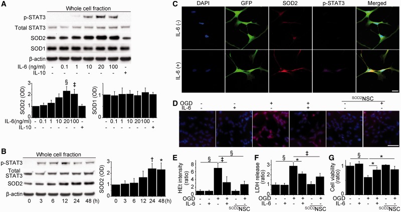
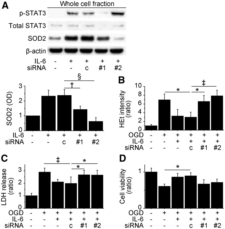
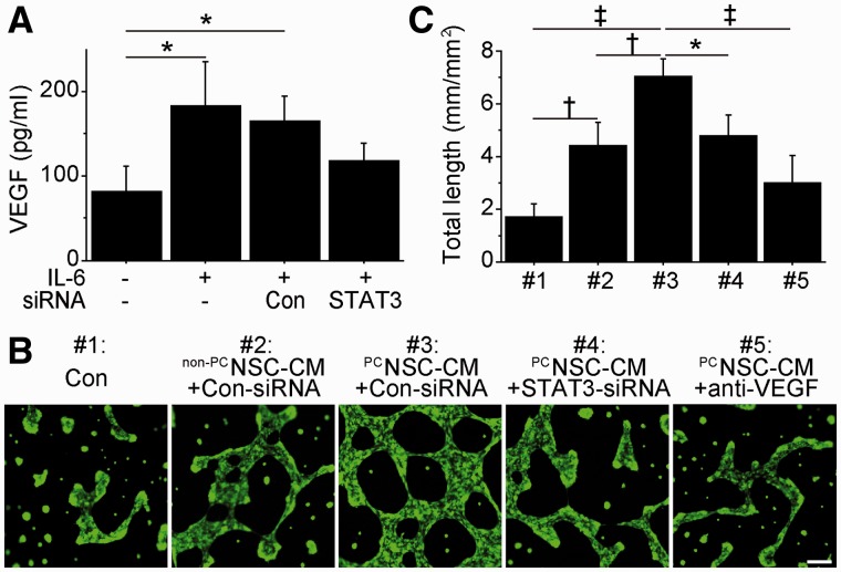
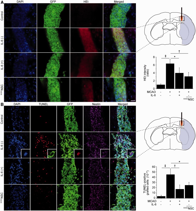
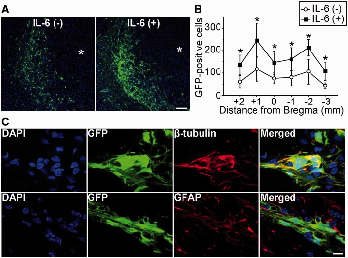
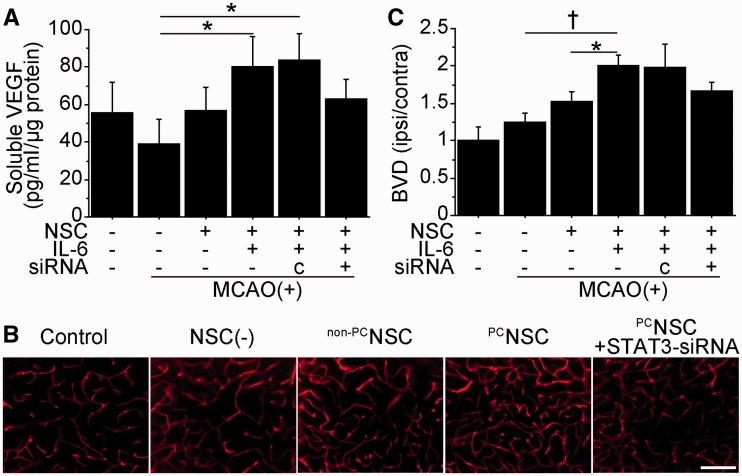
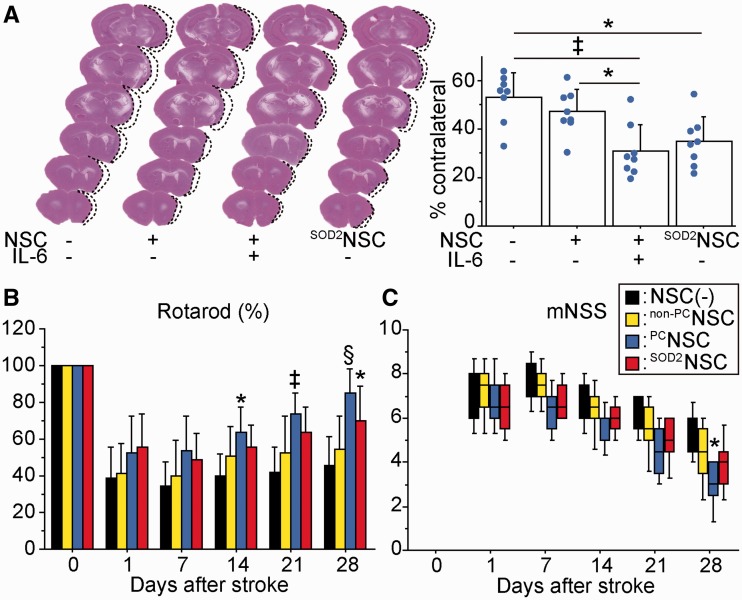
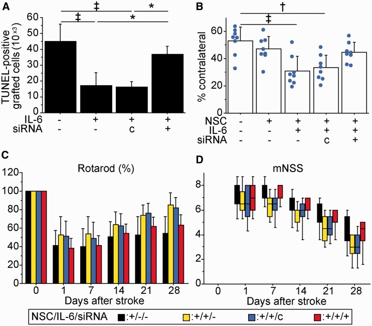
Transplantation of neural stem cells provides a promising therapy for stroke. Its efficacy, however, might be limited because of massive grafted-cell death after transplantation, and its insufficient capability for tissue repair. Interleukin 6 is a pro-inflammatory cytokine involved in the pathogenesis of various neurological disorders. Paradoxically, interleukin 6 promotes a pro-survival signalling pathway through activation of signal transducer and activator of transcription 3. In this study, we investigated whether cellular reprogramming of neural stem cells with interleukin 6 facilitates the effectiveness of cell transplantation therapy in ischaemic stroke. Neural stem cells harvested from the subventricular zone of foetal mice were preconditioned with interleukin 6 in vitro and transplanted into mouse brains 6 h or 7 days after transient middle cerebral artery occlusion. Interleukin 6 preconditioning protected the grafted neural stem cells from ischaemic reperfusion injury through signal transducer and activator of transcription 3-mediated upregulation of manganese superoxide dismutase, a primary mitochondrial antioxidant enzyme. In addition, interleukin 6 preconditioning induced secretion of vascular endothelial growth factor from the neural stem cells through activation of signal transducer and activator of transcription 3, resulting in promotion of angiogenesis in the ischaemic brain. Furthermore, transplantation of interleukin 6-preconditioned neural stem cells significantly attenuated infarct size and improved neurological performance compared with non-preconditioned neural stem cells. This interleukin 6-induced amelioration of ischaemic insults was abolished by transfecting the neural stem cells with signal transducer and activator of transcription 3 small interfering RNA before transplantation. These results indicate that preconditioning with interleukin 6, which reprograms neural stem cells to tolerate oxidative stress after ischaemic reperfusion injury and to induce angiogenesis through activation of signal transducer and activator of transcription 3, is a simple and beneficial approach for enhancing the effectiveness of cell transplantation therapy in ischaemic stroke.


Figures

Figure 1
Figure 1
Upregulation of SOD2 and reduced NSC death with IL-6 preconditioning in vitro. (A) Western blot analysis of NSCs pretreated with various concentrations of IL-6 or IL-10 (10 ng/ml) for 24 h. IL-6 preconditioning (20 and 100 ng/ml) significantly upregulated SOD2 expression in association with STAT3 activation. No change was observed in copper/zinc-superoxide dismutase (SOD1) expression. β-Actin was used as an internal control (n = 4). OD = optical density. (B) Western blot analysis of NSCs treated with IL-6 (20 ng/ml) and harvested at the indicated time points. IL-6 immediately phosphorylated STAT3 and induced SOD2 at later time points. β-Actin was used as an internal control (n = 4). (C) Fluorescent staining with DAPI (blue), GFP (green), SOD2 (red) and phosphorylated STAT3 (p-STAT3) (magenta) in NSCs with or without IL-6 preconditioning (20 ng/ml) for 24 h revealed the distribution of phosphorylated STAT3, predominantly in the nucleus, and increased SOD2 signals in mitochondria in preconditioned NSCs. Scale bar = 20 μm. (D and E) Fluorescent staining of NSCs with hydroethidine (red) and DAPI (blue). The increase in hydroethidine (HEt) signals after 8 h of oxygen–glucose deprivation (OGD), and 15 min of reoxygenation was reduced in preconditioned NSCs and SOD2-NSCs (n = 4). Scale bar = 50 μm. Lactate dehydrogenase (LDH); (F) and water soluble tetrazolium salts 1 (G) assays showed a significant reduction in NSC death and increased cell viability in preconditioned NSCs and SOD2-NSCs after 8 h of oxygen–glucose deprivation and 24 h of reoxygenation (n = 4). *P < 0.05, †P < 0.01, ‡P < 0.005, §P < 0.001.
Figure 2
Figure 2
Inhibition of STAT3 and SOD2 abolished IL-6-induced cytoprotection in vitro. Non-preconditioned NSCs and preconditioned NSCs were pretreated with control small interfering RNA (c), STAT3 small interfering RNA (#1) or SOD2 small interfering RNA (#2) before the experiments. (A) Western blot analysis demonstrated that SOD2 expression in the preconditioned NSCs was downregulated by STAT3 or SOD2 small interfering RNA transfection (n = 4). β-Actin was used as an internal control. (B) IL-6-induced reduction in hydroethidine (HEt) signals after 8 h of oxygen-glucose deprivation (OGD) and 15 min of reoxygenation was reversed by STAT3 or SOD2 small interfering RNA (n = 4). Death and viability of the NSCs analysed by lactate dehydrogenase (C) and water soluble tetrazolium salts 1 (D) assays after 8 h of OGD and 24 h of reoxygenation. STAT3 and SOD2 small interfering RNA abolished IL-6-induced cytoprotection (n = 4). *P < 0.05, †P < 0.01, ‡P < 0.005, §P < 0.001. LDH = lactate dehydrogenase.
Figure 3
Figure 3
IL-6-induced VEGF enhanced capillary-like tube formation in vitro. (A) In vitro ELISA of the conditioned medium from NSCs. IL-6 preconditioning increased VEGF levels, which were suppressed by STAT3-small interfering RNA transfection (n = 4). Con = control. (B and C) Capillary-like tube formation assay performed on mouse cerebral endothelial cells cultured with regular NSC culture medium (#1), conditioned medium (PC) from non-preconditioned (non-PC) NSCs transfected with control-small interfering RNA (#2), conditioned medium from preconditioned NSCs transfected with control-small interfering RNA (#3), conditioned medium from preconditioned NSCs transfected with STAT3-small interfering RNA (#4) or conditioned medium from preconditioned NSCs with a goat anti-mouse neutralizing antibody to VEGF (anti-VEGF) (#5). Preconditioned NSCs-conditioned medium with control small interfering RNA transfection resulted in the greatest increase in total tube length. This was abolished by STAT3 small interfering RNA transfection or by addition of an anti-VEGF neutralizing antibody (n = 4). Scale bar = 100 μm. *P < 0.05, †P < 0.01, ‡P < 0.001.
Figure 4
Figure 4
Reduced grafted-cell death with IL-6 preconditioning in vivo. NSCs were transplanted into the brain 6 h after stroke. (A) Fluorescent staining with DAPI (blue), GFP (green) and hydroethidine (HEt, red) in brain sections 1 h after transplantation. Hydroethidine signals increased in the non-preconditioned NSCs under ischaemic reperfusion injury, but this signal increase was reduced in the preconditioned NSCs and SOD2-NSCs (n = 4). MCAO = middle cerebral artery occlusion. (B) Fluorescent staining with DAPI (blue), TUNEL (red), GFP (green) and nestin (magenta) 2 days after transplantation. IL-6 preconditioning and SOD2 overexpression significantly reduced the number of TUNEL-positive grafted cells in the ischaemic brain. The insets represent high magnification images showing the co-localization of TUNEL with a nestin- and GFP-positive grafted cell (n = 4). Scale bars = 20 μm. *P < 0.05, †P < 0.005, ‡P < 0.001.
Figure 5
Figure 5
Increased survival of grafted cells with IL-6 preconditioning in vivo. NSCs were transplanted into the brain 6 h after stroke. (A) Fluorescent staining with GFP (green) and DAPI (blue) revealed migration of the grafted cells towards the lesion border zone 28 days after stroke and transplantation. Asterisk indicates ischaemic lesion. Scale bar = 100 μm. (B) Quantification of the number of surviving grafted cells 28 days after stroke and transplantation. Specimens were picked up every 1 mm, and the number of GFP-positive cells was counted. IL-6 preconditioning significantly increased survival of the grafted cells (n = 4). (C) Fluorescent staining with GFP (green) and β-tubulin or glial fibrillary acidic protein (GFAP) (red) revealed that the grafted NSCs differentiated into neurons (β-tubulin+) and astrocytes (GFAP+) 28 days after stroke and transplantation. Nuclei were counterstained with DAPI. Scale bar = 10 μm. *P < 0.05.
Figure 6
Figure 6
Transplantation of preconditioned NSCs promoted angiogenesis in vivo. NSCs were transplanted into the brain 6 h after stroke. (A) In vivo ELISA of the cortex revealed a significant elevation of VEGF in the preconditioned NSC group 2 days after transplantation. Transfection with STAT3 small interfering RNA abolished this increase (n = 4). c = control small interfering RNA. Representative images of lectin-perfused vessels (B) and quantification of blood vessel density (BVD) (C) in the peri-infarct cortex 14 days after transplantation. Blood vessel density was significantly increased in the preconditioned NSC group, which was suppressed by STAT3 small interfering RNA transfection (n = 4). Scale bar = 100 μm. *P < 0.05, †P < 0.001.
Figure 7
Figure 7
Effects of preconditioned NSCs on infarct size and behavioural performance. NSCs were transplanted into the brain 6 h after stroke. (A) Measurement of the infarct size by haematoxylin and eosin staining 28 days after stroke and transplantation. Cortical infarct size was significantly attenuated in the preconditioned NSC and SOD2-NSC groups compared with the non-transplanted control group (n = 4). Indices of behavioural performance using the rotarod test (B) and modified neurologic severity scores (mNSS) (C). Transplantation of preconditioned NSCs resulted in the greatest functional recovery 28 days after stroke and transplantation (n = 8). Black bars denote non-transplanted control group; yellow bars denote non-preconditioned NSC group; blue bars denote preconditioned NSC group; red bars denote SOD2-NSC group. The labels show P-value compared with the non-transplanted control group. *P < 0.05, ‡P < 0.005; §P < 0.001.
Figure 8
Figure 8
Inhibition of STAT3 abolished IL-6-induced amelioration of ischaemic stroke. Preconditioned NSCs or non-preconditioned NSCs, transfected with control or STAT3 small interfering RNA, were transplanted 6 h after stroke. (A) Stereological counting of TUNEL-positive grafted cells 2 days after stroke and transplantation. STAT3 small interfering RNA suppressed IL-6-induced cytoprotection (n = 4). (B) Cortical infarct size evaluated by haematoxylin and eosin staining 28 days after stroke and transplantation. The preconditioned NSCs, which also underwent STAT3 small interfering RNA pretreatment, had no attenuation of lesion size (n = 4). Behavioural performance analysed by the rotarod test (C) and mNSS (D). STAT3-small interfering RNA diminished the behavioural improvement observed in the preconditioned NSC group (n = 8). Black bars denote non-preconditioned NSC group; yellow bars denote preconditioned NSC group; blue bars denote preconditioned NSC group with control small interfering RNA transfection; red bars denote preconditioned NSC group with STAT3 small interfering RNA transfection. *P < 0.05, †P < 0.01, ‡P < 0.005.

Similar articles

Cited by

References

    1. Andres RH, Horie N, Slikker W, Keren-Gill H, Zhan K, Sun G, et al. Human neural stem cells enhance structural plasticity and axonal transport in the ischaemic brain. Brain. 2011;134:1777–89. - PMC - PubMed
    1. Bacigaluppi M, Pluchino S, Jametti LP, Kilic E, Kilic Ü, Salani G, et al. Delayed post-ischaemic neuroprotection following systemic neural stem cell transplantation involves multiple mechanisms. Brain. 2009;132:2239–51. - PubMed
    1. Banerjee S, Williamson D, Habib N, Gordon M, Chataway J. Human stem cell therapy in ischaemic stroke: a review. Age Ageing. 2011;40:7–13. - PubMed
    1. Beamer NB, Coull BM, Clark WM, Hazel JS, Silberger JR. Interleukin-6 and interleukin-1 receptor antagonist in acute stroke. Ann Neurol. 1995;37:800–4. - PubMed
    1. Bliss TM, Andres RH, Steinberg GK. Optimizing the success of cell transplantation therapy for stroke. Neurobiol Dis. 2010;37:275–83. - PMC - PubMed

Publication types

MeSH terms