. 2012 Nov 20;3(6):e00224-11.
doi: 10.1128/mBio.00224-11.

Interaction of lipocalin 2, transferrin, and siderophores determines the replicative niche of Klebsiella pneumoniae during pneumonia

Affiliations

Interaction of lipocalin 2, transferrin, and siderophores determines the replicative niche of Klebsiella pneumoniae during pneumonia

Michael A Bachman et al. mBio. .

Abstract

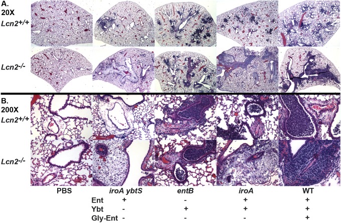
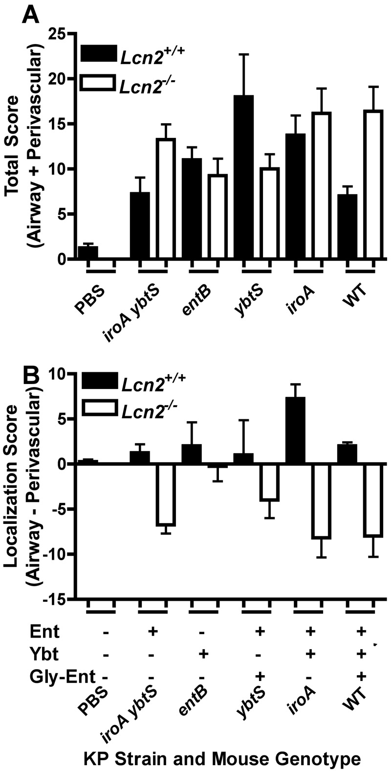
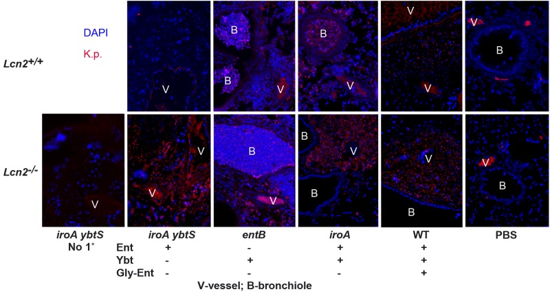
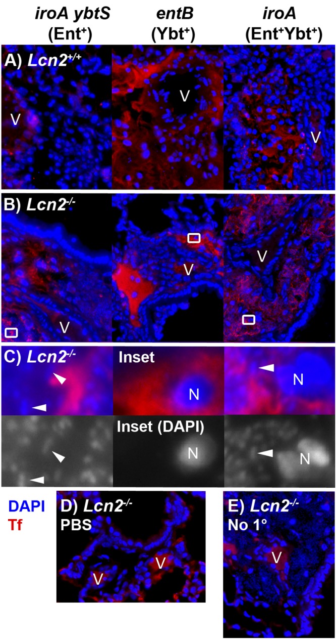
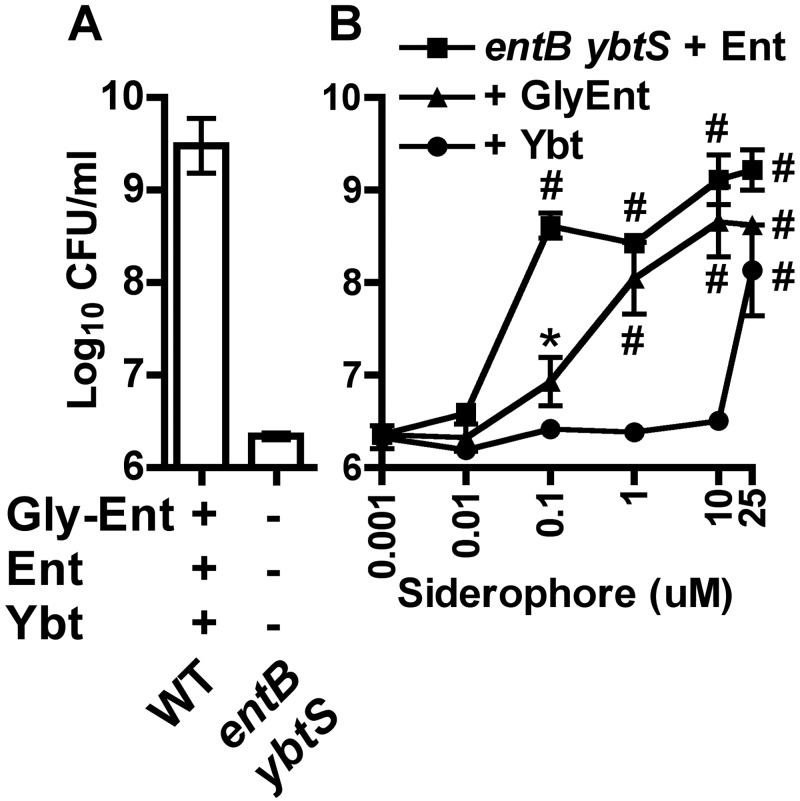
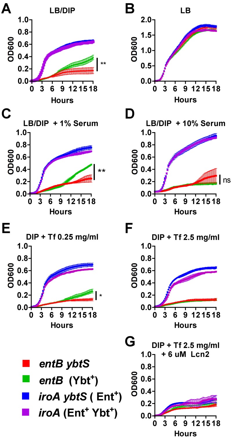
Pathogenic bacteria require iron for replication within their host. Klebsiella pneumoniae and other Gram-negative pathogens produce the prototypical siderophore enterobactin (Ent) to scavenge iron in vivo. In response, mucosal surfaces secrete lipocalin 2 (Lcn2), an innate immune protein that binds Ent to disrupt bacterial iron acquisition and promote acute inflammation during colonization. A subset of K. pneumoniae isolates attempt to evade Lcn2 by producing glycosylated Ent (Gly-Ent, salmochelin) or the alternative siderophore yersiniabactin (Ybt). However, these siderophores are not functionally equivalent and differ in their abilities to promote growth in the upper respiratory tract, lungs, and serum. To understand how Lcn2 exploits functional differences between siderophores, isogenic mutants of an Ent(+) Gly-Ent(+) Ybt(+) K. pneumoniae strain were inoculated into Lcn2(+/+) and Lcn2(-/-) mice, and the pattern of pneumonia was examined. Lcn2 effectively protected against the iroA ybtS mutant (Ent(+) Gly-Ent(-) Ybt(-)). Lcn2(+/+) mice had small foci of pneumonia, whereas Lcn2(-/-) mice had many bacteria in the perivascular space. The entB mutant (Ent(-) Ybt(+) Gly-Ent(-)) caused moderate bronchopneumonia but did not invade the transferrin-containing perivascular space. Accordingly, transferrin blocked Ybt-dependent growth in vitro. The wild type and the iroA mutant, which both produce Ent and Ybt, had a mixed phenotype, causing a moderate bronchopneumonia in Lcn2(+/+) mice and perivascular overgrowth in Lcn2(-/-) mice. Together, these data indicate that Lcn2, in combination with transferrin, confines K. pneumoniae to the airways and prevents invasion into tissue containing the pulmonary vasculature.

Importance: Gram-negative bacteria are a common cause of severe hospital-acquired infections. To cause disease, they must obtain iron and secrete the small molecule enterobactin to do so. Animal models of pneumonia using Klebsiella pneumoniae indicate that enterobactin promotes severe disease. Accordingly, the host defense protein lipocalin 2 exploits this common target by binding enterobactin and disrupting its function. However, pathogenic bacteria often make additional siderophores that lipocalin 2 cannot bind, such as yersiniabactin, which could make this host defense ineffective. This work compares the pattern and severity of pneumonia caused by K. pneumoniae based on which siderophores it produces. The results indicate that enterobactin promotes growth around blood vessels that are rich in the iron-binding protein transferrin, but yersiniabactin does not. Together, transferrin and lipocalin 2 protect this space against all types of K. pneumoniae tested. Therefore, the ability to acquire iron determines where bacteria can grow in the lung.


Figures

FIG 1
FIG 1
Lcn2-siderophore interactions determine the pattern of pneumonia caused by K. pneumoniae. Hematoxylin-and-eosin-stained formalin-fixed paraffin-embedded histological sections of lungs from Lcn2+/+ and Lcn2/ mice 3 days after retropharyngeal inoculation of 1 × 10⁴ CFU/ml of wild-type (WT) or mutant K. pneumoniae producing the siderophores indicated (+) are shown at magnification 20× (A) or 200× (B).
FIG 2
FIG 2
Severity and localization of lung pathology depends on the interaction of Lcn2 with enterobactin- and yersiniabactin-producing K. pneumoniae. Histological sections of lungs from Lcn2+/+ and Lcn2−/− mice 3 days after retropharyngeal inoculation of 1 × 10⁴ CFU/ml of K. pneumoniae were scored by a pathologist for severity and extent of involvement of the airways and perivascular spaces; n ≥ 4 mice per group. The mean total (A) or localization (B) score is shown for each mouse genotype infected with wild-type (WT) or mutant K. pneumoniae producing the siderophores indicated (+). A positive localization score indicates airway predominance; a negative score indicates perivascular predominance.
FIG 3
FIG 3
In the absence of Lcn2, enterobactin-producing K. pneumoniae infects the perivascular space, whereas a yersiniabactin-dependent mutant cannot. Images show immunofluorescence staining of formalin-fixed paraffin-embedded histological sections of lungs from Lcn2+/+ and Lcn2−/− mice 3 days after retropharyngeal inoculation of 1 × 10⁴ CFU/ml of WT or mutant K. pneumoniae encoding the siderophores indicated (+), using rabbit anti-Klebsiella serum and Cy3-conjugated goat anti-rabbit secondary antibody (red) and the nucleic acid stain DAPI (blue), except for “No 1°,” in which anti-Klebsiella serum was omitted. Vessels (V) and bronchioles (B) are shown; magnification, ×200.
FIG 4
FIG 4
The perivascular spaces contain serum exudate as indicated by transferrin. Merged immunofluorescence images of formalin-fixed paraffin-embedded histological sections of lungs from Lcn2+/+ (A) or Lcn2−/− (B) mice 3 days after retropharyngeal inoculation of 1 × 10⁴ CFU/ml of K. pneumoniae siderophore mutants, stained using antitransferrin rabbit polyclonal IgG (Tf) (red) and the nucleic acid stain DAPI (blue), are shown. Magnified merged and DAPI-only images (C) of insets from Lcn2−/− mice in panel B demonstrate nuclei (N) and collections of bacteria (representative bacilli are indicated by arrowheads) in the perivascular space. Merged images of Lcn2−/− mice mock infected with PBS (D) or infected with iroA mutant K. pneumoniae but lacking antitransferrin antibody (E) are shown as controls. Magnification, ×400, except panel C (×4,000); V, vessels.
FIG 5
FIG 5
Ybt is less efficient than Ent and Gly-Ent at iron acquisition in serum. Bacterial densities after overnight growth in RPMI supplemented with 10% human serum of the wild type and entB ybtS mutant (A) or the entB ybtS mutant plus purified enterobactin (squares), glycosylated enterobactin (triangles), or yersiniabactin (circles) at the concentrations indicated (B) are shown. Means ± SEM from at least two independent experiments are shown. *, P < 0.05; #, P < 0.01, as measured by one-way analysis of variance (ANOVA) with Tukey’s posttest compared to results for the entB ybtS mutant without added siderophore.
FIG 6
FIG 6
Serum or the serum component transferrin inhibits Ybt-dependent growth. K. pneumoniae growth was measured by optical density at 600 nm (OD600) over 18 h in LB broth iron depleted with addition of 350 µM 2,2′-dipyridyl (DIP) (A), LB alone (B), LB-DIP with 1% (C) or 10% (D) pooled heat-inactivated human serum, LB-DIP with 0.25 (E) or 2.5 mg/ml (F) purified human transferrin, or 2.5 mg/ml transferrin plus 6 µM recombinant human Lcn2 (G) during incubation at 37°C with intermittent agitation. Means ± SEM are shown for three replicates and are representative of two independent experiments. Final OD600s between strains were compared using one-way ANOVA with Tukey’s multiple-comparison test (*, P < 0.05; **, P < 0.01; ns, not significant).

Similar articles

Cited by

References

    1. Earhart CF. 1996. Uptake and metabolism of iron and molybdenum. In Neidhart F, E. coli and salmonella. ASM Press, Washington, DC
    1. Nairz M, Schroll A, Sonnweber T, Weiss G. 2010. The struggle for iron—a metal at the host-pathogen interface. Cell. Microbiol. 12:1691–1702 - PubMed
    1. Skaar EP. 2010. The battle for iron between bacterial pathogens and their vertebrate hosts. PLoS Pathog. 6:e1000949 http://dx.doi.org/doi:10.1371/journal.ppat.1000949 - PMC - PubMed
    1. Lin H, Fischbach MA, Liu DR, Walsh CT. 2005. In vitro characterization of salmochelin and enterobactin trilactone hydrolases IroD, IroE, and Fes. J. Am. Chem. Soc. 127:11075–11084 - PMC - PubMed
    1. Hantke K, Nicholson G, Rabsch W, Winkelmann G. 2003. Salmochelins, siderophores of Salmonella enterica and uropathogenic Escherichia coli strains, are recognized by the outer membrane receptor IroN. Proc. Natl. Acad. Sci. U. S. A. 100:3677–3682 - PMC - PubMed

Publication types

MeSH terms