. 2012 Oct 1;1(7):1017-1026.
doi: 10.4161/onci.21098.

Primary sterile necrotic cells fail to cross-prime CD8(+) T cells

Affiliations

Primary sterile necrotic cells fail to cross-prime CD8(+) T cells

Jaba Gamrekelashvili et al. Oncoimmunology. .

Abstract

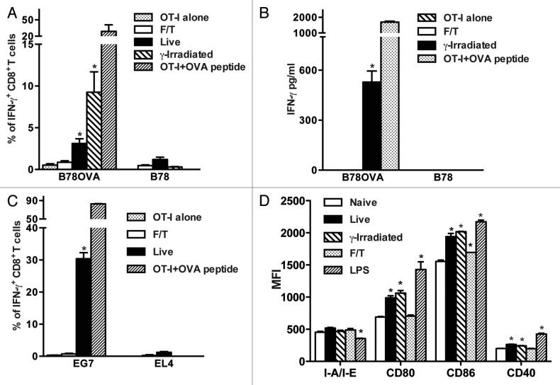
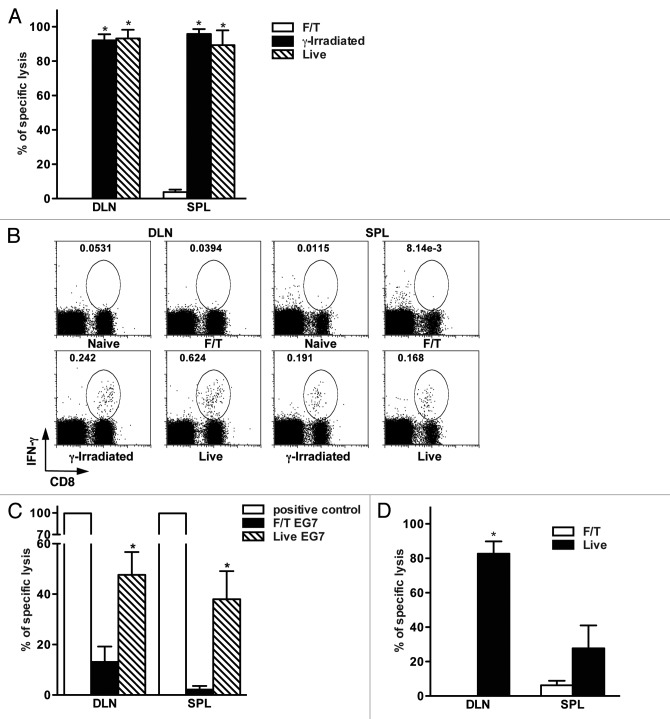
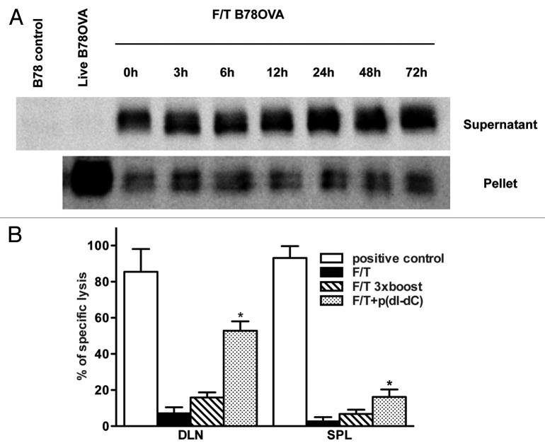
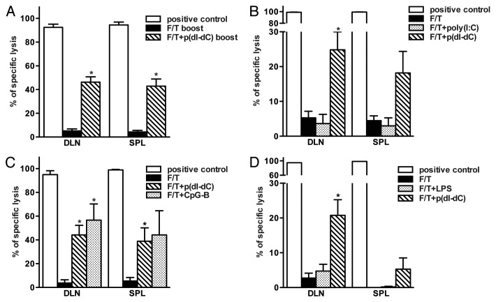
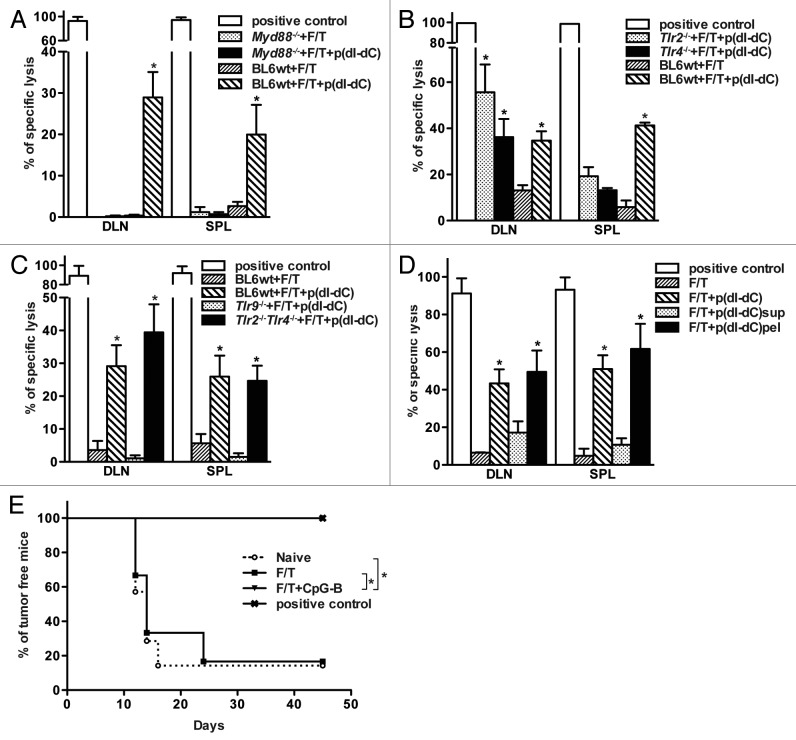
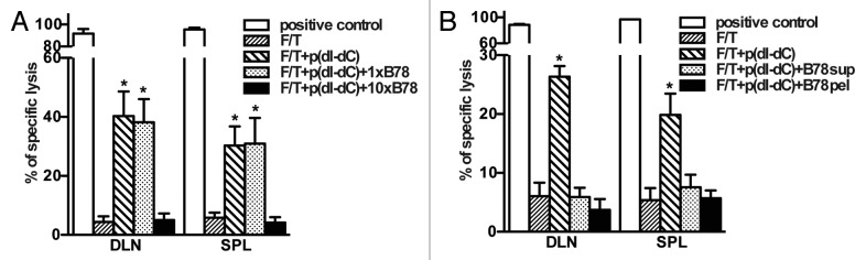
Necrotic cells are known to activate the innate immune system and trigger inflammation by releasing damage associated molecular patterns (DAMPs). However, how necrotic cells influence the induction of antigen-specific CD8(+) T cell-mediated adaptive immune responses under sterile conditions, in the absence of pathogen associated molecular patterns (PAMPs), remains poorly understood. Here, we examined antigen-specific CD8(+) T-cell responses to primary sterile necrotic tumor cells both in vitro and in vivo. We found that primary necrotic cells alone fail to generate CD8(+) T cell-dependent immune responses toward cell-associated antigens. We show that necrotic cells trigger CD8(+) T-cell immunity only in the presence of PAMPs or analogs, such as p(dI-dC) and/or unmethylated CpG DNA. The electroporation of tumor cells with these PAMPs prior to necrosis induction triggered antigen-specific CD8(+) T-cell responses through a TLR9/MyD88-dependent pathway. In addition, we found that necrotic cells contain factors that can block the cross-priming of CD8(+) T cells even under non-sterile conditions and can serve as a possible mechanism of immunosuppression. These results suggest that antigen-specific CD8(+) T-cell responses to primary necrotic tumor cells can be induced in the presence of PAMPs and thus have a substantial impact on the development of antitumor vaccination strategies.


Figures

None
Figure 1. Primary sterile necrotic tumor cells fail to stimulate OVA-specific CD8+ T cells in vitro. (A and B) 5 × 10⁴ F/T, live or γ-irradiated B78OVA or B78 cells were co-cultured in vitro with 5 × 10⁵ OT-I splenocytes for 48 h and interferon γ (IFNγ)-secreting CD8+ T cells (A) or IFNγ concentration (B) were analyzed using IFNγ intracellular staining and IFNγ ELISA respectively. Data are pooled results of 3 experiments. (C) As in (A), OT-I splenocytes were co-cultured for 48 h with F/T or live EG7 cells and % of IFNγ producing cells was determined. EL4 cells were used as an OVA-negative control cell line. Data are representative of 3 independent experiments. (D) 5 × 10⁴ F/T, live or γ-irradiated B78 cells were co-cultured with unsorted splenocytes. After 24 h, CD11c+ cells were stained and analyzed for I-A/I-E, CD80, CD86 and CD40 expression. As a positive control, DCs were stimulated with 100 ng/mL lipopolysaccharide (LPS). Data are representative of 2 independent experiments. *p < 0.05 as compared with OT-I cells alone (Student’s t test).
None
Figure 2. Primary sterile necrosis does not induce antigen-specific CD8+ T cell mediated immune responses in vivo. (A) Mice were vaccinated with 2 × 10⁶ F/T, γ-irradiated or live B78OVA cells and antigen-specific lytic activity was determined using in vivo CTL assay after seven days in draining lymph nodes (DLNs) and spleens. Figure A shows an experiment representative of at least 3 independent experiments with 3 mice per group. (B) Lymphocytes from the DLNs and spleens of naïve, F/T, γ-irradiated or live tumor cell vaccinated mice were isolated 7 d after vaccination. Cells were restimulated in vitro with 0.1 μg/mL OVA peptide for 4 h and the frequency of interferon γ (IFNγ)-secreting CD8+ T cells was analyzed. Data are from one experiment representative of 2 independent experiments. (C). Mice were vaccinated with 5 × 10⁶ F/T or live EG7 cells and antigen-specific lysis was measured as in (A). Pooled data of 2 independent experiments with 3 mice per group are shown. (D) C57BL/6 mice were vaccinated with 2 × 10⁶ allogeneic H2Kd-positive live or F/T CT26OVA cells and the lytic activity of H2Kb-restricted, OVA-specific CD8+ T cells was evaluated. Data are representative of at least 3 independent experiments with 3 mice per group. *p < 0.05 as compared with F/T (necrotic) group (Student’s t test).
None
Figure 3. Antigens are present in sterile necrotic cells but cannot be a substrate for cross-priming of naïve CD8+ T cells. (A) F/T cells were cultured at 37°C and the soluble and particulate fractions were prepared at indicated time points. After subsequent denaturation, electrophoresis and immunoblotting, the model antigen (OVA protein) was detected in both fractions. Samples were normalized for the cell number used in necrosis induction. (B) Mice were primed on day 0 and boosted on days 1, 2, 3 with 5 × 10⁶ F/T cells. Seven days after the last vaccination, on day 10, antigen-specific lysis was tested in the spleen and draining lymph nodes (DLNs). As a positive control in this experiment and in all subsequent experiments, mice were vaccinated with 2 × 10⁶ γ-irradiated B78OVA cells. Pooled data of 2 experiments with 3 mice per group are shown. *p < 0.05 as compared with F/T group (Student’s t test).
None
Figure 4. Failure of sterile necrosis to induce cross-priming of CD8+ T cells in vivo can be reversed using model DNA p(dI-dC) or CpG-B. (A–C) Mice were primed on day 0 and boosted on day 3 with 1 × 10⁶ F/T B78OVA cells electroporated with p(dI-dC) (A), poly(I:C) (B) or CpG-B (C). In vivo CTL analysis was performed on day 10. (D) Mice were primed on day 0 and boosted on day 3 with 2 × 10⁶ F/T cells mixed with p(dI-dC) or LPS. In vivo CTL was performed as in (A) on day 10. Pooled data of 3 (A), 5 (B), or 2 experiments (C and D) with 3 mice per group are shown. *p < 0.05 as compared with F/T group (Student’s t test).
None
Figure 5. Reversal of immunogenicity is TLR9-dependent and relies on MyD88 signaling. (A-C) Wild type, Myd88−/− (A), Tlr2−/−, Tlr4−/−(B), Tlr2−/−Tlr4−/− or Tlr9−/− (C) C57BL/6 mice were primed on day 0 and boosted on day 3 with 1 × 10⁶ p(dI-dC)-electroporated F/T B78OVA cells. Antigen-specific immune responses were analyzed by in vivo CTL assay on day 10. (D) 1 × 10⁶ whole cells, soluble or particulate fractions from p(dI-dC)-electroporated F/T cells were injected into mice on days 0 and 3 and killing activity of CD8+ T cells was evaluated on day 10 as shown in (A). Pooled data from 2 independent experiments are shown with minimum of 3 mice per group. *p < 0.05 as compared with F/T group (Student’s t test). E. B78OVA cells were electroporated with CpG-B or with PBS as in Figure 4C and were subjected to F/T to induce necrosis. Mice were vaccinated with 1 × 10⁶ F/T cells on days 0, 3 and were challenged on the opposite flank with 1 × 10⁴ live B16OVA cells on day 7. As a control, naïve mice or mice vaccinated with γ-irradiated B78OVA cells were used and tumor-free survival was monitored. Data are pooled from 2 independent experiments. *p < 0.05 Log-Rank (Mantel-Cox) test.
None
Figure 6. Necrotic cells release factors, which block cross-priming of antigen-specific CD8+ T cells in vivo. (A and B) Necrotic B78OVA cells (F/T) electroporated with p(dI-dC) were mixed with whole B78 necrotic cells (A), or with soluble/particulate fractions from B78 necrotic cells (B) and injected into mice. In vivo CTL was performed 7 d after boost. Pooled data of 3 (A) or 2 experiments (B) with 3 mice per group are shown. *p < 0.05 as compared with 3 × F/T group (Student’s t test).

References

    1. Ma Y, Conforti R, Aymeric L, Locher C, Kepp O, Kroemer G, et al. How to improve the immunogenicity of chemotherapy and radiotherapy. Cancer Metastasis Rev. 2011;30:71–82. doi: 10.1007/s10555-011-9283-2. - DOI - PubMed
    1. Gallucci S, Lolkema M, Matzinger P. Natural adjuvants: endogenous activators of dendritic cells. Nat Med. 1999;5:1249–55. doi: 10.1038/15200. - DOI - PubMed
    1. Matzinger P. Tolerance, danger, and the extended family. Annu Rev Immunol. 1994;12:991–1045. doi: 10.1146/annurev.iy.12.040194.005015. - DOI - PubMed
    1. Sauter B, Albert ML, Francisco L, Larsson M, Somersan S, Bhardwaj N. Consequences of cell death: exposure to necrotic tumor cells, but not primary tissue cells or apoptotic cells, induces the maturation of immunostimulatory dendritic cells. J Exp Med. 2000;191:423–34. doi: 10.1084/jem.191.3.423. - DOI - PMC - PubMed
    1. Basu S, Binder RJ, Suto R, Anderson KM, Srivastava PK. Necrotic but not apoptotic cell death releases heat shock proteins, which deliver a partial maturation signal to dendritic cells and activate the NF-kappa B pathway. Int Immunol. 2000;12:1539–46. doi: 10.1093/intimm/12.11.1539. - DOI - PubMed

Publication types