. 2013 Jan 1;14(1):13-9.
doi: 10.2174/1389450111314010003.

Oxidation inhibits iron-induced blood coagulation

Affiliations
Free PMC article

Oxidation inhibits iron-induced blood coagulation

Etheresia Pretorius et al. Curr Drug Targets. .
Free PMC article

Abstract

Blood coagulation under physiological conditions is activated by thrombin, which converts soluble plasma fibrinogen (FBG) into an insoluble clot. The structure of the enzymatically-generated clot is very characteristic being composed of thick fibrin fibers susceptible to the fibrinolytic degradation. However, in chronic degenerative diseases, such as atherosclerosis, diabetes mellitus, cancer, and neurological disorders, fibrin clots are very different forming dense matted deposits (DMD) that are not effectively removed and thus create a condition known as thrombosis. We have recently shown that trivalent iron (ferric ions) generates hydroxyl radicals, which subsequently convert FBG into abnormal fibrin clots in the form of DMDs. A characteristic feature of DMDs is their remarkable and permanent resistance to the enzymatic degradation. Therefore, in order to prevent thrombotic incidences in the degenerative diseases it is essential to inhibit the iron-induced generation of hydroxyl radicals. This can be achieved by the pretreatment with a direct free radical scavenger (e.g. salicylate), and as shown in this paper by the treatment with oxidizing agents such as hydrogen peroxide, methylene blue, and sodium selenite. Although the actual mechanism of this phenomenon is not yet known, it is possible that hydroxyl radicals are neutralized by their conversion to the molecular oxygen and water, thus inhibiting the formation of dense matted fibrin deposits in human blood.


Figures
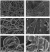
Fig. (1)
Fig. (1)
a) 10 µl of purified fibrinogen + 5 µl thrombin = regular clot formation (thick fibrin fibers). Scale = 1 µm. b) 5 µl of 15mM FeCl3 +10 µl of purified fibrinogen wait 5 minutes + 5 µl thrombin = thin fibrin fibers mixed with DMDs. Scale = 1 µm. c) 10 µl PRP + 5 µl thrombin = regular clot formation (thick fibrin fibers with thin fibers scattered in between). Scale = 1 µm. d) 5 µl of 15mM FeCl3 +10 µl PRP wait 5 minutes + 5 µl thrombin = dense matted deposits (DMD) formation. Scale = 1 µm. e) 10 µl WB + 5 µl thrombin = RBC surrounded by thick fibrin fibers with thin fibers scattered in between. Scale = 1 µm. f) 5 µl of 15mM FeCl3 + 10 µl WB wait 5 minutes + 5 µl thrombin = RBC fused to DMD. Scale = 1 µm.
Fig. (2)
Fig. (2)
a) 5 µl of 15mM FeCl3 + 10 µl of 10 mM Hydrogen Peroxide wait 5 minutes + 10 µl purified fibrinogen + 5 µl thrombin = thin beaded fibrin fibers. Scale = 1 µm. b) 5 µl of 15mM FeCl3 + 10 µl of 10 mM Hydrogen Peroxide wait 5 minutes + 10 µl PRP + 5 µl thrombin = regular clot formation (thick fibrin fibers with scattered thin fibers). Scale = 1 µm. c) 5 µl of 15mM FeCl3 + 10 µl of 10 mM Hydrogen Peroxide wait 5 minutes + 10 µl WB + 5 µl thrombin = normal shape RBC. Scale = 1 µm.
Fig. (3)
Fig. (3)
a) 5 µl of 15mM FeCl3 + 10 µl of 10 mM Methylene Blue wait 5 minutes + 10 µl of purified fibrinogen + 5 µl thrombin = thick beaded fibrin fibers. Scale = 1 µm. b) 5 µl of 15mM FeCl3 + 10 µl of 10 mM Methylene Blue wait 5 minutes + 10 µl PRP + 5 µl thrombin = amorphous clot formation. Scale = 1 µm. c) 5 µl of 15mM FeCl3 + 10 µl of 10 mM Methylene Blue wait 5 minutes + 10 µl WB + 5 µl thrombin = deformed RBC shape. Scale = 1 µm.
Fig. (4)
Fig. (4)
a) 5 µl of 15mM FeCl3 + 10 µl of 10 mM Sodium Selenite wait 5 minutes + 10 µl of purified fibrinogen + 5 µl thrombin = thin fibrin fibers. Scale = 1 µm. b) 5 µl of 15mM FeCl3 + 10 µl of 10 mM Sodium Selenite wait 5 minutes + 10 µl PRP + 5 µl thrombin = thin fibrin fibers. Scale = 1 µm. c) 5 µl of 15mM FeCl3 + 10 µl of 10 mM Sodium Selenite wait 5 minutes + 10 µl WB + 5 µl thrombin = normal RBC shape. Scale = 1 µm.
Fig. (5)
Fig. (5)
a) 5 µl of 15mM FeCl3 + 10 µl of 10 mM Salicylate wait 5 minutes + 10 µl of purified fibrinogen + 5 µl thrombin = thin fibrin fibers. Scale = 1 µm. b) 5 µl of a 15mM FeCl3 + 5 µl of 10 mM Salicylate wait 5 minutes + 10 µl PRP + 5 µl thrombin = dense beaded fibrin fibers. Scale = 1 µm. c) 5 µl of a 15mM FeCl3 + 4 µl of 10 mM Salicylate wait 5 minutes + 10 µl WB + 5 µl thrombin = normal RBC shape. Scale = 1 µm.

References

    1. Kannel WB, Wolf PA, Castelli WP, D'Agnostino RB. Fibrinogen and risk of cardiovascular disease. The Framingham Study. JAMA. 1987;258(9 ):1183–6. - PubMed
    1. Duguid JB. Thrombosis as a factor in the pathogenesis of coronary atherosclerosis. Pathol Bacteriol. 1946;58:207–12. - PubMed
    1. Smith EB. Fibrin deposition and fibrin degradation products in atherosclerotic plaques. Thromb Res. 1994;75:329–35. - PubMed
    1. Undas A, Kolarz M, Kopec G, Tracz W. Altered fibrin clot properties onlong-term haemodialysis relation to cardiovascular mortality. Nephrol Dial Transplant. 2008;23:2010–5. - PubMed
    1. Longstaff C, Thelwell C, Williams SC, Silva MMCG, Szabo L, Kolev K. The interplay between tissue plasminogen activator domains and fibrin structures in the regulation of fibrinolysis kinetic and microscopic studies. Blood. 2011;117:661–8. - PMC - PubMed

MeSH terms

LinkOut - more resources