. 2012 Nov 21:12:540.
doi: 10.1186/1471-2407-12-540.

Detection of lipid-rich prostate circulating tumour cells with coherent anti-Stokes Raman scattering microscopy

Affiliations

Detection of lipid-rich prostate circulating tumour cells with coherent anti-Stokes Raman scattering microscopy

Ranjana Mitra et al. BMC Cancer. .

Abstract

Background: Circulating tumour cells (CTC) are an important indicator of metastasis and associated with a poor prognosis. Detection sensitivity and specificity of CTC in the peripheral blood of metastatic cancer patient remain a technical challenge.

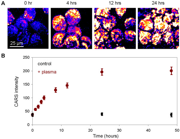
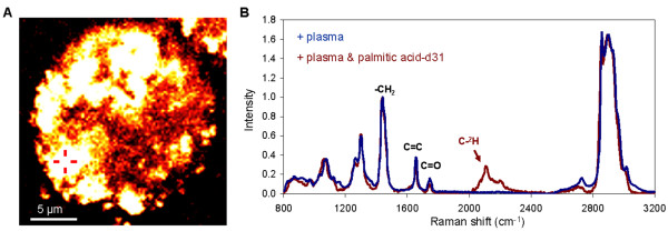
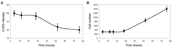
Methods: Coherent anti-Stokes Raman scattering (CARS) microscopy was employed to examine the lipid content of CTC isolated from the peripheral blood of metastatic prostate cancer patients. CARS microscopy was also employed to evaluate lipid uptake and mobilization kinetics of a metastatic human prostate cancer cell line.

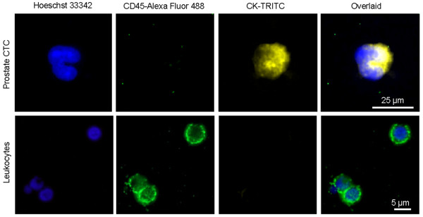
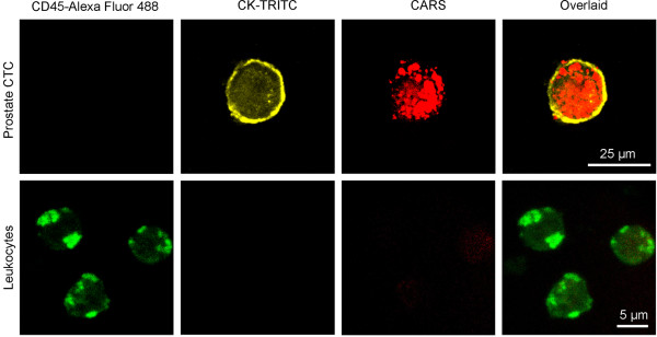
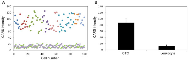
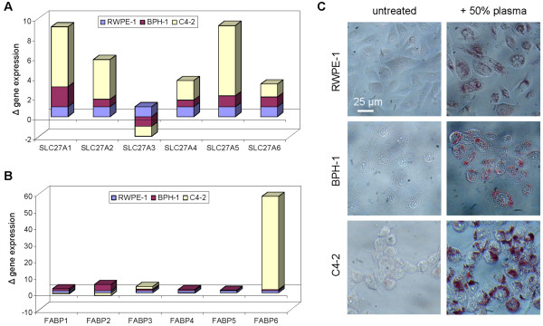
Results: One hundred CTC from eight metastatic prostate cancer patients exhibited strong CARS signal which arose from intracellular lipid. In contrast, leukocytes exhibited weak CARS signal which arose mostly from cellular membrane. On average, CARS signal intensity of prostate CTC was 7-fold higher than that of leukocytes (P<0.0000001). When incubated with human plasma, C4-2 metastatic human prostate cancer cells exhibited rapid lipid uptake kinetics and slow lipid mobilization kinetics. Higher expression of lipid transport proteins in C4-2 cells compared to non-transformed RWPE-1 and non-malignant BPH-1 prostate epithelial cells further indicated strong affinity for lipid of metastatic prostate cancer cells.

Conclusions: Intracellular lipid could serve as a biomarker for prostate CTC which could be sensitively detected with CARS microscopy in a label-free manner. Strong affinity for lipid by metastatic prostate cancer cells could be used to improve detection sensitivity and therapeutic targeting of prostate CTC.


Figures

Figure 1
Figure 1
Detection of circulating tumour cells in the peripheral blood of metastatic prostate cancer patients. Upper row: CTC were identified as Hoechst33342+CD45-CK+ cells. Lower row: leukocytes were identified as Hoechst33342+CD45+CK- cells. Images were taken with widefield fluorescent microscopy
Figure 2
Figure 2
Detection of lipid-rich prostate CTC with CARS microscopy. Upper row: Prostate CTC exhibited strong CARS signal arising from intracellular lipid accumulation. Lower row: Leukocytes exhibited weak CARS signal arising mainly from cellular membrane.
Figure 3
Figure 3
Average CARS signal intensity of prostate CTC versus leukocytes. (A) CARS intensity, average pixel intensity (0-255) of individual cells, as a function of 100 prostate CTC (circles) and 100 leukocytes (purple squares) in eight metastatic prostate cancer patients and 100 leukocytes from healthy volunteers (green squares). CTC from each individual patient were color-coded (same color circles). (B) Average CARS intensity of 100 prostate CTC from cancer patients and 200 leukocytes from both cancer patient and healthy volunteers. Error bars are the standard deviations.
Figure 4
Figure 4
Gene expression of lipid transport proteins and lipid uptake ability. Real-time PCR gene expression profiling of (A) fatty acid transport proteins (FATPs encoded by SLC27A genes) and (B) fatty acid binding proteins (FABPs) of non-transformed prostate epithelial cell line RWPE-1, benign prostatic hyperplasia epithelial cell line BPH-1, and metastatic prostate cancer cell line C4-2. Gene expression levels were normalized to 1 for RWPE-1 cell line and respectively for other cell lines. (C) Oil Red O staining to evaluate the uptake of plasma lipid by RWPE-1, BPH-1, and C4-2 cells after 24 hours of incubation with 50% human plasma.
Figure 5
Figure 5
Plasma lipid uptake kinetics of C4-2 metastatic prostate cancer cells. (A) CARS images of C4-2 cells incubated with 50% human plasma as a function of time. (B) CARS intensity of individual C4-2 cells as a function of incubation time with 50% human plasma (red circles) versus control C4-2 cells in growth media (black squares). Error bars represent the standard deviations across 50 cells analyzed per time point.
Figure 6
Figure 6
Tracking the uptake of deuterated palmitic acid with Raman microspectroscopy. (A) CARS image of a C4-2 cell at 24 hours after incubation with 50% human plasma spiked with 50 μM of palmitic acid d-31. Cross-hair indicates a representative location for Raman microspectroscopy analysis. (B) Representative of Raman signatures of C4-2 cells incubated in 50% human plasma (blue) or in 50% human plasma spiked with deuterated palmitic acid d-31 (red) for 24 hours.
Figure 7
Figure 7
Lipid mobilization kinetics of C4-2 metastatic prostate cancer cells. (A) CARS intensity of individual C4-2 cells as a function of time. C4-2 cells were first incubated with 50% human plasma for 4 hours, then plasma was removed and replaced with growth media. Error bars represent the standard deviations across 50 cells analyzed per time point. (B) Average C4-2 cell number per analysis area of 3 mm2 as a function of time after the removal of human plasma. Error bars are standard deviations across 10 areas evaluated at each time point.

References

    1. Pantel K, Brakenhoff RH, Brandt B. Detection, clinical relevance and specific biological properties of disseminating tumour cells. Nat Rev Cancer. 2008;8(5):329–340. doi: 10.1038/nrc2375. - DOI - PubMed
    1. Cristofanilli M, Budd GT, Ellis MJ, Stopeck A, Matera J, Miller MC, Reuben JM, Doyle GV, Allard WJ, Terstappen LW. et al.Circulating tumor cells, disease progression, and survival in metastatic breast cancer. N Engl J Med. 2004;351(8):781–791. doi: 10.1056/NEJMoa040766. - DOI - PubMed
    1. Goodman OB Jr, Fink LM, Symanowski JT, Wong B, Grobaski B, Pomerantz D, Ma Y, Ward DC, Vogelzang NJ. Circulating tumor cells in patients with castration-resistant prostate cancer baseline values and correlation with prognostic factors. Cancer Epidemiol Biomarkers Prev. 2009;18(6):1904–1913. doi: 10.1158/1055-9965.EPI-08-1173. - DOI - PubMed
    1. Kaiser J. Medicine. Cancer's circulation problem. Science. 2010;327(5969):1072–1074. doi: 10.1126/science.327.5969.1072. - DOI - PubMed
    1. Fidler IJ. The pathogenesis of cancer metastasis: the 'seed and soil' hypothesis revisited. Nat Rev Cancer. 2003;3(6):453–458. doi: 10.1038/nrc1098. - DOI - PubMed

Publication types

MeSH terms

Substances