. 2013 Feb;87(3):509-25.
doi: 10.1111/mmi.12109. Epub 2012 Dec 11.

The response regulator SypE controls biofilm formation and colonization through phosphorylation of the syp-encoded regulator SypA in Vibrio fischeri

Affiliations

The response regulator SypE controls biofilm formation and colonization through phosphorylation of the syp-encoded regulator SypA in Vibrio fischeri

Andrew R Morris et al. Mol Microbiol. 2013 Feb.

Abstract

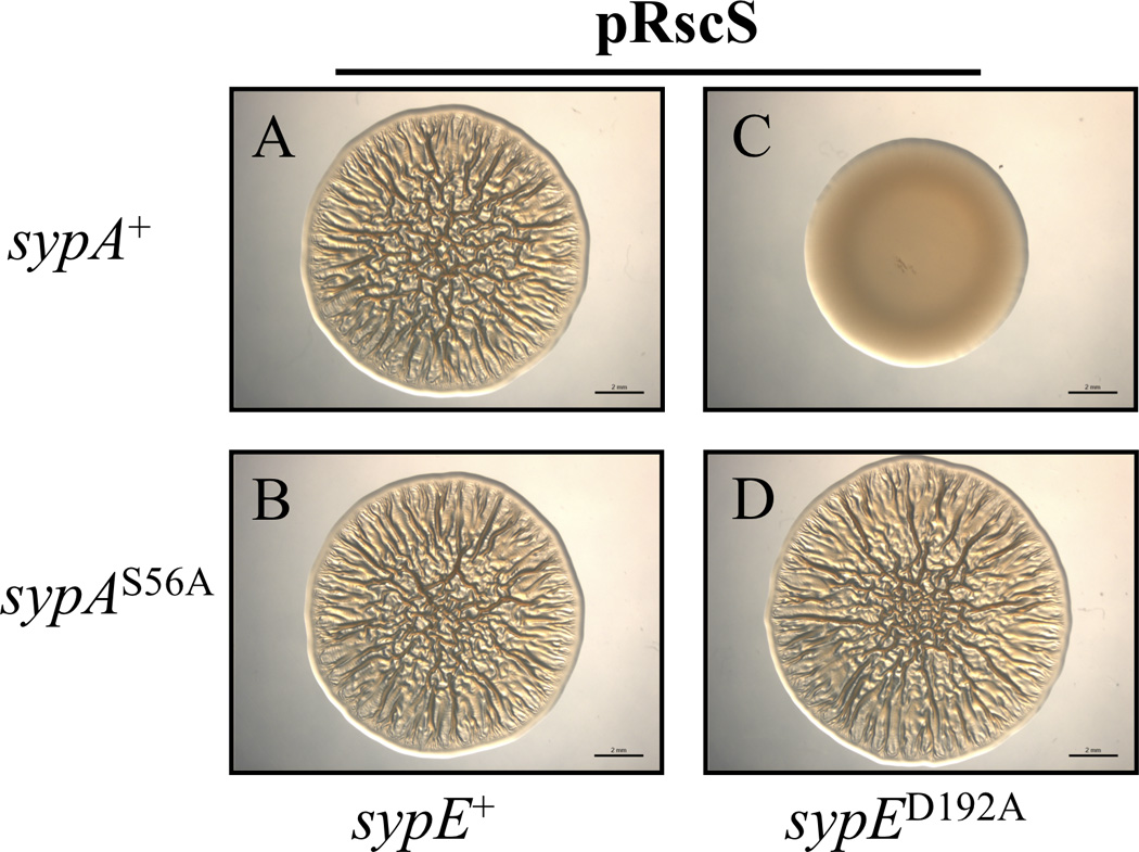
Bacteria utilize multiple regulatory systems to modulate gene expression in response to environmental changes, including two-component signalling systems and partner-switching networks. We recently identified a novel regulatory protein, SypE, that combines features of both signalling systems. SypE contains a central response regulator receiver domain flanked by putative kinase and phosphatase effector domains with similarity to partner-switching proteins. SypE was previously shown to exert dual control over biofilm formation through the opposing activities of its terminal effector domains. Here, we demonstrate that SypE controls biofilms in Vibrio fischeri by regulating the activity of SypA, a STAS (sulphate transporter and anti-sigma antagonist) domain protein. Using biochemical and genetic approaches, we determined that SypE both phosphorylates and dephosphorylates SypA, and that phosphorylation inhibits SypA's activity. Furthermore, we found that biofilm formation and symbiotic colonization required active, unphosphorylated SypA, and thus SypA phosphorylation corresponded with a loss of biofilms and impaired host colonization. Finally, expression of a non-phosphorylatable mutant of SypA suppressed both the biofilm and symbiosis defects of a constitutively inhibitory SypE mutant strain. This study demonstrates that regulation of SypA activity by SypE is a critical mechanism by which V. fischeri controls biofilm development and symbiotic colonization.


Figures

Figure 1
Figure 1. Model of V. fischeri biofilm regulation by SypE and SypA
(A) Model of the Bacillus subtilis partner switching system that regulates the activity of σB in the general stress response pathway (see text for full description). Shown are the relevant domains for partner-switching regulation. (B) SypE contains a central RR REC domain flanked by an N-terminal RsbW-like serine kinase domain and a C-terminal PP2C-like serine phosphatase domain. SypA contains a single STAS-domain conserved in anti-sigma factor antagonists. Under conditions in which SypE is unphosphorylated, its N-terminal kinase domain is active, resulting in the phosphorylation of SypA (on conserved serine residue S56) and inhibition of biofilm formation. When phosphorylated (presumably on conserved residue D192), SypE functions as a phosphatase (Morris et al., 2011). This results in the dephosphorylation of SypA, activating SypA to promote biofilm formation. A D192A mutation “locks” SypE into a constitutive kinase, resulting in SypA phosphorylation and the inhibition of biofilms and colonization. Protein phosphatase 2C (PP2C) domain (black box); Serine/threonine kinase (RsbW) domain (white box); anti-sigma factor antagonist (STAS) domain (grey box); response regulator receiver (REC) domain (light grey box).
Figure 2
Figure 2. SypE interacts with SypA
(A) Soluble lysates from V. fischeri ΔsypA ΔsypE cells [KV4716] carrying plasmids expressing FLAG-SypE (pARM80), HA-SypA (pARM36), or untagged SypE (pCLD48) and SypA (pARM13) control plasmids were used in immunoprecipation assays with non-specific anti-rabbit IgG antibody (Lanes 1 and 5), anti-FLAG antibody (Lanes 2–4), or anti-HA antibody (Lanes 6–8). The samples were resolved using SDS-PAGE and subjected to western blot analysis with anti-FLAG (top panel) or anti-HA (bottom panel) antibodies. (+) indicates V. fischeri cells carrying the epitope tagged SypE (pARM80) and/or SypA (pARM36) plasmids. (−) indicates V. fischeri cells carrying the control plasmids expressing untagged SypE (pCLD48) or SypA (pARM13). (B) Soluble lysates from V. fischeri ΔsypA ΔsypE cells [KV4716] carrying plasmids expressing HA-SypA (pARM36) and either FLAG-SypEΔNTD (pARM162) [Lanes 1 and 3] or FLAG-SypENTD (pARM136) [Lanes 2 and 4] were used in immunoprecipation assays with anti-FLAG (or anti-HA antibody. Lanes 5 and 6, lysates from ΔsypA ΔsypE cells [KV4716] carrying both pARM36 and either pARM162 (lane5) or pARM136 (lane 6) immunoprecipitated with non-specific, anti-rabbit IgG. Samples were resolved using SDS-PAGE and subjected to western blot analysis with anti-FLAG (top panel) or anti-HA (bottom panel) antibodies.
Figure 3
Figure 3. SypE phosphorylates SypA in vitro
(A) In vitro phosphorylation of SypA by SypE. Purified SypA-FLAG (3 µg) and/or SypE proteins (2 µg) were incubated in kinase buffer in the presence or absence of ATP. Reaction samples were resolved by SDS-PAGE on a 25 µM Phos-tag™ acrylamide gel and the proteins were detected via western blot analysis using an anti-FLAG antibody. Lane 1, SypE incubated in kinase buffer. Lanes 2 and 3, wild-type SypA-FLAG protein incubated in kinase buffer alone (Lane 2) or with SypE (Lane 3). Lane 4, SypAS56A-FLAG protein incubated with SypE in kinase buffer. Lane 5, wild-type SypA-FLAG protein incubated with SypE in kinase buffer lacking ATP. (+) indicates reactions containing wild-type SypA-FLAG or SypE protein. (−) indicates reactions not containing purified protein. (S56A) indicates reactions containing the SypAS56A–FLAG protein. (B) SypE-mediated phosphorylation of SypA in E. coli cells. SypA-FLAG protein was purified from E. coli cells carrying both pARM157 (GST-SypA-FLAG) and either empty vector, pVSV105, (Lane 1) or plasmid pCLD64, which expresses the N-terminal, serine kinase domain of SypE (Lane 2). Samples were resolved using SDS-PAGE on a 25 µM Phos-tag™ acrylamide gel and proteins were detected by anti-FLAG western blot analysis. SypA~P denotes phosphorylated SypA.
Figure 4
Figure 4. SypE dephosphorylates SypA in vitro
Western blot analysis of in vitro SypA dephosphorylation samples analyzed on Phos-tag™ acrylamide gels. Purified phosphorylated SypA-FLAG protein (SypA~P; 2 µg) was incubated in Mg2+-containing phosphatase buffer in the presence or absence of increasing concentrations of purified SypE C-terminal phosphatase domain (SypECTD; 2–10 µg). The reactions were terminated at zero or 30 minutes and the samples resolved by SDS-PAGE on a 25 µM Phos-tag™ acrylamide gel. SypA proteins were detected by western blot analysis using an anti-FLAG antibody. Lane 1, SypECTD incubated in buffer alone. Lane 2, non-phosphorylated SypA incubated in buffer alone. Lanes 3 and 7, phosphorylated SypA (SypA~P) incubated in buffer alone for zero (Lane 3) and 30 (Lane 7) minutes. Lanes 4–6, phosphorylated SypA (SypA~P) incubated for 30 minutes in buffer containing 2 µg (Lane 4), 5 µg (Lane 5), or 10 µg (Lane 6) of purified SypECTD. (+) indicates reactions containing SypA-FLAG or SypECTD protein. (−) indicates reactions not containing the indicated purified protein.
Figure 5
Figure 5. Active SypA is required for RscS-induced biofilm formation
The RscS plasmid (pARM7) was introduced into wild-type (WT) or indicated sypA strains. Cultures of the following strains were spotted onto LBS medium at 24°C and wrinkled colony formation was assessed at 48 h post spotting: WT cells containing empty Tn7 cassette (EC) [KV4389] and carrying either empty vector pKV282 (A) or pARM7 (B); pARM7-carrying ΔsypA cells containing EC [KV5079] (C), or complemented with wild-type sypA+ [KV5479] (D), sypAS56D [KV5480] (E), or sypAS56A [KV5481] (F). Images are representative of at least three independent experiments. Black bar represents 2 mm.
Figure 6
Figure 6. SypA functions downstream of SypE
Assessment of RscS-induced wrinkled colony formation. Cultures of the following strains were spotted onto LBS medium at 24°C and wrinkled colony formation was assessed at 48 h post-spotting: Wild-type (WT) cells containing empty Tn7 cassette (EC) [KV4389] and carrying empty vector pKV282 (A) or pRscS plasmid pARM7 (B); ΔsypE cells containing EC [KV4390] and carrying pARM7 (C); ΔsypA cells containing EC [KV5079] and carrying pARM7 (D); pARM7-carrying ΔsypA ΔsypE cells containing either EC [KV6392] (E) or wild-type sypA+ [KV6393] (F). Images are representative of at least three independent experiments. Black bar represents 2 mm.
Figure 7
Figure 7. A sypAS56A mutant suppresses the sypED192A biofilm defect
Assessment of RscS-induced wrinkled colony formation. The pRscS plasmid pARM7 was introduced into ΔsypE cells complemented with either wild-type sypE+ or the inhibitory sypED192A allele and expressing either wild-type sypA+ or sypAS56A. Cultures of the following strains were spotted onto LBS medium at 24°C and wrinkled colony formation was assessed at 48 h post spotting: sypE+ cells expressing wild-type sypA [KV6213] (A) or sypAS56A [KV6215] (B); sypED192A cells expressing either wild-type sypA [KV6214] (C) or sypAS56A [KV6216] (D). Images are representative of at least three independent experiments. Black bar represents 2 mm.
Figure 8
Figure 8. SypE promotes SypA phosphorylation in vivo
Western blot analysis of V. fischeri cell lysates analyzed on Phos-tag™ acrylamide gels. Soluble lysates from indicated V. fischeri strains were resolved by SDS-PAGE on 25 µM Phos-tag™ acrylamide gels and the proteins were detected by western blot analysis using anti-HA antibody. ΔsypA ΔsypE cells containing wild-type sypE+ [KV6424] and carrying pRscS plasmid (pCLD46) and plasmids expressing either untagged sypA+ (pARM13) (Lane 1), HA-tagged sypA+ (pARM36) (Lane 2), or HA-tagged sypAS56A (pARM78) (Lane 4); ΔsypA ΔsypE cells containing inhibitory sypED192A [KV6425] and carrying pCLD46 and plasmids expressing either HA-tagged sypA+ (pARM36) (Lane 3), or HA-tagged sypAS56A (pARM78) (Lane 5). (+) indicates cells expressing wild-type sypA-HA and/or sypE. (S56A) indicates cells expressing the sypAS56A mutant. (D192A) indicates cells expressing the sypED192A mutant. (−) indicates cells expressing untagged sypA. Images are representative of at least three independent experiments.
Figure 9
Figure 9. Active SypA is required for host colonization
(A and B) Competitive colonization with wild-type (WT) V. fischeri and select sypA strains. Newly hatched squid were exposed to a mixed inoculum of WT cells and either ΔsypA [KV5079] cells (A) or ΔsypA cells complemented with wild-type sypA+ [KV5479] (B). The Log RCI is plotted on the x-axis. The position of the circles on the y-axis is merely for spacing. Each circle represents a single animal. Closed circles indicate animals containing no sypA mutant cells. The black diamond and errors bars indicate the average Log RCI ± SD for the indicated data set. Data shown are representative of at least three independent experiments. (C) Single-strain colonization by WT and sypA mutant strains. Newly hatched squid were exposed for 18 h to WT cells carrying empty vector (EC) [KV4389] or ΔsypA cells carrying empty vector (EC) [KV5079] or complemented with either wild-type sypA+ [KV5479] or sypAS56D [KV5480]. As a negative control, aposymbiotic (APO) juvenile squid were maintained in bacteria free water. Each circle represents the number of V. fischeri cells recovered from an individual animal. The dashed line indicates the limit of detection (14 CFU/squid). The black bar indicates the average CFU for 10 animals. Data shown are from one experiment and are representative of at least three independent experiments.
Figure 10
Figure 10. SypE inhibits colonization through inactivation of SypA
Single-strain colonization by wild-type (WT) V. fischeri and select mutant strains. Newly hatched squid were exposed for 18 h to either WT cells containing empty Tn7 cassette (EC) [KV4389] or sypED192A mutant cells expressing wild-type sypA [KV6214] or sypAS56A [KV6216]. As a negative control, aposymbiotic (APO) juvenile squid were maintained in bacteria free water. Each circle represents the number of V. fischeri cells recovered from an individual animal. The dashed line indicates the limit of detection. The black bar indicates the average CFU for 10 animals. Data shown are from one experiment and are representative of at least three independent experiments.

Similar articles

Cited by

References

    1. Benson AK, Haldenwang WG. Bacillus subtilis sigma B is regulated by a binding protein (RsbW) that blocks its association with core RNA polymerase. Proc Natl Acad Sci U S A. 1993a;90:2330–2334. - PMC - PubMed
    1. Benson AK, Haldenwang WG. Regulation of sigma B levels and activity in Bacillus subtilis. J Bacteriol. 1993b;175:2347–2356. - PMC - PubMed
    1. Bhuwan M, Lee HJ, Peng HL, Chang HY. Histidine-containing phosphotransfer protein-B (HptB) regulates swarming motility through partner-switching system in Pseudomonas aeruginosa PAO1 strain. J Biol Chem. 2012;287:1903–1914. - PMC - PubMed
    1. Boettcher KJ, Ruby EG. Depressed light emission by symbiotic Vibrio fischeri of the sepiolid squid Euprymna scolopes. J Bacteriol. 1990;172:3701–3706. - PMC - PubMed
    1. Bourret RB. Receiver domain structure and function in response regulator proteins. Curr Opin Microbiol. 2010;13:142–149. - PMC - PubMed

Publication types

MeSH terms

Substances

LinkOut - more resources