

Gene therapy for colorectal cancer by an oncolytic adenovirus that targets loss of the insulin-like growth factor 2 imprinting system
- PMID: 23171475
- PMCID: PMC3546838
- DOI: 10.1186/1476-4598-11-86
Gene therapy for colorectal cancer by an oncolytic adenovirus that targets loss of the insulin-like growth factor 2 imprinting system
Abstract
Background: Colorectal cancer is one of the most common malignant tumors worldwide. Loss of imprinting (LOI) of the insulin-like growth factor 2 (IGF2) gene is an epigenetic abnormality observed in human colorectal neoplasms. Our aim was to investigate the feasibility of using the IGF2 imprinting system for targeted gene therapy of colorectal cancer.
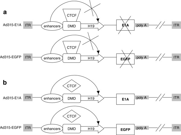
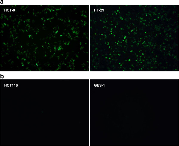
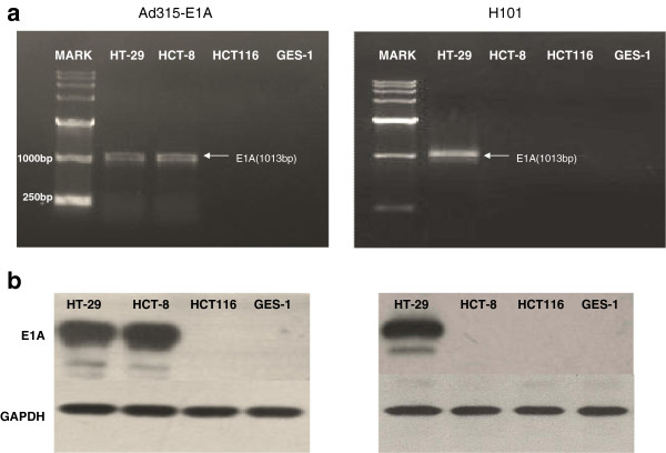
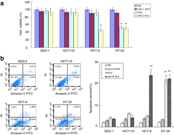
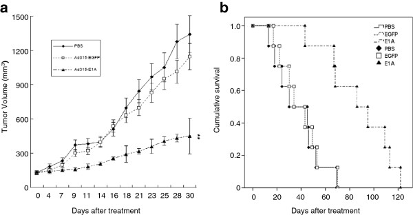
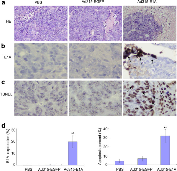
Results: We constructed a novel oncolytic adenovirus, Ad315-E1A, and a replication-deficient recombinant adenovirus, Ad315-EGFP, driven by the IGF2 imprinting system by inserting the H19 promoter, CCCTC binding factor, enhancer, human adenovirus early region 1A (E1A) and enhanced green fluorescent protein (EGFP) reporter gene into a pDC-315 shuttle plasmid. Cell lines with IGF2 LOI (HCT-8 and HT-29), which were infected with Ad315-EGFP, produced EGFP. However, no EGFP was produced in cell lines with maintenance of imprinting (HCT116 and GES-1). We found that Ad315-E1A significantly decreased cell viability and induced apoptosis only in LOI cell lines in vitro. In addition, mice bearing HCT-8-xenografted tumors, which received intratumoral administration of the oncolytic adenovirus, showed significantly reduced tumor growth and enhanced survival.
Conclusions: Our recombinant oncolytic virus targeting the IGF2 LOI system inhibits LOI cell growth in vitro and in vivo, and provides a novel approach for targeted gene therapy.
Figures
Similar articles
-
Insulin-Like Growth Factor 2 (IGF2) Signaling in Colorectal Cancer-From Basic Research to Potential Clinical Applications.Int J Mol Sci. 2019 Oct 3;20(19):4915. doi: 10.3390/ijms20194915. Int J Mol Sci. 2019. PMID: 31623387 Free PMC article. Review.
-
Gene therapy for human colorectal cancer cell lines with recombinant adenovirus 5 based on loss of the insulin-like growth factor 2 imprinting.Int J Oncol. 2015 Apr;46(4):1759-67. doi: 10.3892/ijo.2015.2852. Epub 2015 Jan 26. Int J Oncol. 2015. PMID: 25625919
-
Gene therapy for cancer through adenovirus vector‑mediated expression of the Ad5 early region gene 1A based on loss of IGF2 imprinting.Oncol Rep. 2013 Oct;30(4):1814-22. doi: 10.3892/or.2013.2646. Epub 2013 Jul 30. Oncol Rep. 2013. PMID: 23900345 Free PMC article.
-
Gene therapy for colorectal cancer by adenovirus-mediated siRNA targeting CD147 based on loss of the IGF2 imprinting system.Int J Oncol. 2015 Nov;47(5):1881-9. doi: 10.3892/ijo.2015.3181. Epub 2015 Sep 23. Int J Oncol. 2015. PMID: 26397886
-
Loss of IGF2 imprinting: mechanisms and consequences.Novartis Found Symp. 2004;262:108-21; discussion 121-4, 265-8. Novartis Found Symp. 2004. PMID: 15562825 Review.
Cited by
-
Systems Biology-Based Investigation of Cellular Antiviral Drug Targets Identified by Gene-Trap Insertional Mutagenesis.PLoS Comput Biol. 2016 Sep 15;12(9):e1005074. doi: 10.1371/journal.pcbi.1005074. eCollection 2016 Sep. PLoS Comput Biol. 2016. PMID: 27632082 Free PMC article.
-
Insulin-Like Growth Factor 2 (IGF2) Signaling in Colorectal Cancer-From Basic Research to Potential Clinical Applications.Int J Mol Sci. 2019 Oct 3;20(19):4915. doi: 10.3390/ijms20194915. Int J Mol Sci. 2019. PMID: 31623387 Free PMC article. Review.
-
Therapeutic effect of the treatment for colorectal cancer with adenoviral vectors mediated estrogen receptor β gene therapy combined with thermotherapy.J Cancer Res Clin Oncol. 2014 Apr;140(4):623-32. doi: 10.1007/s00432-014-1611-9. Epub 2014 Feb 15. J Cancer Res Clin Oncol. 2014. PMID: 24531912 Free PMC article.
-
Precision oncolytic viral therapy in colorectal cancer: Genetic targeting and immune modulation for personalized treatment (Review).Int J Mol Med. 2025 Jul;56(1):104. doi: 10.3892/ijmm.2025.5545. Epub 2025 May 9. Int J Mol Med. 2025. PMID: 40342021 Free PMC article. Review.
-
Distinct allelic expression patterns of imprinted IGF2 in adenocarcinoma and squamous cell carcinoma of the lung.Oncol Lett. 2014 Dec;8(6):2561-2564. doi: 10.3892/ol.2014.2572. Epub 2014 Sep 29. Oncol Lett. 2014. PMID: 25364428 Free PMC article.
References
-
- World Health Organization. The global burden of disease: 2004 update. 2008. http://www.who.int/healthinfo/global_burden_disease/2004_report_update/e....
Publication types
MeSH terms
Substances
LinkOut - more resources
Full Text Sources
Medical
Miscellaneous
