. 2012 Nov 21;4(1):21.
doi: 10.1186/2036-7902-4-21.

Focused assessment with sonography for HIV-associated tuberculosis (FASH): a short protocol and a pictorial review

Affiliations

Focused assessment with sonography for HIV-associated tuberculosis (FASH): a short protocol and a pictorial review

Tom Heller et al. Crit Ultrasound J. .

Abstract

Background: Ultrasound can rapidly identify abnormal signs, which in high prevalence settings, are highly suggestive of extra-pulmonary tuberculosis (EPTB). Unfortunately experienced sonographers are often scarce in these settings.

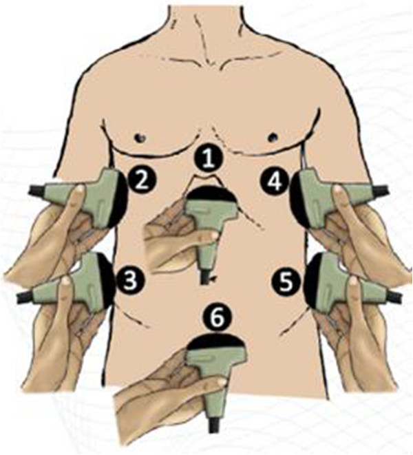
Methods: A protocol for focused assessment with sonography for HIV-associated tuberculosis (FASH) which can be used by physicians who are relatively inexperienced in ultrasound was developed.

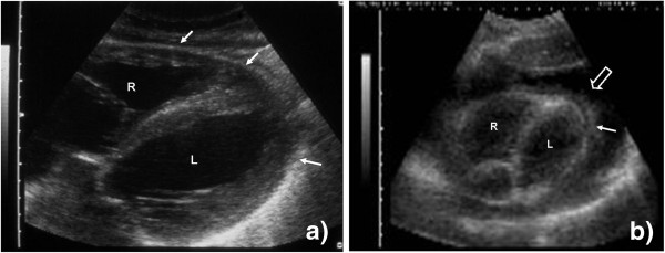
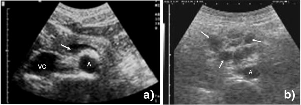
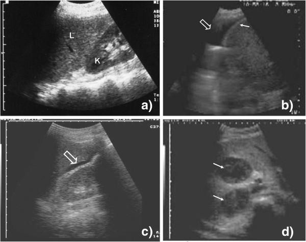
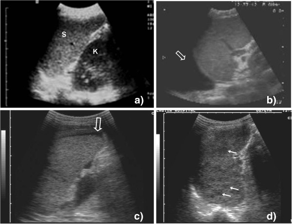
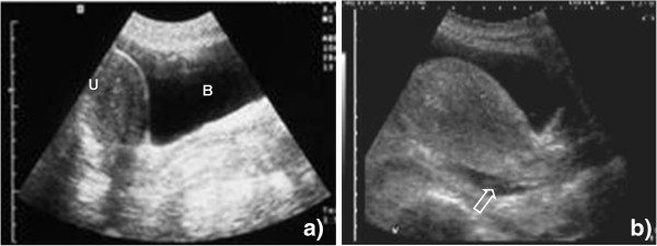
Results: The technique as well as normal and pathological findings are described and the diagnostic and possible therapeutic reasoning explained. The protocol is intended for settings where the prevalence of HIV/TB co-infected patients is high.

Conclusion: FASH is suitable for more rapid identification of EPTB even at the peripheral hospital level where other imaging modalities are scarce and most of the HIV and TB care will be delivered in the future.


Figures

Figure 1
Figure 1
Schematic drawing of the ultrasound probe positions during the FASH examination.
Figure 2
Figure 2
Probe position 1a. (a) Right (R) and left (L) ventricle of the heart are visible. The pericardium surrounds the heart as an echogenic rim (filled arrow). (b) Right (R) and left (L) ventricle of the heart are visible. The heart is surrounded by a large echo-free rim (open arrow), the pericardial effusion. On the visceral side, echogenic fibrinous material (filled arrow) is visible inside the effusion.
Figure 3
Figure 3
Probe position 1b. (a) Vascular structures (abdominal aorta (A), inferior vena cava (VC), splenic vein (arrow)) of the upper abdomen are visible. (b) Multiple round hypoechoic structures are visible (arrow). These represent pathologically enlarged lymph nodes close to the aorta (A).
Figure 4
Figure 4
Probe position 2, 3a, and 3b. (a) Liver (L) and right kidney (K) are visible; there is no echo-free fluid above or below the liver. (b) An anechoic fluid collection is visible above the liver and the echogenic diaphragm (filled arrow) representing pleural effusion (open arrow) on the right side. (c) A small anechoic fluid collection is visible between the liver and the right kidney (open arrow) (Morrison's pouch). Free abdominal fluid can be diagnosed. (d) Two hypoechoic large lesions (filled arrow) can be seen in the parenchyma of the liver.
Figure 5
Figure 5
Probe position 4, 5a, and 5b. (a) Spleen (S) and left kidney (K) are visible; there is no echo-free fluid above or below the spleen. (b) An anechoic fluid collection is visible above the spleen representing pleural effusion (open arrow) on the left side. (c) An anechoic fluid collection is visible around the lower pole of the spleen (open arrow). Free abdominal fluid can be diagnosed. (d) Hypoechoic lesions can be seen inside the spleen (filled arrow). Micro-abscesses due to disseminated TB are a probable explanation.
Figure 6
Figure 6
Probe position 6. (a) The pear-shaped uterus (U) is visible behind the fluid-filled bladder (B). There are no extra-vesical fluid collections, especially no collections in the Douglas' pouch behind the uterus. (b) Small, anechoic collection behind the uterus (open arrow), free abdominal fluid can be diagnosed.

Similar articles

Cited by

References

    1. World Health Organization. Global tuberculosis control: epidemiology, strategy, financing. World Health Organization, Geneva; 2009.
    1. World Health Organization. Improving the diagnosis and treatment of smear-negative pulmonary and extrapulmonary tuberculosis among adults and adolescents. Recommendations for HIV-prevalent and resource-constrained settings. World Health Organization, Geneva; 2007.
    1. Reuter H, Burgess LJ, Doubell AF. Epidemiology of pericardial effusions at a large academic hospital in South Africa. Epidemiol Infect. 2005;133:393–399. doi: 10.1017/S0950268804003577. - DOI - PMC - PubMed
    1. Heller T, Lessells RJ, Wallrauch C, Brunetti E. TB pericarditis with cardiac tamponade: management in the resource-limited setting. AJTMH. 2010;83:1311–14. - PMC - PubMed
    1. Luzze H, Elliott AM, Joloba ML, Odida M, Oweka-Onyee J, Nakiyingi J, Quigley M, Hirsch C, Mugerwa RD, Okwera A, Johnson JL. Evaluation of suspected tuberculous pleurisy: clinical and diagnostic findings in HIV-1-positive and HIV-negative adults in Uganda. Int J Tuberc Lung Dis. 2001;5:746–753. - PubMed

LinkOut - more resources