. 2013 Jan 15;304(2):L92-102.
doi: 10.1152/ajplung.00272.2012. Epub 2012 Nov 21.

Differential susceptibility of inbred mouse strains to chlorine-induced airway fibrosis

Affiliations

Differential susceptibility of inbred mouse strains to chlorine-induced airway fibrosis

Yiqun Mo et al. Am J Physiol Lung Cell Mol Physiol. .

Abstract

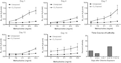
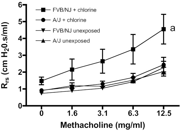
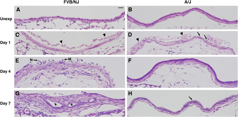
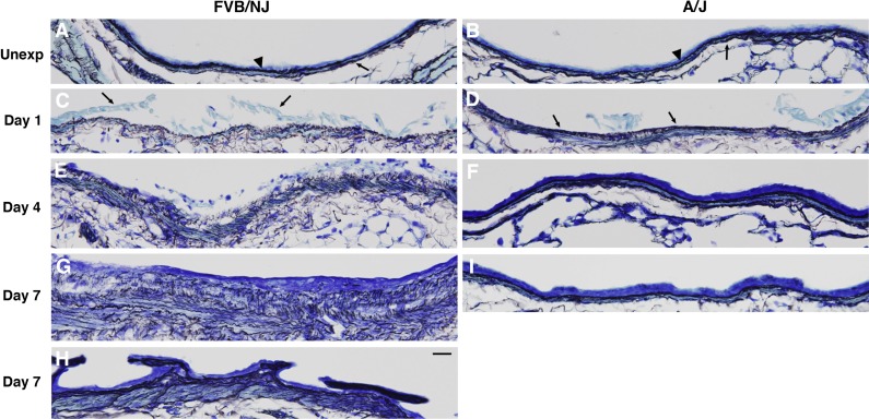
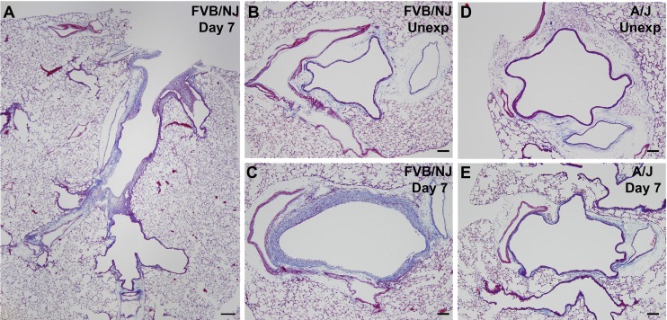
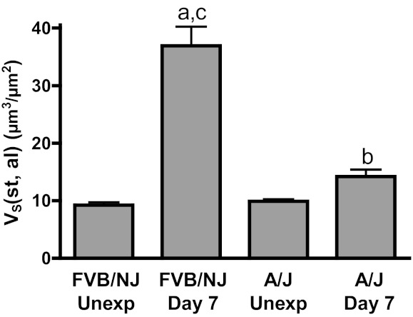
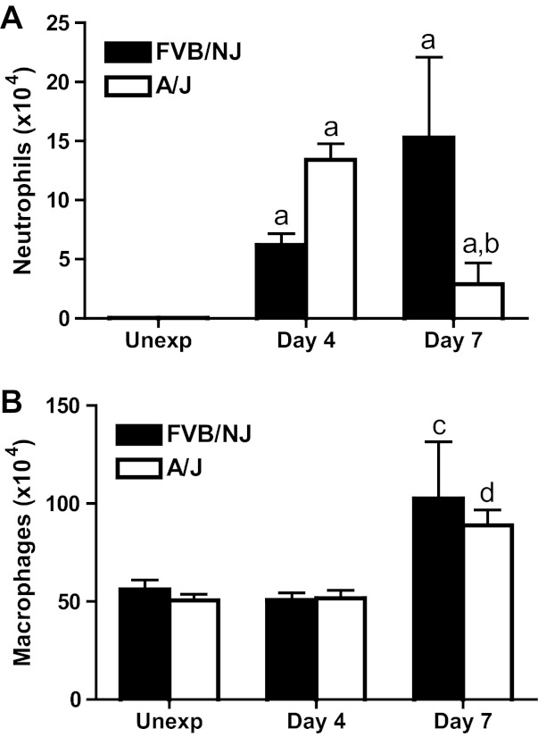
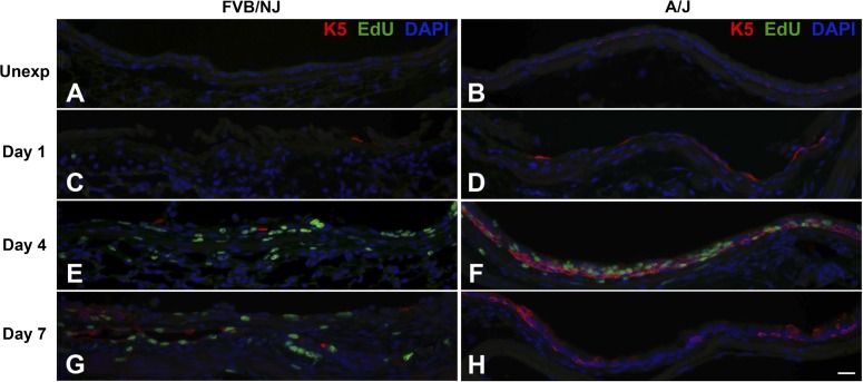
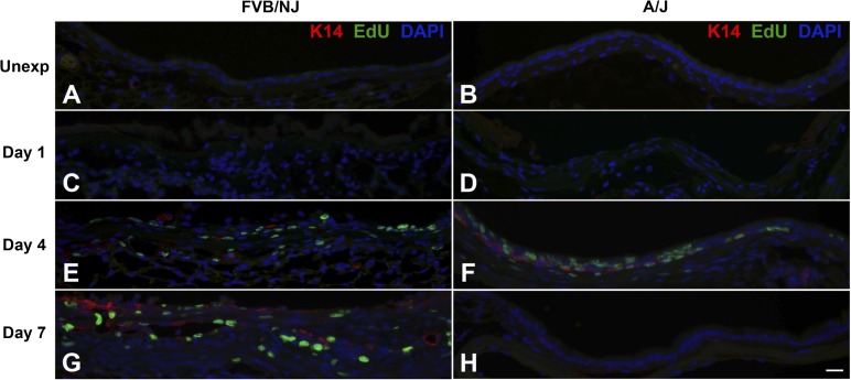
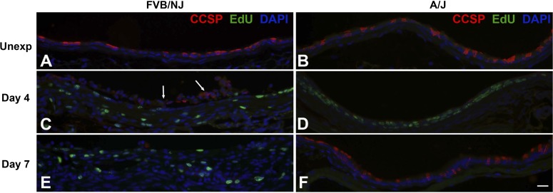
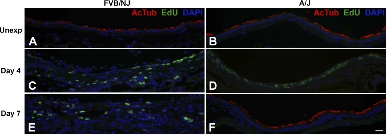
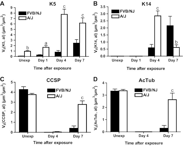
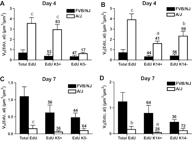
Chlorine is a reactive gas that is considered a chemical threat agent. Humans who develop acute lung injury from chlorine inhalation typically recover normal lung function; however, a subset can experience chronic airway disease. To examine pathological changes following chlorine-induced lung injury, mice were exposed to a single high dose of chlorine, and repair of the lung was analyzed at multiple times after exposure. In FVB/NJ mice, chlorine inhalation caused pronounced fibrosis of larger airways that developed by day 7 after exposure and was associated with airway hyperreactivity. In contrast, A/J mice had little or no airway fibrosis and had normal lung function at day 7. Unexposed FVB/NJ mice had less keratin 5 staining (basal cell marker) than A/J mice in large intrapulmonary airways where epithelial repair was poor and fibrosis developed after chlorine exposure. FVB/NJ mice had large areas devoid of epithelium on day 1 after exposure leading to fibroproliferative lesions on days 4 and 7. A/J mice had airways covered by squamous keratin 5-stained cells on day 1 that transitioned to a highly proliferative reparative epithelium by day 4 followed by the reappearance of ciliated and Clara cells by day 7. The data suggest that lack of basal cells in the large intrapulmonary airways and failure to effect epithelial repair at these sites are factors contributing to the development of airway fibrosis in FVB/NJ mice. The observed differences in susceptibility to chlorine-induced airway disease provide a model in which mechanisms and treatment of airway fibrosis can be investigated.


Figures

Fig. 1.
Fig. 1.
Lung function and survival in chlorine-exposed FVB/NJ mice. Mice were exposed to chlorine, and respiratory system resistance (Rrs) at baseline and in response to inhaled methacholine was analyzed at multiple times after exposure. One group of unexposed mice was measured; the same data were plotted in each graph for ease of comparison with exposed mice. Values are means ± SE (n = 3–6 mice per group); methacholine dose-response curves were significantly different (P < 0.05) between chlorine-exposed and unexposed mice at each time point. Lethality data are shown as the percentage of remaining animals that died during each time period.
Fig. 2.
Fig. 2.
Lung function in FVB/NJ and A/J mice 7 days after chlorine exposure. Mice were exposed to chlorine, and airway reactivity to inhaled methacholine was measured 7 days after exposure. Values are means ± SE (n = 6–10 mice per group); amethacholine dose-response curve for chlorine-exposed FVB/NJ mice significantly different (P < 0.05) from FVB/NJ unexposed.
Fig. 3.
Fig. 3.
Histological changes in lobar bronchi of chlorine-exposed FVB/NJ and A/J mice. Mice were exposed to chlorine, and lung sections collected from mice 1, 4, and 7 days after exposure were analyzed by hematoxylin and eosin (H&E) staining. A and B are from unexposed mice (Unexp), showing normal structure of bronchi. C and D are from mice at day 1 after exposure, showing massive sloughing of bronchial epithelium in both FVB/NJ and A/J mice (arrowheads) and a thin and flattened layer of epithelial cells remaining in bronchi of A/J mice (arrows in D). E and F are from mice at day 4 after exposure, showing subepithelial thickening with inflammatory cells in the bronchial lumen of FVB/NJ mice (E) and a pluristratified, reparative epithelium in A/J mice (F). N, neutrophil; M, macrophage. G and H are from mice at day 7 after exposure, showing the development of fibroproliferative tissue with epithelial tube formation (asterisks) in FVB/NJ mice (G) and the repair of an injured bronchus by hyperplastic epithelium (arrow) in A/J mice (H). Bar in A represents 20 μm for all panels.
Fig. 4.
Fig. 4.
Effects of chlorine inhalation on epithelial basement membrane in mouse bronchi. FVB/NJ and A/J mice were exposed to chlorine, and lung sections collected from mice 1, 4, and 7 days after exposure were analyzed by reticulin staining. A and B are from unexposed mice, showing normal structure of dense, compact basement membrane underlying the bronchial epithelium (arrowheads) and a thin layer of reticulin fibers beneath smooth muscle (arrows). C and D are from mice at day 1 after exposure, showing a loose and disorganized basement membrane and massive detachment of bronchial epithelium (arrows in C) in FVB/NJ mice (C). In A/J mice, although the basement membrane is disrupted, it is more compact and organized than in FVB/NJ mice (arrows in D) (D). E and F are from mice at day 4 after exposure, showing disorganized basement membrane in FVB/NJ mice (E) and repaired dense basement membrane in A/J mice (F). G and H are from FVB/NJ mice at day 7 after exposure, showing disorganized basement membrane, increased amount of reticulin fibers in the subepithelial area (G), and abnormal epithelial structures growing into the airway lumen (H). I is from an A/J mouse at day 7 after exposure showing repaired smooth and dense basement membrane. Bar in H represents 20 μm for all panels.
Fig. 5.
Fig. 5.
Trichrome staining in chlorine-exposed FVB/NJ and A/J mice. Mice were exposed to chlorine, and lung sections collected from mice 7 days after exposure were analyzed by trichrome staining to reveal collagen deposition (blue staining). Bar in A represents 200 μm and bars in BE represent 100 μm.
Fig. 6.
Fig. 6.
Morphometric analysis of airway fibrosis. Lung sections collected from mice 7 days after chlorine exposure or from unexposed mice were evaluated by trichrome staining. The volume of subepithelial tissue normalized to airway lumen surface area [VS(st,al)] in the lobar bronchi of left lung and the inferior lobe of right lung was measured as described in materials and methods. Values are means ± SE (n = 5–8 mice per group); aP < 0.001 vs. FVB/NJ unexposed; bP < 0.05 vs. A/J unexposed; cP < 0.001 vs. A/J day 7.
Fig. 7.
Fig. 7.
Neutrophils and macrophages in lung lavage fluid. Mice were exposed to chlorine, and neutrophils (A) and macrophages (B) in lung lavage fluid collected 4 and 7 days after exposure were counted. Values are means ± SE (n = 6–12 mice per group); aP < 0.001 vs. corresponding unexposed; bP < 0.05 vs. FVB/NJ day 7; cP < 0.01 vs. corresponding unexposed; dP < 0.05 vs. corresponding unexposed.
Fig. 8.
Fig. 8.
Dual fluorescence staining for 5-ethynyl-2′-deoxyuridine (EdU) and basal cell marker keratin 5 (K5). FVB/NJ and A/J mice were exposed to chlorine, and lung sections collected from mice 1, 4, and 7 days after exposure were analyzed by dual staining. Nuclei in proliferating cells are stained green (EdU staining) and K5 is stained red. Tissues were counterstained with DAPI (blue). Bar in H represents 20 μm for all panels.
Fig. 9.
Fig. 9.
Dual fluorescence staining for EdU and basal cell marker keratin 14 (K14). FVB/NJ and A/J mice were exposed to chlorine, and lung sections collected from mice 1, 4, and 7 days after exposure were analyzed by dual staining. Nuclei in proliferating cells are stained green (EdU staining) and K14 is stained red. Tissues were counterstained with DAPI (blue). Bar in H represents 20 μm for all panels.
Fig. 10.
Fig. 10.
Dual fluorescence staining for EdU and Clara cell marker Clara cell secretory protein (CCSP). FVB/NJ and A/J mice were exposed to chlorine, and lung sections collected from mice 4 and 7 days after exposure were analyzed by dual staining. Nuclei in proliferating cells are stained green (EdU staining) and CCSP is stained red. Tissues were counterstained with DAPI (blue). Arrows in C show detached epithelial cells, some of which retain CCSP immunoreactivity. Bar in F represents 20 μm for all panels.
Fig. 11.
Fig. 11.
Dual fluorescence staining for EdU and ciliated cell marker acetylated tubulin (AcTub). FVB/NJ and A/J mice were exposed to chlorine, and lung sections collected from mice 4 and 7 days after exposure were analyzed by dual staining. Nuclei in proliferating cells are stained green (EdU staining) and AcTub is stained red. Tissues were counterstained with DAPI (blue). Bar in F represents 20 μm for all panels.
Fig. 12.
Fig. 12.
Morphometric analysis of K5, K14, CCSP, and AcTub staining in bronchial epithelium. The volume of K5- (A) and K14-expressing (B) cells and of CCSP (C) and AcTub (D) staining normalized to airway lumen (al) surface area (VS) in the bronchial epithelium of the left lung and the inferior lobe of the right lung was measured as described in materials and methods. Values are means ± SE (n = 5–8 mice per group); aP < 0.05 vs. FVB/NJ; bP < 0.01 vs. FVB/NJ; cP < 0.001 vs. FVB/NJ.
Fig. 13.
Fig. 13.
Morphometric analysis of EdU staining in K5- and K14-expressing cells in bronchial epithelium. The volume of EdU staining normalized to airway lumen surface area (VS) in the bronchial epithelium of the left lung and the inferior lobe of right lung was measured as described in materials and methods. Values are means ± SE (n = 5–8 mice per group). The numbers above the bars indicate the percentage of total EdU staining that was in K5+ and K5− cells (A and C) or in K14+ and K14- cells (B and D). aP < 0.05 vs. FVB/NJ; bP < 0.01 vs. FVB/NJ; cP < 0.001 vs. FVB/NJ.

Similar articles

Cited by

References

    1. Boulet LP, Laviolette M, Turcotte H, Cartier A, Dugas M, Malo JL, Boutet M. Bronchial subepithelial fibrosis correlates with airway responsiveness to methacholine. Chest 112: 45–52, 1997 - PubMed
    1. Brooks SM, Weiss MA, Bernstein IL. Reactive airways dysfunction syndrome (RADS). Persistent asthma syndrome after high level irritant exposures. Chest 88: 376–384, 1985 - PubMed
    1. Caniggia I, Tseu I, Rolland G, Edelson J, Tanswell AK, Post M. Inhibition of fibroblast growth by epithelial cells in fetal rat lung. Am J Respir Cell Mol Biol 13: 91–98, 1995 - PubMed
    1. Chang-Yeung M, Lam S, Kennedy SM, Frew AJ. Persistent asthma after repeated exposure to high concentrations of gases in pulpmills. Am J Respir Crit Care Med 149: 1676–1680, 1994 - PubMed
    1. Chang W, Chen J, Schlueter CF, Rando RJ, Pathak YV, Hoyle GW. Inhibition of chlorine-induced lung injury by the type 4 phosphodiesterase inhibitor rolipram. Toxicol Appl Pharmacol 263: 251–258, 2012 - PMC - PubMed

Publication types

LinkOut - more resources