. 2012 Dec;135(Pt 12):3567-83.
doi: 10.1093/brain/aws275. Epub 2012 Nov 20.

Myelin is dependent on the Charcot-Marie-Tooth Type 4H disease culprit protein FRABIN/FGD4 in Schwann cells

Affiliations

Myelin is dependent on the Charcot-Marie-Tooth Type 4H disease culprit protein FRABIN/FGD4 in Schwann cells

Michael Horn et al. Brain. 2012 Dec.

Abstract

Studying the function and malfunction of genes and proteins associated with inherited forms of peripheral neuropathies has provided multiple clues to our understanding of myelinated nerves in health and disease. Here, we have generated a mouse model for the peripheral neuropathy Charcot-Marie-Tooth disease type 4H by constitutively disrupting the mouse orthologue of the suspected culprit gene FGD4 that encodes the small RhoGTPase Cdc42-guanine nucleotide exchange factor Frabin. Lack of Frabin/Fgd4 causes dysmyelination in mice in early peripheral nerve development, followed by profound myelin abnormalities and demyelination at later stages. At the age of 60 weeks, this was accompanied by electrophysiological deficits. By crossing mice carrying alleles of Frabin/Fgd4 flanked by loxP sequences with animals expressing Cre recombinase in a cell type-specific manner, we show that Schwann cell-autonomous Frabin/Fgd4 function is essential for proper myelination without detectable primary contributions from neurons. Deletion of Frabin/Fgd4 in Schwann cells of fully myelinated nerve fibres revealed that this protein is not only required for correct nerve development but also for accurate myelin maintenance. Moreover, we established that correct activation of Cdc42 is dependent on Frabin/Fgd4 function in healthy peripheral nerves. Genetic disruption of Cdc42 in Schwann cells of adult myelinated nerves resulted in myelin alterations similar to those observed in Frabin/Fgd4-deficient mice, indicating that Cdc42 and the Frabin/Fgd4-Cdc42 axis are critical for myelin homeostasis. In line with known regulatory roles of Cdc42, we found that Frabin/Fgd4 regulates Schwann cell endocytosis, a process that is increasingly recognized as a relevant mechanism in peripheral nerve pathophysiology. Taken together, our results indicate that regulation of Cdc42 by Frabin/Fgd4 in Schwann cells is critical for the structure and function of the peripheral nervous system. In particular, this regulatory link is continuously required in adult fully myelinated nerve fibres. Thus, mechanisms regulated by Frabin/Fgd4-Cdc42 are promising targets that can help to identify additional regulators of myelin development and homeostasis, which may crucially contribute also to malfunctions in different types of peripheral neuropathies.


Figures

Figure 1
Figure 1
Loss of Frabin/Fgd4 leads to electrophysiological characteristics of demyelinating peripheral neuropathies. (A) Ablation of exon 4 in the Fgd4 locus generates a premature stop codon in exon 5 because of a frame shift (filled triangles = introduced loxP sites; START = translational start codon; STOP = conventional translational stop codon; STOP (bold) = premature translational stop codon generated after Fgd4 exon 4 ablation), resulting in B, loss of Frabin/Fgd4 protein (western blot; asterisk: unspecific signal). (C and D) At 60 weeks, Fgd4/ mice show longer distal latency, disperse compound muscle action potentials with mild reduction in amplitude, longer F-wave latency and reduced nerve conduction velocity in sciatic nerve compared with wild-type mice; in C, a representative original recording is shown. Arrowhead indicates stimulus artefact; open arrow indicates onset of compound muscle action potential (distal latency); filled arrow indicates onset of F-wave. Data represent the mean ± standard error of the mean. Wild-type mice, n = 7; Fgd4/ mice, n = 10. P-values in D: *P < 0.05; ***P < 0.001; Student’s t-test (two-tailed).
Figure 2
Figure 2
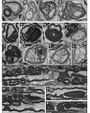
Frabin/Fgd4-deficient mice form aberrant PNS myelin. Fgd4/ mice display aberrant myelin features during early steps of myelination (A–D: post-natal Day 5; sciatic nerve) and in myelin maintenance (E–G, I and K: 80 weeks old mice; H, J and M–P: 60 weeks old mice; L: 10 weeks old mice; plantaris nerve), including simple myelin outfoldings (A), redundant myelin (B), complex myelin outfoldings (E) and highly complex myelin outfoldings (C and D), redundant myelin loops outside (F) and protruding into the axon (G), degradation of myelin (I), signs of demyelination (K) and remyelination (H and J) and rarely polyaxonal myelination (L). Aberrant myelin features tend to be located in the vicinity of nodes of Ranvier and Schmidt–Lanterman incisures (M, N and P). (A–L) Cross-sections. (M–P) Longitudinal sections. Scale bars = 1 µm (A–L); 5 µm (M–P).
Figure 3
Figure 3
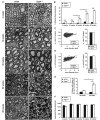
Aberrant development of PNS myelination in Fgd4/ mice. (A) Aberrant myelin structures of post-natal Day 5 (P5) sciatic nerves were categorized in four sets (simple, complex, very complex outfoldings and infoldings) and quantified at electron microscopy level. Very complex outfoldings were significantly increased in Fgd4/ animals. (B) Overall number of fibres showing aberrant myelin features was also increased in Frabin/Fgd4-deficient mice. (C) Total number of myelinated fibres was not significantly altered between Fgd4/ and wild-type (wt/wt) animals. Three mice per genotype were analysed. Scale bars = 1 µm. *P < 0.05, **P < 0.01 Student’s t-test (two-tailed).
Figure 4
Figure 4
Fgd4/ mice develop a progressive demyelinating neuropathy with aberrant myelin formation. (A) Transverse sections of plantaris nerves show a temporally progressive accumulation of aberrant myelin features in Fgd4/ mice compared with wild-type (wt/wt) mice aged 2–80 weeks. (B) Quantification of aberrant myelin features on entire transversal nerve reconstructions at electron microscopy level reveals a significant and progressive increase in the amount of affected fibres in Fgd4/ mice aged 2–80 weeks. (C and D) G-ratio is not significantly changed between wild-type and Fgd4/ mice aged 10 and 60 weeks (at least 100 fibres quantified per animal). (E) Signs of demyelination and remyelination accumulate significantly between the age of 60 and 80 weeks in Fgd4/ mice, the difference to wild-type (wt/wt) mice reaching significance by 80 weeks. (F) No loss in the overall number of myelinated fibres was detectable up to the age of 80 weeks. Arrows indicate fibres displaying myelin alterations. Arrowhead indicates demyelinated fibre. Scale bars = 5 µm; *P < 0.05, **P < 0.01, ***P < 0.001; Student’s t-test (two-tailed). Three animals were analysed for each genotype per time point.
Figure 5
Figure 5
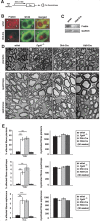
Cell type-specific gene ablation reveals that Schwann cells require Frabin/Fgd4 for proper myelination. (A) Schwann cell- or motor neuron-specific ablation of Fgd4 by Dhh- or Hb9-gene regulatory elements-driven Cre recombinase (Dhh- or Hb9-Cre/Fgd4 flox/flox animals) results in B, Schwann cell-specific (Dhh-Cre) loss of detectable Frabin/Fgd4 on cryosections of sciatic nerves (white arrows mark Schwann cells, arrowheads mark axons) or (C) strongly reduced Frabin/Fgd4 expressed by motor neurons (Hb9-Cre) as shown by western blot analysis of mutant ventral roots lysates compared with wild-type (wt/wt). (D) Schwann cell-specific loss of Frabin/Fgd4 (Dhh-Cre) in plantaris nerves of 60-week-old mice or quadriceps and saphenous nerve of 30-week-old mice leads to aberrant myelin formation, similar to that seen in Fgd4/ mice (white arrows mark aberrant myelin features). Motor neuron-specific ablation of Frabin/Fgd4 (Hb9-Cre), however, does not result in a detectable pathological phenotype at electron microscopy level in plantaris, quadriceps or saphenous nerves at the corresponding ages. (E) Quantification of aberrant myelin features shown in D. Note the similar numbers of fibres with aberrant myelin features (affected fibres) present in DhhCre/Fgd4 flox/flox mice (Dhh-Cre) compared with Fgd4/ mice. Both are significantly increased compared with wild-type mice (wt/wt). The numbers in Hb9Cre/Fgd4 flox/flox mice (Hb9-Cre), however, were not different from wild-type mice. Total numbers of myelinated fibres were not changed in all genotypes. Three mice were analysed for each genotype, time point and type of nerve. Scale bars = 5 µm; n.s. = not significant. *P > 0.05, **P < 0.01, ***P < 0.001; Student’s t-test (two-tailed). GADPH = glyceraldehyde-3-phosphate dehydrogenase.
Figure 6
Figure 6
Inducible Schwann cell-specific gene ablation reveals that myelin maintenance depends on Frabin/Fgd4. (A) Tamoxifen-mediated induction of Frabin/Fgd4 ablation in 10-week-old Fgd4 flox/flox mice through activation of Plp promotor-driven Cre recombinase (Plp-CreERT2) results in (B) Schwann cell-specific loss of Frabin/Fgd4 protein in peripheral nerves of adult mice, shown on cryosections of sciatic nerve, 4 months after tamoxifen injections (arrow: Schwann cell; arrowhead: axon). (C) Aberrant myelin formation in plantaris nerves of 30-week-old Plp-CreERT2/Fgd4 flox/flox (Plp-CreERT2) mice, tamoxifen-treated at 10 weeks of age as control mice, compared with age-matched Fgd4/ and wild-type (wt/wt) mice. (D) Quantification of myelinated fibres displaying aberrant myelin features (affected fibres) of identically obtained nerves as shown in C, revealing significantly increased numbers of affected fibres in Plp-CreERT2/Fgd4 flox/flox mice (Plp-CreERT2) compared with wild-type or tamoxifen-injected control mice. Comparison of Plp-CreERT2 with Fgd4/ mice shows only a slight reduction in affected fibres. Total numbers of myelinated fibres were unchanged between the groups. Three mice were analysed for each group in all experiments. Scale bars = 5 µm. White arrows indicate affected fibres. **P < 0.01, ***P < 0.001; Student’s t-test (two-tailed).
Figure 7
Figure 7
Ablation of Frabin/Fgd4 reduces activation of the RhoGTPase Cdc42 in peripheral nerves in vivo. (A and B) Western blot analyses demonstrating that total and active levels of AKT, ErbB2 receptor and JNK, and total levels of MBP and Dlg1, are not changed in sciatic nerve of Fgd4/ mice compared with wild-type (wt/wt) mice at the age of 10 weeks. (C and D) Active, but not total levels of Cdc42 are significantly reduced in sciatic nerves of adult Fgd4/ mice compared with age-matched wild-type mice. Tissues from four wild-type and Fgd4/ mice were analysed. ***P < 0.001; Student’s t-test (two-tailed). GADPH = glyceraldehyde-3-phosphate dehydrogenase.
Figure 8
Figure 8
Inducible gene ablation reveals that loss of Cdc42 in adult myelinating Schwann cells causes histopathological aberrations phenocopying loss of Frabin/Fgd4. (A) Tamoxifen-mediated induction of Cdc42 ablation in 10-week-old Cdc42 flox/flox mice through activation of Plp promotor-driven Cre recombinase (Plp-CreERT2) results in (B) strongly reduced Cdc42 protein as shown by western blot analysis of sciatic nerve lysates obtained 7 months post-tamoxifen injection. (C) Aberrant myelin formations, including outfoldings and redundant myelin, in sciatic nerves of 10 months old PlpCreERT2/Cdc42 flox/flox (Cdc42 mutant) mice (electron microscopy cross-sections). (D) Representative FIB-SEM-derived longitudinal section of Cdc42 mutant sciatic nerves prepared as in C (see also Supplementary Videos 1 and 2) showing myelin outfoldings in the vicinity of nodes of Ranvier. Scale bars = 5 µm. White arrows indicate fibres with aberrant myelin features.
Figure 9
Figure 9
Knock-down of Frabin/Fgd4 leads to reduced activation of the RhoGTPase Cdc42 and impaired endocytosis. (A and B) Active, but not total levels of Cdc42 are significantly reduced in RT4 cells transfected with small interfering RNA (siRNA) targeting Frabin/Fgd4 in comparison with control-transfected cells. Four independent experiments were quantified. (C) Frabin/Fgd4-silenced RT4 cells qualitatively display reduced transferrin uptake ability compared with control-transfected cells. (D) Fluorescence activated cell sorting quantifications of Frabin/Fgd4-silenced RT4 cells reveal significant reductions in levels of incorporated transferrin. Transferrin fluorescence levels in cells transfected with small interfering RNA (upper panel) or short hairpin RNA (lower panel) targeting Frabin/Fgd4 were compared with transferrin fluorescence levels in cells transfected with control small interfering RNA or short hairpin RNA. Three independent experiments were quantified with small interfering RNA-transfected cells and four independent experiments with short hairpin RNA-transfected cells. Scale bars = 10 µm; **P < 0.01; ***P < 0.001; Student’s t-test (two-tailed). GADPH = glyceraldehyde-3-phosphate dehydrogenase.

References

    1. Arber S, Han B, Mendelsohn M, Smith M, Jessell TM, Sockanathan S. Requirement for the homeobox gene Hb9 in the consolidation of motor neuron identity. Neuron. 1999;23:659–74. - PubMed
    1. Azzedine H, Bolino A, Taieb T, Birouk N, Di Duca M, Bouhouche A, et al. Mutations in MTMR13, a new pseudophosphatase homologue of MTMR2 and Sbf1, in two families with an autosomal recessive demyelinating form of Charcot-Marie-Tooth disease associated with early-onset glaucoma. Am J Hum Genet. 2003;72:1141–53. - PMC - PubMed
    1. Baets J, Deconinck T, De Vriendt E, Zimon M, Yperzeele L, Van Hoorenbeeck K, et al. Genetic spectrum of hereditary neuropathies with onset in the first year of life. Brain. 2011;134:2664–76. - PMC - PubMed
    1. Benninger Y, Thurnherr T, Pereira JA, Krause S, Wu X, Chrostek-Grashoff A, et al. Essential and distinct roles for cdc42 and rac1 in the regulation of Schwann cell biology during peripheral nervous system development. J Cell Biol. 2007;177:1051–61. - PMC - PubMed
    1. Boerkoel CF, Takashima H, Stankiewicz P, Garcia CA, Leber SM, Rhee-Morris L, et al. Periaxin mutations cause recessive Dejerine-Sottas neuropathy. Am J Hum Genet. 2001;68:325–33. - PMC - PubMed

Publication types

MeSH terms

Supplementary concepts