. 2012 Nov 21:9:278.
doi: 10.1186/1743-422X-9-278.

Isolation and molecular characterization of type I and type II feline coronavirus in Malaysia

Affiliations

Isolation and molecular characterization of type I and type II feline coronavirus in Malaysia

Alazawy Amer et al. Virol J. .

Abstract

Background: Feline infectious peritonitis virus (FIPV) and feline enteric coronavirus (FECV) are two important coronaviruses of domestic cat worldwide. Although FCoV is prevalent among cats; the fastidious nature of type I FCoV to grow on cell culture has limited further studies on tissue tropism and pathogenesis of FCoV. While several studies reported serological evidence for FCoV in Malaysia, neither the circulating FCoV isolated nor its biotypes determined. This study for the first time, describes the isolation and biotypes determination of type I and type II FCoV from naturally infected cats in Malaysia.

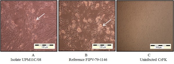
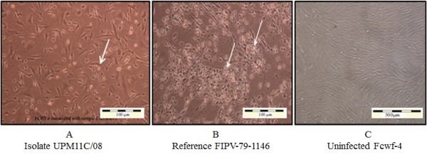
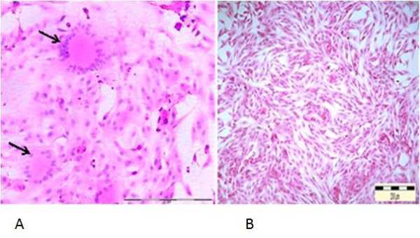
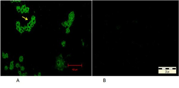
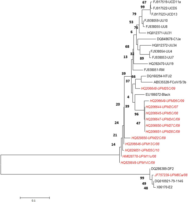
Findings: Of the total number of cats sampled, 95% (40/42) were RT-PCR positive for FCoV. Inoculation of clinical samples into Crandell feline kidney cells (CrFK), and Feline catus whole fetus-4 cells (Fcwf-4), show cytopathic effect (CPE) characterized by syncytial cells formation and later cell detachment. Differentiation of FCoV biotypes using RT-PCR assay revealed that, 97.5% and 2.5% of local isolates were type I and type II FCoV, respectively. These isolates had high sequence homology and phylogenetic similarity with several FCoV isolates from Europe, South East Asia and USA.

Conclusions: This study reported the successful isolation of local type I and type II FCoV evident with formation of cytopathic effects in two types of cell cultures namely the CrFK and Fcwf-4 , where the later cells being more permissive. However, the RT-PCR assay is more sensitive in detecting the antigen in suspected samples as compared to virus isolation in cell culture. The present study indicated that type I FCoV is more prevalent among cats in Malaysia.


Figures

Figure 1
Figure 1
CrFK-4 cell cultures showing cytopathic changes following infection with reference and local FCoV isolate UPM 11C/08. Morphology of cells infected with local FCoV UPM 11C/08 isolate at passage 3 (A). where the initial CPE was observed at passage 2. At fifth viral passages, complete CPE occurred within 48-72hours PI. Morphology of cells infected with reference FIPV-79-1146 strain (B). Infected cells show CPE characterized by cells rounding, clumped and detachment (arrows). Note that the changes in cells infected with UPM11C/08 are comparable to reference strain. Control uninfected cells remained normal (C). Unstained.
Figure 2
Figure 2
Fcwf-4 cell cultures showing cytopathic changes following infection with reference and local FCoV isolate UPM 11C/08. Morphology of cells infected with local FCoV UPM 11C/08 isolate at passage 3 (A). where the initial CPE was observed at passage 1. At third viral passages, complete CPE occurred within 24 hours PI. Morphology of cells infected with reference FIPV-79-1146 strain. (B). The infected cells show CPE characterized by cells rounding, clumped and detachment (arrows). Note that the changes in cells infected with UPM11C/08 are comparable to reference strain. Control uninfected cells remained normal (C). Unstained.
Figure 3
Figure 3
Syncytial cells formation following infection of Fcwf-4 cell culture with FCoV UPM 11C/08 isolate. Infected cells show loss of plasma membrane and nuclear aggregation, resulting in formation of syncytial cells containing more than 20 nuclei (arrows), 36 hours PI. 20x Mag. Scale bar, 100 μ m. Normal uninfected Fcwf-4 cell culture at 72 hours. H&E staining (A). Cells appeared as spindle to stellate morphology and regularly arranged. 10x Mag. Scale bar, 200 μm (B).
Figure 4
Figure 4
Immunofluorescent antibodies staining of infected CrFK cells following infection with local FCoV UPM11C/08 isolate. The fluorescence signal appeared as granules covering areas within the cytoplasm at 24 hours PI (arrow). No signal is present in the nucleus. 100x Mag. Scale bar, 20μm (A). Absence of fluorescence signal in control uninfected cell cultures. 10x Mag. Bar = 200μm (B).
Figure 5
Figure 5
Phylogenetic tree depicting relationships between Malaysian FCoV and reference FCoV isolates based on partial S gene sequence. The tree was generated using MEGA5 program. The tree reliability was calculated using 1000 bootstrapped replicates. Malaysian FCoVs are indicated in red italic.

References

    1. Addie DD, Jarrett JO. A study of naturally occurring feline coronavirus infection in kittens. Vet Rec. 1992;130:133–137. doi: 10.1136/vr.130.7.133. - DOI - PubMed
    1. Boyle JF, Pedersen NC, Evermann JF, McKeirnan AJ, Ott RL, Black JW. Plaque assay, polypeptide composition and immunochemistry of feline infectious peritonitis virus and feline enteric coronavirus isolates. Adv Exp Med Biol. 1984;173:133–147. doi: 10.1007/978-1-4615-9373-7_12. - DOI - PubMed
    1. Motokawa K, Hohdatsu T, Aizawa C, Koyama H, Hashimoto H. Molecular cloning and sequence determination of the peplomer protein gene of feline infectious peritonitis virus type I. Arch Virol. 1995;140:469–480. doi: 10.1007/BF01718424. - DOI - PMC - PubMed
    1. Pedersen NC. A review of feline infectious peritonitis virus infection: 1963–2008. J Feline Med Surg. 2009;11:225–258. doi: 10.1016/j.jfms.2008.09.008. - DOI - PMC - PubMed
    1. Arshad SS, Lee WW, Hassan L, Kamarudin AIM, Siti-Farawahida AW, Cheng NABY. Serological survey of catteries for cats infected with feline coronavirus. J Vet Malaysia. 2004;17:19–22.

Publication types

Substances