. 2013 Jan 18;288(3):1939-52.
doi: 10.1074/jbc.M112.428409. Epub 2012 Nov 21.

Melanoma antigen-A11 (MAGE-A11) enhances transcriptional activity by linking androgen receptor dimers

Affiliations

Melanoma antigen-A11 (MAGE-A11) enhances transcriptional activity by linking androgen receptor dimers

John T Minges et al. J Biol Chem. .

Abstract

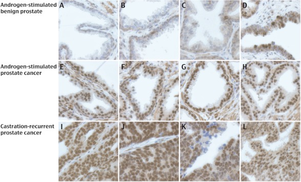
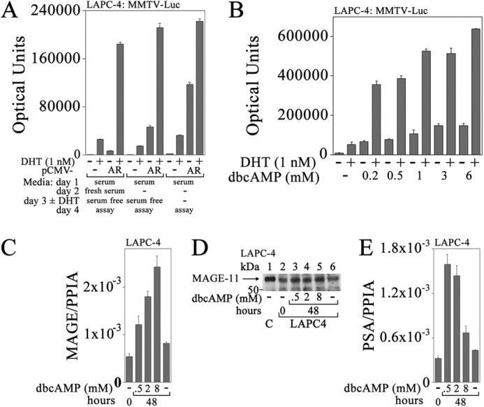
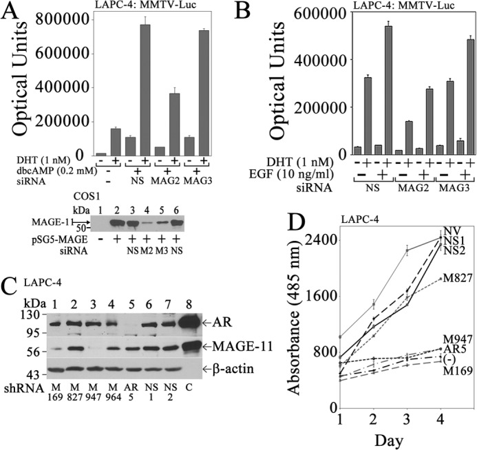
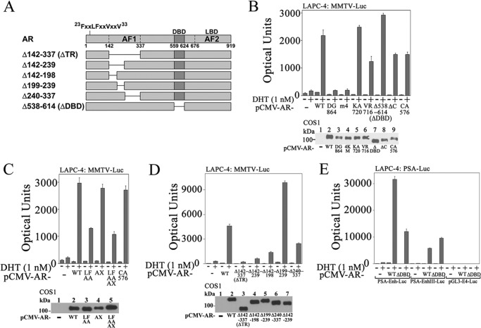
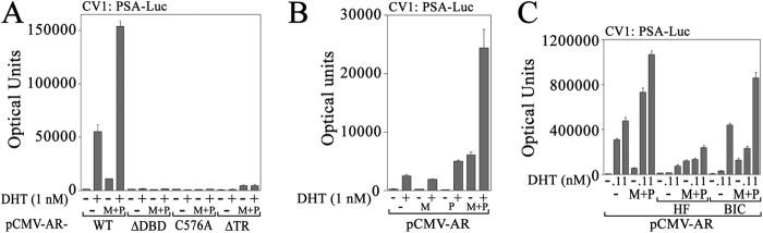
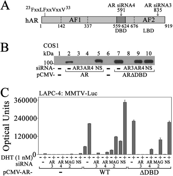
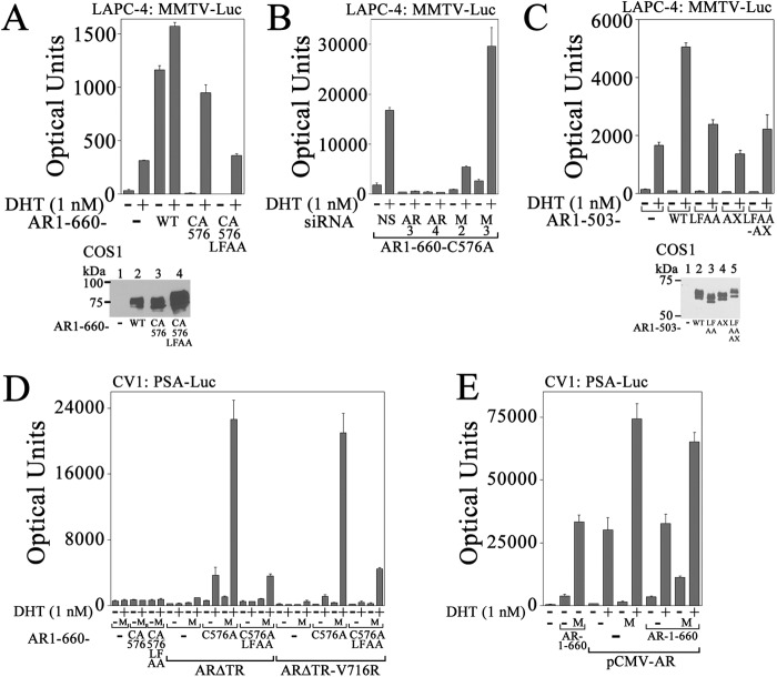
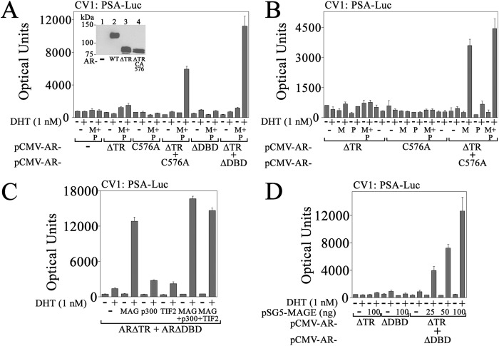
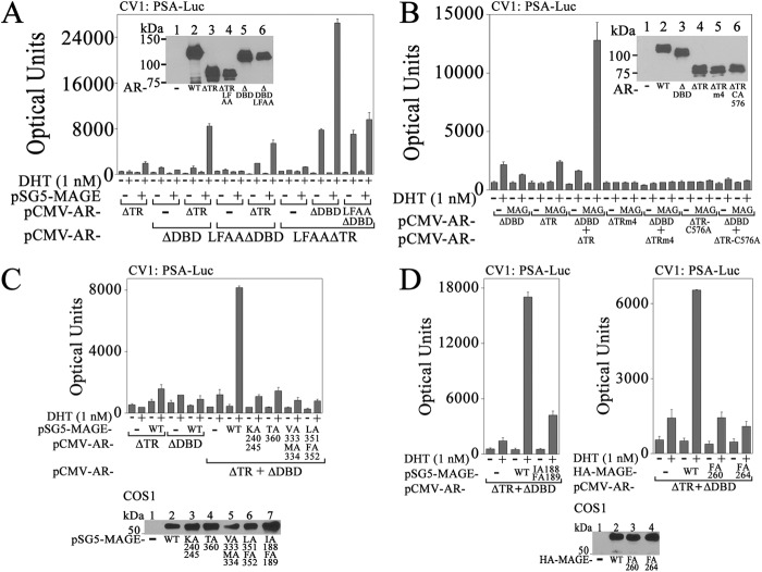
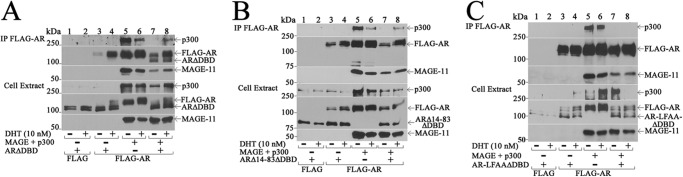
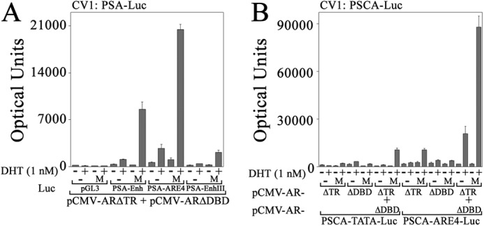
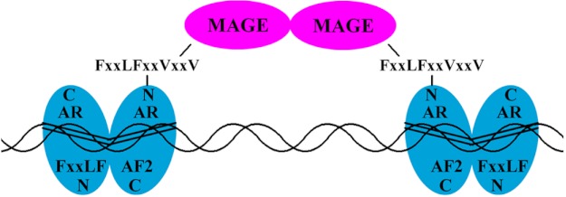
Prostate cancer growth and progression depend on androgen receptor (AR) signaling through transcriptional mechanisms that require interactions with coregulatory proteins, one of which is the primate-specific steroid receptor coregulator melanoma antigen-A11 (MAGE-A11). In this report, we provide evidence how increased expression of MAGE-A11 during prostate cancer progression enhances AR signaling and prostate cancer growth. MAGE-A11 protein levels were highest in castration-recurrent prostate cancer. The cyclic AMP-induced increase in androgen-dependent and androgen-independent AR transcriptional activity correlated with an increase in MAGE-A11 and was inhibited by silencing MAGE-A11 expression. MAGE-A11 mediated synergistic AR transcriptional activity in LAPC-4 prostate cancer cells. The ability of MAGE-A11 to rescue transcriptional activity of complementary inactive AR mutants and promote coimmunoprecipitation between unlike forms of AR suggests that MAGE-A11 links transcriptionally active AR dimers. A model for the AR·MAGE-A11 multidimeric complex is proposed in which one AR FXXLF motif of the AR dimer engages in the androgen-dependent AR NH(2)- and carboxyl-terminal interaction, whereas the second FXXLF motif region of the AR dimer interacts with dimeric MAGE-A11. The AR·MAGE-A11 multidimeric complex accounts for the dual functions of the AR FXXLF motif in the androgen-dependent AR NH(2)- and carboxyl-terminal interaction and binding MAGE-A11 and for synergy between reported AR splice variants and full-length AR. We conclude that the increased expression of MAGE-A11 in castration-recurrent prostate cancer, which is enhanced by cyclic AMP signaling, increases AR-dependent growth of prostate cancer by MAGE-A11 forming a molecular bridge between transcriptionally active AR dimers.


Figures

FIGURE 1.
FIGURE 1.
MAGE-A11 immunostaining in benign prostate and prostate cancer. MAGE-A11 was immunostained using 3 μg/ml MAGE-(94–108) antibody as described under “Experimental Procedures.” The four samples shown are representative of 17 specimens of androgen-stimulated benign prostate (A–D), 16 specimens of androgen-stimulated prostate cancer (E–H), and 11 specimens of castration-recurrent prostate cancer (I–L). The brown positive reaction product is shown against background staining using hematoxylin. Initial magnification, 400×.
FIGURE 2.
FIGURE 2.
Cyclic AMP-dependent increase in MAGE-A11 correlates with increased AR transactivation of PSA in LAPC-4 cells. A, empty vector pCMV5 (−) or pCMV-AR (0.1 μg) was expressed in LAPC-4 cells (2 × 10⁵/well) in 12-well plates with 0.2 μg of MMTV-Luc/well. The next day, medium was exchanged or unchanged, and 24 h later, it was replaced with serum-free medium or unchanged. Cells were then incubated for 24 h with or without 1 nm DHT, and luciferase activity was measured. B, LAPC-4 cells (4 × 10⁵/well) in 6-well plates were transfected using Lipofectamine 2000 with 0.25 μg of MMTV-Luc. The next day, cells were placed in serum-free medium with or without 0.2, 0.5, 1, 3, or 6 mm dibutyryl-cAMP (dbcAMP) with or without 1 nm DHT, and luciferase activity was measured. C, the day after plating LAPC-4 cells (2 × 10⁶/6-cm dish), cells were transferred to medium containing 10% charcoal-stripped serum, and the next day, cells were treated with or without 0.5, 2, or 8 mm dibutyryl-cAMP for 48 h. RNA was extracted and analyzed using quantitative RT-PCR for MAGE-A11 as described under “Experimental Procedures.” D, the day after plating LAPC-4 cells as in C, cells were transferred to medium containing 5% charcoal-stripped serum and treated with or without 0.5, 2, or 8 mm dibutyryl-cAMP for 48 h. Cells were harvested in immunoblot lysis buffer, and 40 μg of protein/lane was probed using FLAG-MAGE antibody (10 μg/ml). Lane 1 contains 0.1 μg of COS1 cell extract control (C) from cells expressing pSG5-MAGE. E, complementary DNA used in C was analyzed using quantitative RT-PCR for PSA as described under “Experimental Procedures.” PPIA, peptidylprolyl isomerase A. Data in A–C and E are the mean ± S.D. (error bars) and are representative of three independent experiments.
FIGURE 3.
FIGURE 3.
Requirement for MAGE-A11 in cyclic AMP-dependent AR transcriptional activity and LAPC-4 prostate cancer cell growth. A, top, LAPC-4 cells (3 × 10⁵/well) in 6-well plates were transfected with 0.25 μg of MMTV-Luc and 5 nm nonspecific (NS) or MAGE-A11 siRNA-2 or siRNA-3 using Lipofectamine 2000. The next day, cells were incubated for 24 h with or without 1 nm DHT and 0.2 mm dibutyryl-cAMP (dbcAMP) before assaying for luciferase. Bottom, empty vector pSG5 (−) (lane 1) or pSG5-MAGE (1 μg; lanes 2–5) was expressed in COS1 cells using Lipofectamine 2000 with or without a 5 nm concentration of two nonspecific siRNAs (NS), MAGE-A11 siRNA-2 (M2), or MAGE-A11 siRNA-3 (M3). Cell extracts (10 μg of protein/lane) were probed on the transblot using 10 μg/ml MAGE-(94–108) antibody incubated for 72 h at 4 °C. B, LAPC-4 cells were transfected with 0.5 μg of MMTV-Luc and 5 nm nonspecific siRNA (NS), MAGE-A11 siRNA-2, or MAGE-A11 siRNA-3. Cells were incubated with or without 1 nm DHT and 10 ng/ml EGF before assaying for luciferase. C, LAPC-4 cells (3 × 10⁶/well) in 6-well plates were transduced with 150 μl of lentivirus for MAGE-A11 shRNA-169 (M169), shRNA-827 (M827), shRNA-947 (M947), and shRNA-964 (M964), AR shRNA-5, empty vector, or 18-bp nonspecific (NS) shRNA. Cells were selected using puromycin as described under “Experimental Procedures.” Cell extracts (100 μg of protein/lane) were analyzed on immunoblots using AR32 (1 μg/ml) and AR52 (10 μg/ml) antibodies, 10 μg/ml of FLAG-MAGE antibody, 5 μg/ml MAGE-(94–108) antibody, and a 1:5000 dilution of β-actin antibody. Lane 8 contains extracts of COS1 cells expressing pCMV-AR (5 μg of protein/lane) or pSG5-MAGE (0.1 μg of protein/lane) as control (C). D, LAPC-4 cells transduced with lentivirus and selected as in C using puromycin were grown in the presence of 0.1 nm DHT and 10 ng/ml EGF in 24-well plates. Shown also is the no-hormone control of LAPC-4 cells transduced with nonspecific lentivirus shRNA and grown in the absence of DHT and EGF (−). Cell growth was assayed as described under “Experimental Procedures.” NV, no virus. Data in A, B, and D are the mean ± S.D. (error bars) representative of at least three independent experiments.
FIGURE 4.
FIGURE 4.
AR requirements for transcriptional activity in LAPC-4 cells. A, schematic diagram of AR deletion mutants expressed in LAPC-4 cells. LBD, ligand binding domain. B, top, pCMV5 (−) or pCMV-AR wild-type (WT) or mutants (0.1 μg) were expressed in LAPC-4 cells with 25 ng of MMTV-Luc. Cells were incubated with or without 1 nm DHT. Bottom, pCMV5 (−) or pCMV-AR WT or mutants (5 μg) were expressed in COS1 cells. The transblot of cell extracts (15 μg of protein/lane) was probed using AR32 antibody. C, top, pCMV5 (−) or pCMV-AR WT or mutants (0.1 μg) were expressed in LAPC-4 cells with 25 ng of MMTV-Luc. Cells were incubated with or without 1 nm DHT. Bottom, pCMV5 (−) or pCMV-AR WT or mutants (5 μg) were expressed in COS1 cells as in B. The transblot of cell extracts (15 μg of protein/lane) was probed using AR32 antibody. D, top, pCMV5 (−) or pCMV-AR WT or AR deletion mutants (0.1 μg) were expressed in LAPC-4 cells with 25 ng of MMTV-Luc. Cells were incubated with or without 1 nm DHT. Bottom, pCMV5 (−) or pCMV-AR WT or AR deletion mutants (6 μg) were expressed in COS1 cells. The transblot of cell extracts (20 μg of protein/lane) was probed using AR32 antibody. E, pCMV5 (−), pCMV-AR WT, or pCMV-ARΔDBD (0.1 μg) was expressed in LAPC-4 cells with 0.1 μg of PSA-Enh-Luc, PSA-Enh-SIII-Luc, or pGL3-E4-Luc. Cells were incubated with or without 1 nm DHT. Shown in B–E are the mean ± S.D. (error bars) representative of three independent experiments.
FIGURE 5.
FIGURE 5.
AR requirements for transactivation in CV1 cells. A, pCMV-AR WT, ΔDBD, C576A, or ΔTR (25 ng) was expressed in CV1 cells with 0.25 μg of PSA-Enh-Luc with or without 0.1 μg of pSG5 (−) or 50 ng of pSG5-MAGE (M) and 50 ng of pSG5-HA-p300 (P). Cells were incubated with or without 1 nm DHT. B, pCMV-AR (50 ng) was expressed in CV1 cells (3 × 10⁴/well) in 12-well plates with 0.1 μg of PSA-Enh-Luc and 0.1 of μg pSG5 (−) or 50 ng of pSG5-MAGE (M) and/or 50 ng of pSG5-HA-p300 (P). Cells were incubated with or without 1 nm DHT. C, CV1 cells were plated in phenol red-free medium containing 5% charcoal-stripped fetal calf serum, and the medium was not changed. pCMV-AR (25 ng) was expressed with 0.25 μg of PSA-Enh-Luc and 0.1 μg of pSG5 (−) or 50 ng of pSG5-MAGE (M) and 50 ng of pSG5-HA-p300 (P). Twenty-four hours before harvest, DHT was added to 0.1 or 1 nm with or without 0.5 μm hydroxyflutamide (HF) or 1 μm bicalutamide (BIC). Data shown are mean ± S.D. (error bars) representative of three independent experiments.
FIGURE 6.
FIGURE 6.
Dependence of ARΔDBD activity on endogenous AR and MAGE-A11 in LAPC-4 cells. A, schematic diagram of human AR (hAR) with the 23FXXLFXXVXXV33 interaction site for MAGE-A11, AF1, DBD, ligand binding domain (LBD), and AF2. The target position is indicated for AR siRNA-3 in the ligand binding domain and for AR siRNA-4 in the DBD. B, pCMV5 (−) (lanes 1 and 6), pCMV-AR (lanes 2–5), or pCMV-ARΔDBD (lanes 7–10) (0.5 μg) was expressed in COS1 cells using Lipofectamine 2000 with or without 10 nm AR siRNA-3, AR siRNA-4, or nonspecific (NS) siRNA. Cells were incubated without DHT, and cell extracts (15 μg of protein/lane) were probed on transblots using AR32 antibody. C, pCMV5 (−) or pCMV-AR WT or ΔDBD (25 ng) was expressed in LAPC-4 cells (4 × 10⁵/well) in 6-well plates using Lipofectamine with 25 ng of MMTV-Luc with or without 10 nm AR siRNA-3 (AR3), AR siRNA-4 (AR4), MAGE-A11 siRNA-2 (MAG2), or nonspecific (NS) siRNA. Cells were incubated with or without 1 nm DHT. Data are the mean ± S.D. (error bars) and are representative of three independent experiments.
FIGURE 7.
FIGURE 7.
Cooperativity between endogenous AR and AR-(1–660) in LAPC-4 cells. A, top, pCMV5 (−) or pCMV-AR-(1–660) WT, C576A, or L26A,F27A (LFAA)-C576A (0.1 μg) was expressed with 25 ng of MMTV-Luc in LAPC-4 cells. Cells were incubated with or without 1 nm DHT. Bottom, pCMV5 (−) or pCMV-AR-(1–660) WT, C576A, or LFAA-C576A (5 μg) was expressed in COS1 cells. Cell extracts (20 μg of protein/lane) were probed on transblots using AR32 antibody. B, pCMV-AR-(1–660)-C576A (25 ng) was expressed in LAPC-4 cells using Lipofectamine 2000 with 25 ng of MMTV-Luc and 5 nm nonspecific (NS) siRNA, AR siRNA-3 (AR3), AR siRNA-4 (AR4), MAGE-A11 siRNA-2 (M2), or MAGE-A11 siRNA-3 (M3). Cells were incubated with or without 1 nm DHT. C, top, pCMV5 (−) or pCMV-AR-(1–503) WT, LFAA, W433A,L436A,F437A (AX), or LFAA-AX combined mutant (0.1 μg) was expressed in LAPC-4 cells with 25 ng of MMTV-Luc. Cells were incubated with or without 1 nm DHT. Bottom, pCMV5 (−) or pCMV-AR-(1–503) WT, LFAA, AXXAA (AX), or combined mutant (5 μg) was expressed in COS1 cells. The transblot of cell extracts (10 μg of protein/lane) was probed using AR32 antibody. D, pCMV5 (−) or pCMV-AR-(1–660)-C576A or -AR-(1–660)-LFAA-C576A (10 ng) was expressed with 0.25 μg of PSA-Enh-Luc with or without 25 ng of pCMV-ARΔTR or ARΔTR-V716R with 50 ng of pSG5 (−) or pSG5-MAGE (M). Cells were incubated with or without 1 nm DHT. E, 10 ng of pCMV5 (−) or 2 ng of pCMV-AR-(1–660) was expressed in CV1 cells with 0.25 μg of PSA-Enh-Luc with or without 10 ng of pCMV-AR and 50 ng of pSG5 (−) or pSG5-MAGE (M). Cells were incubated with or without 1 nm DHT. Data in A–E are the mean ± S.D. (error bars) and are representative of three independent experiments.
FIGURE 8.
FIGURE 8.
MAGE-A11 rescues complementary inactive AR mutants. A, pCMV5 (−), pCMV-ARΔTR, -C576A, and/or -ΔDBD (25 ng) were expressed in CV1 cells with 0.25 μg of PSA-Enh-Luc and 0.1 μg of pSG5 (−) or 50 ng of pSG5-MAGE (M) and 50 ng of pSG5-HA-p300 (P). Cells were incubated with or without 1 nm DHT. Inset, pCMV5 (−) or pCMV-AR WT, ΔTR, or ΔTR-C576A (8 μg) was expressed in COS1 cells. Cells were incubated with 10 nm DHT. The transblot of cell extracts (20 μg of protein/lane) was probed using AR32 antibody. B, pCMV-ARΔTR and -AR-C576A alone (first dash, 25 ng; second dash, 50 ng) or ARΔTR and AR-C576A (25 ng each) were expressed in CV1 cells with 0.25 μg of PSA-Enh-Luc with or without 0.1 μg of pSG5 or 50 ng of pSG5-MAGE (M) and/or 50 ng of pSG5-HA-p300 (P). Cells were incubated with or without 1 nm DHT. C, pCMV-ARΔTR and -ARΔDBD (25 ng) were expressed in CV1 cells with 0.25 μg of PSA-Enh-Luc and 50 ng of pSG5 (−), pSG5-MAGE (MAG), pSG5-HA-p300, and/or pSG5-TIF2. Cells were incubated with or without 1 nm DHT. D, pCMV-ARΔTR and/or -ARΔDBD (25 ng) were expressed in CV1 cells with 0.25 μg of PSA-Enh-Luc and 100 ng of pSG5 (−) or 25, 50, or 100 ng of pSG5-MAGE. Cells were incubated with or without 1 nm DHT. Data in A–D are the mean ± S.D. (error bars) and are representative of three independent experiments.
FIGURE 9.
FIGURE 9.
Site-specific requirements for synergistic effects of AR and MAGE-A11. A, pCMV-ARΔTR, -ΔDBD, -LFAAΔDBD, and/or -LFAAΔTR (25 ng) were expressed in CV1 cells with 0.25 μg of PSA-Enh-Luc and 50 ng of pSG5 (−) or pSG5-MAGE. Cells were incubated with or without 1 nm DHT. Inset, pCMV5 (−) or pCMV-AR WT, ΔTR, LFAAΔTR, ΔDBD, or LFAAΔDBD (5 μg) was expressed in COS1 cells. Cells were incubated with 10 nm DHT. Cell extracts (20 μg of protein/lane) were probed on the transblot using AR32 antibody. B, pCMV-ARΔDBD, -ΔTR, -ΔTRm4, and/or -ΔTR-C576A (25 ng) was expressed in CV1 cells with 0.25 μg of PSA-Enh-Luc and 50 ng of pSG5 (−) or pSG5-MAGE (MAG). Cells were incubated with or without 1 nm DHT. Inset, pCMV5 (−) or pCMV-AR WT or mutants (5 μg) were expressed in COS1 cells. Cells were incubated with 2 nm DHT, and extracts (20 μg of protein/lane) were probed on transblots using AR32 antibody. C, top, pCMV-ARΔTR and -ARΔDBD (25 ng) were expressed alone or together with 0.25 μg of PSA-Enh-Luc and 50 of ng pSG5 (−) or pSG5-MAGE WT or mutants. Cells were incubated with or without 1 nm DHT. Bottom, pSG5 (−) or pSG5-MAGE WT or mutants (8 μg) were expressed in COS1 cells. Cell extracts (20 μg of protein/lane) were probed on the transblot using 0.5 μg/ml FLAG-MAGE antibody. D, top left and right, pCMV-ARΔTR and -ARΔDBD (25 ng) were expressed in CV1 cells with 0.25 μg of PSA-Enh-Luc and 50 ng of pSG5 (−), pSG5-MAGE WT or mutants (left), or pSG5-HA-MAGE WT or mutants (right). Cells were incubated with or without 1 nm DHT. Bottom, pSG5 (−) or pSG5-HA-MAGE WT or mutants (8 μg) were expressed in COS1 cells. Cell extracts (20 μg of protein/lane) were probed on transblots using HA antibody. Data in A–D are the mean ± S.D. (error bars) and are representative of three independent experiments.
FIGURE 10.
FIGURE 10.
MAGE-A11 links AR dimers. FLAG empty vector or FLAG-AR (2 μg) was expressed in COS1 cells with 2 μg of pCMV-ARΔDBD (A), pCMV-ARΔ14–83ΔDBD (B), or pCMV-AR-LFAAΔDBD (C) with or without 2 μg of pSG5-MAGE and 4 μg of pSG5-HA-p300. Cells were incubated in the presence of 0.1 μg/ml EGF with or without 10 nm DHT. The cell extract (20 μg of protein/lane) and immunoprecipitate (IP) transblots were probed using AR32, FLAG-MAGE, and p300 antibodies.
FIGURE 11.
FIGURE 11.
Synergy between AR and MAGE-A11 on multiple AREs. pCMV-ARΔTR and/or -ARΔDBD (25 ng) were expressed in CV1 cells with 50 ng of pSG5 (−) or pSG5-MAGE (M) and 0.25 μg of pGL3-E4-Luc, PSA-Enh-Luc, PSA-ARE4-Luc, and PSA-Enh-SIII-Luc (A) or 0.25 μg of PSCA-TATA-Luc or PSCA-ARE4-Luc (B). Cells were incubated with or without 1 nm DHT. Data are the mean ± S.D. (error bars) and are representative of three independent experiments.
FIGURE 12.
FIGURE 12.
Model of synergistic effects of MAGE-A11 bridging AR dimers. The AR dimer undergoes an androgen-dependent interaction between the NH2-terminal 23FXXLF27 motif and AF2 in the ligand binding domain. The AR N/C interaction is important for AR transactivation of androgen-dependent genes (67). MAGE-A11 forms a dimer (14) and interacts through its F box-like region with the extended 23FXXLFXXVXXV33 motif region in the AR dimer. The ability of MAGE-A11 to rescue transcriptional activity of complementary inactive AR mutants that lack activation function 1 or the DNA binding domain provides evidence that MAGE-A11 links AR dimers. This function of MAGE-A11 was supported by coimmunoprecipitation of unlike AR dimers that required MAGE-A11.

Similar articles

Cited by

References

    1. Askew E. B., Gampe R. T., Jr., Stanley T. B., Faggart J. L., Wilson E. M. (2007) Modulation of androgen receptor activation function 2 by testosterone and dihydrotestosterone. J. Biol. Chem. 282, 25801–25816 - PMC - PubMed
    1. Langley E., Zhou Z. X., Wilson E. M. (1995) Evidence for an antiparallel orientation of the ligand activated human androgen receptor dimer. J. Biol. Chem. 270, 29983–29990 - PubMed
    1. He B., Kemppainen J. A., Wilson E. M. (2000) FXXLF and WXXLF sequences mediate the NH2-terminal interaction with the ligand binding domain of the androgen receptor. J. Biol. Chem. 275, 22986–22994 - PubMed
    1. Bai S., He B., Wilson E. M. (2005) Melanoma antigen gene protein MAGE-11 regulates androgen receptor function by modulating the interdomain interaction. Mol. Cell. Biol. 25, 1238–1257 - PMC - PubMed
    1. Su S., Blackwelder A. J., Grossman G., Minges J. T., Yuan L., Young S. L., Wilson E. M. (2012) Primate-specific melanoma antigen-A11 regulates isoform-specific human progesterone receptor-B transactivation. J. Biol. Chem. 287, 34809–34824 - PMC - PubMed

Publication types

MeSH terms