

Choroid plexus papillomas: advances in molecular biology and understanding of tumorigenesis
- PMID: 23172371
- PMCID: PMC3578480
- DOI: 10.1093/neuonc/nos289
Choroid plexus papillomas: advances in molecular biology and understanding of tumorigenesis
Abstract
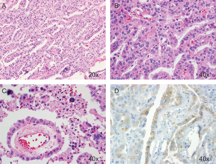
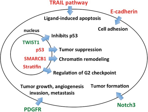
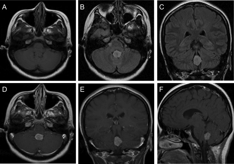
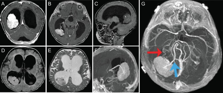
Choroid plexus papillomas are rare, benign tumors originating from the choroid plexus. Although generally found within the ventricular system, they can arise ectopically in the brain parenchyma or disseminate throughout the neuraxis. We sought to review recent advances in our understanding of the molecular biology and oncogenic pathways associated with this disease. A comprehensive PubMed literature review was conducted to identify manuscripts discussing the clinical, molecular, and genetic features of choroid plexus papillomas. Articles concerning diagnosis, treatment, and long-term patient outcomes were also reviewed. The introduction of atypical choroid plexus papilloma as a distinct entity has increased the need for accurate histopathologic diagnosis. Advances in immunohistochemical staining have improved our ability to differentiate choroid plexus papillomas from other intracranial tumors or metastatic lesions using combinations of key markers and mitotic indices. Recent findings have implicated Notch3 signaling, the transcription factor TWIST1, platelet-derived growth factor receptor, and the tumor necrosis factor-related apoptosis-inducing ligand pathway in choroid plexus papilloma tumorigenesis. A combination of commonly occurring chromosomal duplications and deletions has also been identified. Surgical resection remains the standard of care, although chemotherapy and radiotherapy may be considered for recurrent or metastatic lesions. While generally considered benign, these tumors possess a complex biology that sheds insight into other choroid plexus tumors, particularly malignant choroid plexus carcinomas. Improving our understanding of the molecular biology, genetics, and oncogenic pathways associated with this tumor will allow for the development of targeted therapies and improved outcomes for patients with this disease.
Figures
References
-
- Wolburg H, Paulus W. Choroid plexus: biology and pathology. Acta Neuropathol. 2010;119(1):75–88. - PubMed
-
- Cataltepe O, Liptzin D, Jolley L, Smith TW. Diffuse villous hyperplasia of the choroid plexus and its surgical management. J Neurosurg Pediatr. 2010;5(5):518–522. - PubMed
-
- Janisch W, Staneczek W. [Primary tumors of the choroid plexus. Frequency, localization and age] Zentralbl Allg Pathol. 1989;135(3):235–240. - PubMed
-
- Rickert CH, Paulus W. Tumors of the choroid plexus. Microsc Res Tech. 2001;52(1):104–111. - PubMed
