. 2013 Feb;61(2):116-24.
doi: 10.1369/0022155412470455. Epub 2012 Nov 19.

Cell and tissue microarray technologies for protein and nucleic acid expression profiling

Affiliations

Cell and tissue microarray technologies for protein and nucleic acid expression profiling

Marina Cardano et al. J Histochem Cytochem. 2013 Feb.

Abstract

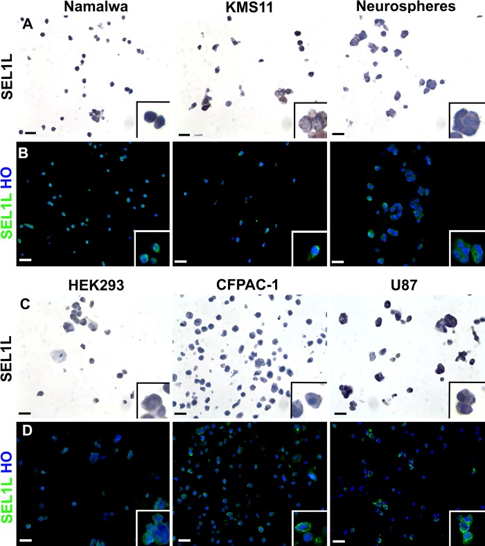
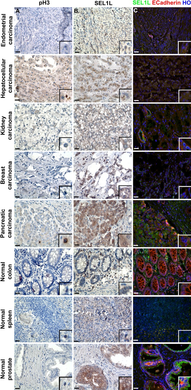
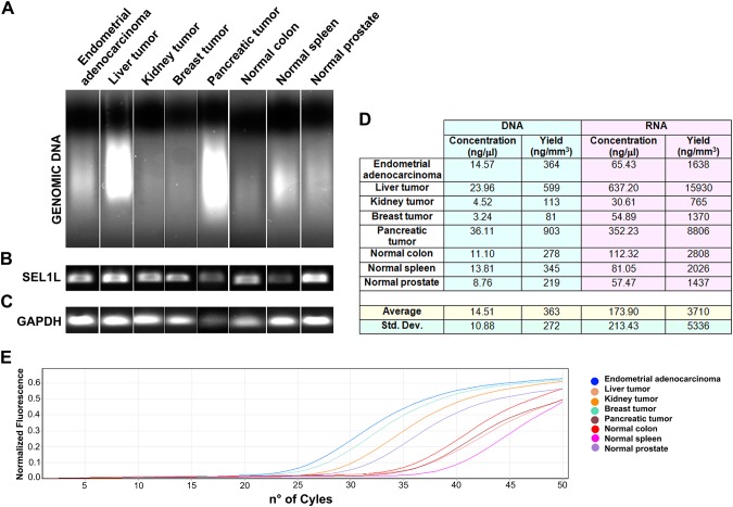
Tissue microarray (TMA) and cell microarray (CMA) are two powerful techniques that allow for the immunophenotypical characterization of hundreds of samples simultaneously. In particular, the CMA approach is particularly useful for immunophenotyping new stem cell lines (e.g., cardiac, neural, mesenchymal) using conventional markers, as well as for testing the specificity and the efficacy of newly developed antibodies. We propose the use of a tissue arrayer not only to perform protein expression profiling by immunohistochemistry but also to carry out molecular genetics studies. In fact, starting with several tissues or cell lines, it is possible to obtain the complete signature of each sample, describing the protein, mRNA and microRNA expression, and DNA mutations, or eventually to analyze the epigenetic processes that control protein regulation. Here we show the results obtained using the Galileo CK4500 TMA platform.


Conflict of interest statement

Declaration of Conflicting Interests: The authors declared a potential conflict of interest (e.g. a financial relationship with the commercial organizations or products discussed in this article) as follows: Pasquale DeBlasio and Maurizio Falavigna work in the company that makes the TMA.

Figures

Figure 1.
Figure 1.
Immunocharacterization of six cell lines by cell microarray technology. After cell collection, pellets deriving from six different cell lines were resuspended in agarose and then embedded in paraffin. Two consecutive sections were analyzed for SEL1L expression by immunohistochemistry (A, C) and immunofluorescence (B, D) techniques (using the monoclonal antibody described by Orlandi et al. 2003), showing the typical perinuclear/cytoplasmic subcellular location of this protein. In the immunohistochemical images, the brown color reflects SEL1L expression and nuclei were counterstained with hematoxylin; differently, in immunofluorescence analysis, SEL1L is represented in green and nuclei in blue (counterstaining with Hoechst 33258, HO). Scale bars = 10 µm.
Figure 2.
Figure 2.
Immunophenotypical analysis of tissue microarray (TMA) slices to identify nuclear, cytoplasmic, and membrane antigens. Five different tumoral and three normal tissues were used to construct a TMA, from which three sequential slices were analyzed for pH3 (A) and SEL1L (B; monoclonal antibody described by Orlandi et al. 2002) expression by immunohistochemistry and concurrently for SEL1L and E-cadherin (C) by immunofluorescence. The immunohistochemical figures show SEL1L and H3 proteins in brown, whereas nuclei were counterstained with hematoxylin. In the immunofluorescence study, SEL1L is depicted in green, E-cadherin in red, and nuclei in blue (counterstaining with Hoechst 33258, HO). Scale bars = 25 µm.
Figure 3.
Figure 3.
Nucleic acids extraction from tissues cored by the Galileo CK4500 Arrayer. Tissue cores were processed to extract both DNA (A) and RNA, showing good yields, but with an expected variability depending on the tissue quality and cellular content (D). The DNA can be used to amplify genes of interest (B), whereas the total RNA is suitable for RT-PCR (C) or to analyze the expression of specific microRNAs (E).

References

    1. Abrahamsen HN, Steiniche T, Nexo E, Hamilton-Dutoit SJ, Sorensen BS. 2003. Towards quantitative mRNA analysis in paraffin-embedded tissues using real-time reverse transcriptase polymerase chain reaction: a methodological study on lymphnodes from melanoma patients. J Mol Diagn. 5:34–41 - PMC - PubMed
    1. Andersson AC, Strömberg S, Bäckvall H, Kampf C, Uhlen M, Wester K, Pontén F. 2006. Analysis of protein expression in cell microarrays: a tool for antibody-based proteomics. J Histochem Cytochem. 54(12):1413–1423 - PMC - PubMed
    1. Ashktorab H, Green W, Finzi G, Sessa F, Nouraie M, Lee EL, Morgano A, Moschetta A, Cattaneo M, Mariani-Costantini R, et al. 2012. SEL1L, an UPR response protein, a potential marker of colonic cell transformation. Dig Dis Sci. 57(4):905–912 - PMC - PubMed
    1. Auerbach C, Moutschen-Dahmen M, Moutschen J. 1977. Genetic and cytogenetical effects of formaldehyde and related compounds. Mutat Res. 39(3–4):317–361 - PubMed
    1. Benavides J, García-Pariente C, Gelmetti D, Fuertes M, Ferreras MC, García-Marín JF, Pérez V. 2006. Effects of fixative type and fixation time on the detection of Maedi Visna virus by PCR and immunohistochemistry in paraffin-embedded ovine lung samples. J Virol Methods. 137(2):317–324 - PubMed

Publication types