. 2013 Aug;70(2):527-36.
doi: 10.1002/mrm.24480. Epub 2012 Nov 21.

Noncontrast-enhanced renal angiography using multiple inversion recovery and alternating TR balanced steady-state free precession

Affiliations

Noncontrast-enhanced renal angiography using multiple inversion recovery and alternating TR balanced steady-state free precession

Hattie Z Dong et al. Magn Reson Med. 2013 Aug.

Abstract

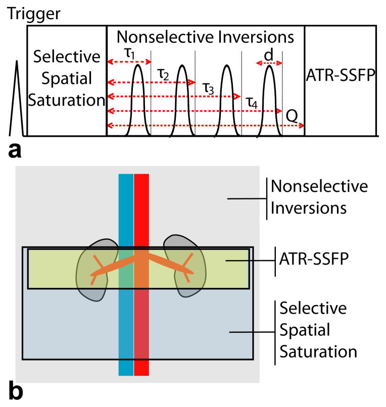
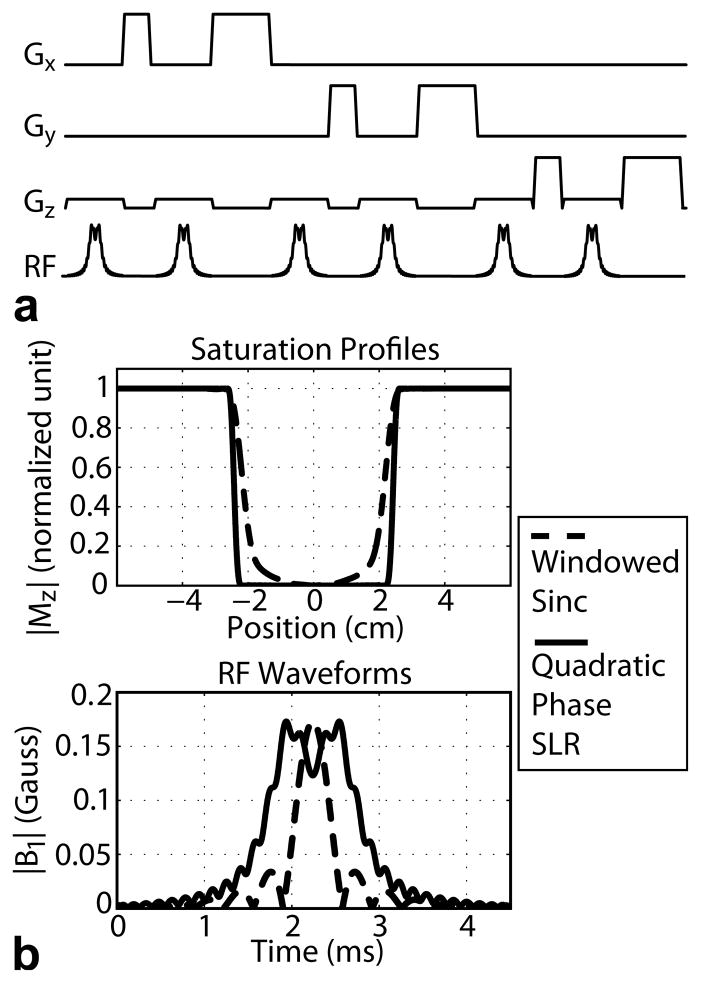
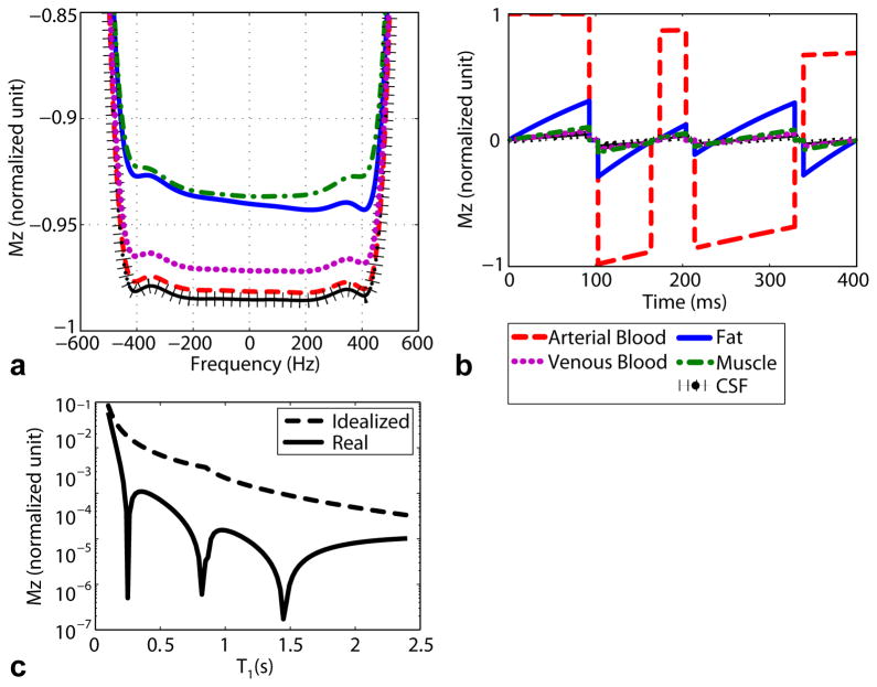
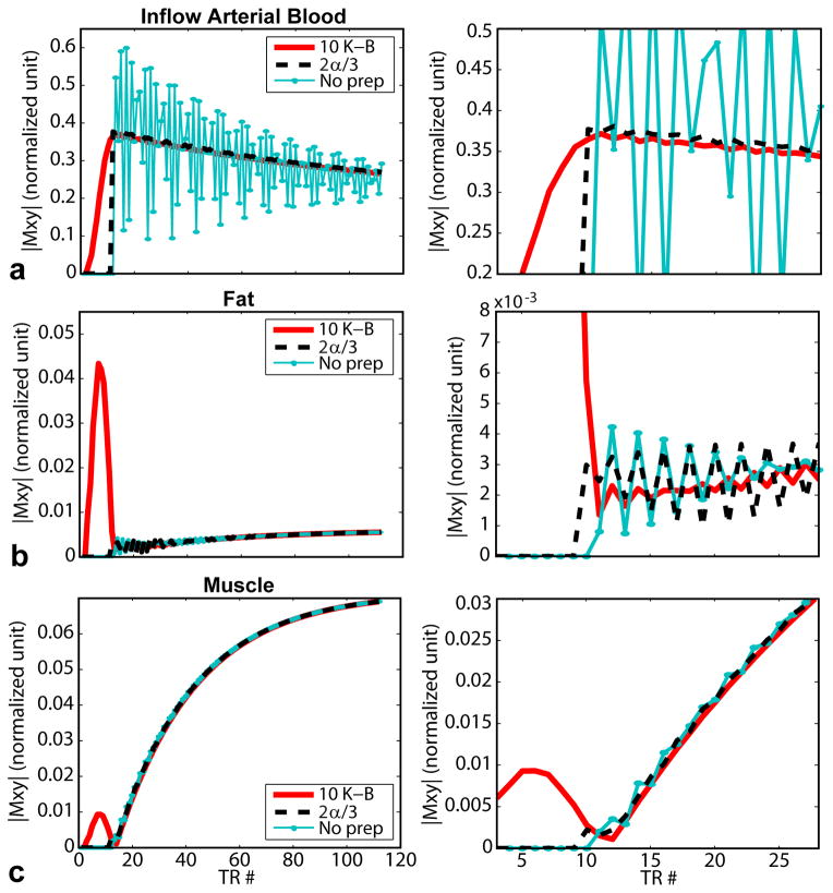
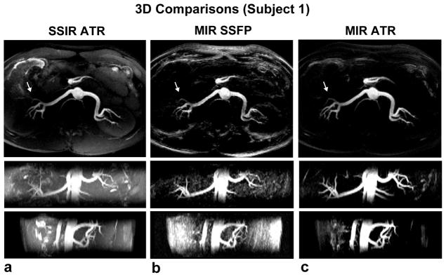
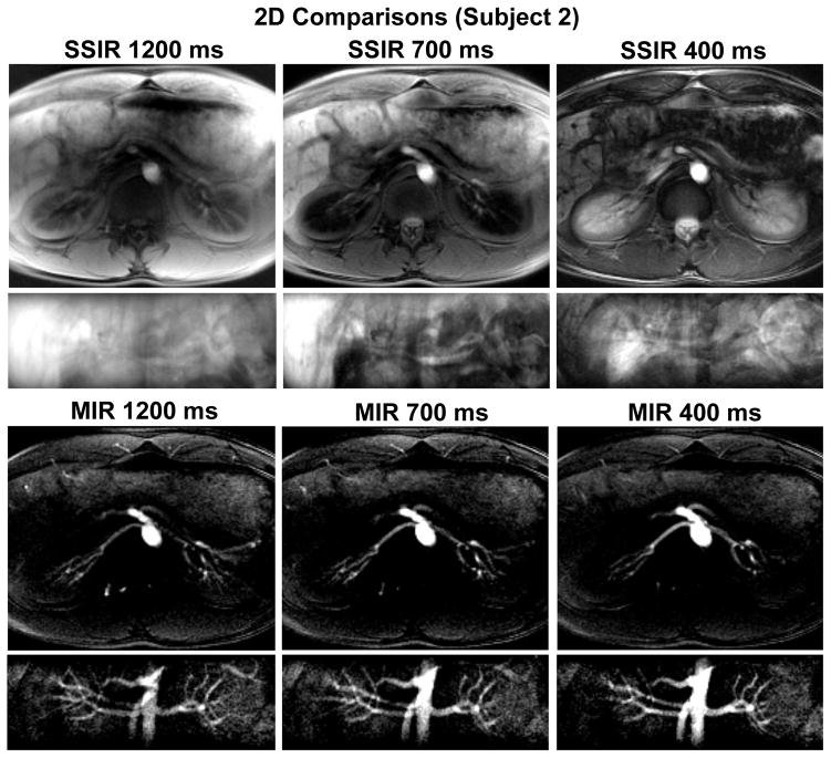
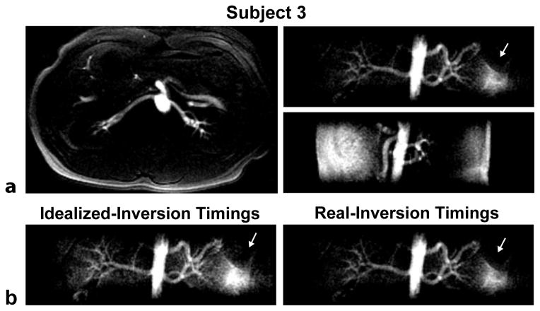
Noncontrast-enhanced renal angiography techniques based on balanced steady-state free precession avoid external contrast agents, take advantage of high inherent blood signal from the T 2 / T 1 contrast mechanism, and have short steady-state free precession acquisition times. However, background suppression is limited; inflow times are inflexible; labeling region is difficult to define when tagging arterial flow; and scan times are long. To overcome these limitations, we propose the use of multiple inversion recovery preparatory pulses combined with alternating pulse repetition time balanced steady-state free precession to produce renal angiograms. Multiple inversion recovery uses selective spatial saturation followed by four nonselective inversion recovery pulses to concurrently null a wide range of background T 1 species while allowing for adjustable inflow times; alternating pulse repetition time steady-state free precession maintains vessel contrast and provides added fat suppression. The high level of suppression enables imaging in three-dimensional as well as projective two-dimensional formats, the latter of which has a scan time as short as one heartbeat. In vivo studies at 1.5 T demonstrate the superior vessel contrast of this technique.

Keywords: SSFPangiography; angiography; multiple inversion recovery; noncontrast-enhanced angiography; projective imaging.


Figures

Figure 1
Figure 1
a) MIR pulse sequence. Spatial saturation resets magnetization. Four nonselective inversions suppress a range of background T1 species, specifically fat, muscle, venous blood, and CSF. ATR-SSFP maintains arterial inflow contrast and suppresses fat. τi = time between the end of spatial saturation and end of the ith inversion; d = adiabatic inversion pulse duration; Q = inflow time. b) The spatial saturation, inversions, and imaging regions of interest.
Figure 2
Figure 2
a) The spatial saturation module consists of six quadratic phase RF pulses interspersed with dephasing gradients of different areas and placed on different axes, to achieve a well-defined spatial profile with maximal signal saturation. b) Quadratic-phase RF pulse (20) designed using the Shinnar-Le Roux framework (21). The pulse achieves a sharper saturation profile (transition width 4 times narrower) than conventional windowed-sinc pulses, while keeping the same peak RF power.
Figure 3
Figure 3
Simulations of suppression levels achievable using MIR preparation. a) An example of relaxation effects during the inversion pulse, for Mz = 1 at the start of the pulse. In Equation 1, k = (Mz at end of inversion pulse)/(Mz at start of inversion pulse). Hence in this case, karterial blood = −0.982, kvenous blood = −0.972, kfat = −0.936, kmuscle = −0.937, and kCSF = −0.985. kfat was obtained at −220 Hz while others were obtained at 0 Hz. Prior to solving Equation 1, we calculated k values corresponding to the different starting Mz’s using Bloch simulation. b) Mz progression over an inflow period of 400 ms with inversions timed at 103, 174, 214, and 339 ms after spatial saturation. At the end of the inflow period, background materials are suppressed while inflow blood retains 69% of its original signal, which is consistent with Equations 1 and 4. c) Suppression level vs. T1 values for idealized- and real-inversion timings. Using real-inversion timings, calculated taking into account inversion pulse duration and k, led to much improved suppression levels. Further, optimizing for the 4 background species achieved good suppression across a wide range of T1 materials.
Figure 4
Figure 4
Simulations of transverse magnetization progression during various ATR-SSFP catalyst periods and the first 100 imaging TRs (inflow time = 400 ms; TE/TR1/TR2 = 1.72/3.44/1.16 ms; TR = TR1 + TR2; flip angle = 50°). TR #11 is the first imaging TR, timed to play out at the end of the inflow period. Zoomed out (left) and zoomed in (right) plots of three species are shown: a) inflow arterial blood, b) fat, and c) muscle. (CSF was also simulated but is not shown.) Kaiser-Bessel catalysts show the best performance with little difference across different numbers of catalyst TRs. (8–12 K-B catalysts were simulated but not all are shown.) Background suppression remains high even after 100 TRs.
Figure 5
Figure 5
Axial, targeted coronal and targeted sagittal MIP renal angiograms using three 3D methods: a) SSIR ATR-SSFP (slab inversion time = 325 ms), b) MIR SSFP without ATR, and c) MIR ATR-SSFP (inflow time = 350 ms, inversion timings = 87, 166, 206, 300 ms). Images acquired using MIR ATR-SSFP show improved depiction of the renal arteries and distal branches (arrows) afforded by greater background suppression than the other two methods. The improved contrast is especially visible in the coronal and sagittal views, as large portions of background materials were present along these intensity projection directions. Scan time was 2:17 minutes for all scans based on 14 breaths per minute.
Figure 6
Figure 6
Axial and coronal projective 2D renal angiograms using SSIR SSFP and MIR ATR-SSFP for inflow times of 1200 ms, 700 ms, and 400 ms. These images illustrate the need for a very high level of background suppression in projective imaging, and that MIR is a suitable candidate. In SSIR SSFP, the renal arteries are heavily confounded by background signals and are nearly indecipherable. The 700-ms inflow time yielded the best suppression of the kidneys, but other background materials still obscured the renal arteries. With MIR ATR-SSFP, good background suppression is consistently achieved across all inflow times. Arterial signal visibly increases as inflow time decreases as expected, i.e. according to Equation 4, inflow blood retains 35%, 54%, and 69% of M0 for 1200, 700, and 400 milliseconds of inflow duration. For all scans, scan time was four heartbeats per image for the axial orientation, and one heartbeat per image for the coronal orientation.
Figure 7
Figure 7
MIR ATR-SSFP projective 2D renal angiograms. a) Good background suppression and vessel contrast are obtained in all three of axial, coronal and sagittal orientations. Arrows indicate signal recovery of short-T1 materials in the bowels. Compared to the 3D MIR images, the 2D projections have visibly lower renal artery signals due to much shorter total readout times (~1 second vs. ~32 seconds) leading to lower SNR, and much larger slice thicknesses (65–320 mm vs. 2 mm) leading to greater background signal accumulation and dynamic range issues. b) Comparison between coronal acquisitions using inversion timings calculated assuming idealized inversions (“idealized-inversion timings”: inflow time = 300 ms; inversion timings = 41, 92, 154, 252 ms), and using inversion timings calculated taking into account inversion pulse’s duration and relaxation effects (“real-inversion timings”: inflow time = 300 ms; inversion timings = 60, 111, 151, 250 ms). The real-inversion timings led to better background suppression. Scan time was 4 heartbeats per image for the axial orientation, and 1 heartbeat per image for the coronal and sagittal orientations.

Similar articles

Cited by

References

    1. Safian RD, Textor SC. Renal-artery stenosis. N Engl J Med. 2001;344:431–442. - PubMed
    1. Prince MR, Narasimham DL, Stanley JC, Chenevert TL, Williams DM, Marx MV, Cho KJ. Breath-hold gadolinium-enhanced MR angiography of the abdominal aorta and its major branches. Radiology. 1995;197:785–792. - PubMed
    1. Hany TF, Debatin JF, Leung DA, Pfammatter T. Evaluation of the aortoiliac and renal arteries: comparison of breath-hold, contrast-enhanced, three-dimensional MR angiography with conventional catheter angiography. Radiology. 1997;204:357–362. - PubMed
    1. Schoenberg SO, Rieger J, Weber CH, Michaely HJ, Waggershauser T, Ittrich C, Dietrich O, Reiser MF. High-spatial-resolution MR angiography of renal arteries with integrated parallel acquisitions: comparison with digital subtraction angiography and US. Radiology. 2005;235:687–698. - PubMed
    1. Marckmann P, Skov L, Rossen K, Dupont A, Damholt MB, Heaf JG, Thomsen HS. Nephrogenic systemic fibrosis: suspected causative role of gadodiamide used for contrast-enhanced magnetic resonance imaging. J Am Soc Nephrol. 2006;17:2359–62. - PubMed

Publication types